21 Jan 2019
Tim Mair discusses the evaluation of horses showing clinical signs, challenges that can arise when choosing the appropriate treatment, and the need to promptly refer surgical cases.
Tim Mair
Job Title
Figure 1. Colic is one of the most common reasons for emergency visits.

Colic is one of the most common emergencies faced by equine practitioners (Figure 1).
For example, the caseload seen out-of-hours by two first-opinion equine practices during a three-year period (2011-13) identified 2,602 presentations – with the three most common reasons for emergency visits being colic (35%), wounds (20%) and lameness (11%; Bowden et al, 2017).
Although most colic cases encountered in the field will respond to simple medical treatments, a proportion – between 10% and 15% – will be affected by serious, life-threatening diseases. As a result, affected horses must be treated promptly – both for the welfare of the animal and to ease the owner’s distress.
Although it can be difficult to reach a definitive diagnosis in every case, especially during the first visit, a thorough examination should be performed to determine what emergency treatments should be initiated and decide whether the horse requires surgery or intensive care.
Many horses with colic will recover spontaneously or respond to the initial treatment. For example, a study of colic episodes in Thoroughbred horses at training premises in the UK revealed 28.7% recovered spontaneously and 63.1% after medical treatment (Hillyer et al, 2001).
Since some horses will require surgery, making the decision about the need for surgery in a timely manner will increase the chance of a successful outcome, minimise patient morbidity and potentially decrease the risk of postoperative complications (Ragle, 1999). Early identification of critically ill horses requiring surgery and rapid implementation of appropriate treatments are paramount for their survival (Ragle, 1999).
Cardiovascular compromise indicative of responses to circulating endotoxins increases the risk for several postoperative complications after an acute abdominal crisis. Therefore, early referral of colic cases before shock develops may minimise the risk of some of these complications (French et al, 2002).
On the other hand, for horses that have no surgical option, recognition of serious diseases where no chance exists of survival without surgery is equally important, to enable euthanasia to be carried out in a timely and humane fashion.
During examination of a horse with colic in the field, the veterinary surgeon should decide whether appropriate treatment can be completed on the owner’s premises, or whether the horse should be transported to a hospital facility for further diagnostic procedures and/or treatment. In some cases, the requirement for intensive treatment/surgery/further diagnostic evaluation will be clear-cut – for example, the horse demonstrating severe pain and signs of shock. However, in many other cases the need for surgery/further evaluation will be less certain; in these cases, repeated evaluations over a period of time, as well as assessing the response to initial medical treatments, will be helpful.
It should be remembered some horses – especially older horses – can appear very stoic and may not demonstrate the signs of severe pain other horses would be expected to show. Additionally, in certain advanced diseases – including strangulating obstructions of the small intestine or small colon – depression may replace pain as the primary sign.
If uncertainty surrounds the need for surgery or the horse’s condition, the horse should be referred to a hospital where the decision for surgery can be made by a veterinary surgeon who makes these decisions frequently (Desrochers and White, 2017).
While the significant improvements in survival rates after colic surgery in the past four decades may be due to advancements in all aspects of colic management, early referral of horses requiring surgery is one of the most important determinants of outcome (Freeman et al, 2014).
The horse’s signalment, as well as accurate information relating to its history, can be extremely helpful in evaluating those with colic. For example, a horse older than 10 to 15 years, with small intestinal distention, is at high risk of having a strangulating lipoma (Freeman and Schaeffer, 2001).
Young foals most commonly have meconium impaction and enteritis as a cause of colic. Shetland ponies and miniature horses are predisposed to small colon impaction and faecaliths (Dart et al, 1992), while standardbreds and some Warmblood breeds have an increased risk of inguinal/scrotal hernias (Schneider et al, 1982).
Three general areas of history need consideration:
For example, exposure to lush grass or grazing on sandy soils may predispose the horse to tympanic colic and sand impactions, respectively.
Changes in management or diet should be established. A sudden decrease in the level of exercise, for example, is commonly associated with pelvic flexure impactions.
Questions that should be asked as appropriate include (White, 1990):
The physical examination should be undertaken in a systematic fashion, paying particular attention to the gastrointestinal and cardiovascular systems, but remembering pain due to so-called “false colics” – such as pleuritis, rhabdomyolysis and laminitis – can sometimes be confused with colic.
The degree of pain the horse is showing is important to assess, in view of the fact more severe pain is more likely to be associated with serious, surgical conditions.
However, it should be noted some horses are more stoic than others, and horses with strangulating small intestinal and small colon conditions may pass from a state of severe pain to a state of depression without pain in the advanced stages of the disease. Acute rupture of the stomach or intestine can also cause a dramatic shift from severe pain to depression.
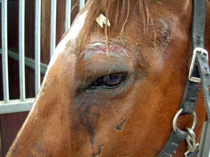
The presence of abrasions around the bony protuberances of the head and pelvis indicate previous severe pain (Figure 2). Classical signs of colic pain include restlessness, bruxism, pawing, stretching, flank watching, kicking at the abdomen, lying down in sternal or lateral recumbency, falling to the ground, and rolling. Horses with severe pain are often difficult to control, and the pain may only respond to analgesics for a few minutes or not at all; sedation with romifidine, detomidine or xylazine – possibly combined with butorphanol – can be helpful to control the pain in such cases to allow a safer examination of the patient.
Simple obstructions are usually characterised by mild to moderate intermittent pain. Most simple large colon obstructions are incomplete; some gas and fluid will pass around the obstruction. However, the horse may become more persistently or severely painful as the obstruction of the intestinal lumen becomes complete. Acute severe pain is often caused by strangulation or severe tympany (White, 1990).
A horse that has mild, intermittent pain or no pain, but has depression, may be suffering from a primary inflammatory disorder, such as peritonitis or enteritis (White, 1990). Therefore, the absence of severe pain does not indicate the horse does not have a severe illness.
If the horse is quiet or depressed, but has evidence of previous trauma and severe pain, the veterinary surgeon should be concerned about the possibility of an advanced strangulating obstruction, or rupture of either the stomach or caecum.
Sweating can also be a sign of severe pain, due to stimulation of the sympathetic nervous system or from endotoxic shock (White, 1990).
A bloated appearance indicates marked distention of the caecum, large colon or, possibly, the entire small intestine. Caecal distention typically results in a bloated right side, whereas a left dorsal displacement of the large colon may result in a bloated left side of the abdomen.
A miniature pony with generalised bloating may have a distended large colon secondary to complete obstruction of the small colon by an impaction or faecalith (Figure 3).

The heart rate of a normal adult horse is usually between 28bpm and 44bpm. The heart rate is an indicator of the severity of the disease and the degree of circulatory shock (Morton and Blikslager, 2002).
Heart rate has been shown to be an important prognostic indicator for survival (Furr et al, 1995; van der Linden et al, 2003) and for complications after colic surgery (French et al, 2002). However, heart rate can be unreliable and variable, and should always be interpreted in conjunction with the other clinical signs.
Pulse quality can be assessed by palpating the facial artery. A weak pulse may indicate low blood pressure secondary to hypovolaemic shock or systemic inflammatory response syndrome.
The colour and moistness of the oral and conjunctival mucous membranes are used to assess perfusion and the horse’s hydration status.
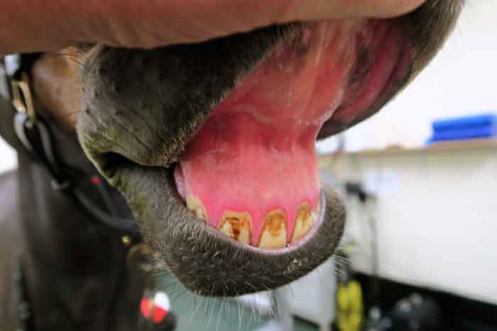
In hypovolaemic/shocked horses, mucous membranes may vary from pale pink to brick red if venous congestion is present. If the horse is endotoxaemic, membranes may have a congested appearance, with a dark “toxic” line around the teeth (Figure 4).
Capillary refill time is a good indicator of tissue perfusion and cardiovascular function. It is determined by pressing on the mucous membranes above the incisors to cause a blanching of colour, then timing how long it takes for the colour to return to normal as the blood returns. The normal capillary refill time is between one and two seconds.
Checking the temperature of extremities – such as the ears, nose and distal limbs – can be helpful. Cold extremities can indicate poor tissue perfusion secondary to vasoconstriction and redistribution of blood to vital organs.
PCV and total plasma protein are the most common tests to evaluate hydration. Both values often increase simultaneously with a decrease in blood volume, as fluid leaks into the extravascular space.
A PCV exceeding 50% usually indicates dehydration and is usually associated with a surgical lesion or loss of fluid into the intestines – for example, due to colitis.
To prevent gastric rupture, a nasogastric tube should be passed wherever possible in any horse that shows moderate or severe pain and/or is tachycardic (more than 60bpm).
Gas in the stomach is easily relieved with a nasogastric tube, but fluids sequestered in the stomach can be more difficult to remove. Draining a large volume of fluid from the stomach suggests an obstruction is present in the small intestine due to physical or functional obstruction, and is a sign the horse may require surgery.
Gastric reflux may also be present with primary gastric lesions or with some diseases of the large colon, such as large colon displacement.
The stethoscope should be placed over four major sites, including the left and right lower and upper paralumbar regions. Sounds will normally be heard on both sides of the abdomen, both high and low.
The characteristic borborygmi – for example, fluid gurgling sounds – are produced by the interface, and mixing of gas and fluid in the large colon and caecum. In most cases of abdominal pain, the propulsive sounds are decreased; however, in spasmodic colic they may be increased intermittently. In cases of severe intestinal disease, such as strangulated bowel, the borborygmi are usually absent.
Rectal examination is one of the most important diagnostic procedures when evaluating the acute abdomen, to determine the location and severity of the condition, and help make a decision for surgery.
It may not be necessary to perform a rectal examination at the first visit for colic if the horse’s physical examination parameters are within normal limits and no evidence exists of pain. However, if possible, rectal examination should always be performed when moderate or severe colic pain is present, or when colic recurs following initial medical treatment.
A survey of 1,965 colic cases from 10 equine referral hospitals in the US found being able to distinguish normal from abnormal rectal findings was the most important factor in deciding the need for surgery (Reeves et al, 1991).
Abnormal rectal examination findings include:
In some cases, rectal examination may be diagnostic of a certain condition – for example, pelvic flexure impaction – while in others, non-specific findings, such as distended loops of small intestine, may help build a picture (in conjunction with the results of other components of the examination) of the likely cause of the colic and the need for surgery.
Peritoneal fluid can be collected using an 18G, 1.5in needle or a teat cannula. Using a needle is the simplest method, through an aseptically prepared site on the ventral midline. The needle is inserted directly into the abdomen through the linea alba, or slightly to the right of the linea alba.
Enterocentesis is, usually, not a serious complication – except for in foals, in which the intestine may not seal adequately.
Peritoneal fluid should be evaluated grossly for volume, colour, turbidity and food particles. It can be examined microscopically for leukocyte and erythrocyte counts, as well as total protein determination. Normal peritoneal fluid is clear or straw coloured, with a protein concentration up to 25g/L and total white blood cell count lower than 5 × 10⁹/L, consisting mostly of macrophages and neutrophils.
The presence of food particles or bacteria in peritoneal fluid can indicate loss of bowel integrity and a poor prognosis. Prior to euthanasia, abdominocentesis findings should be confirmed by repeating the technique in at least one different site to rule out enterocentesis.
Blood-tinged fluid is consistent with advanced intestinal disease, such as intestinal strangulation (Figure 5). Neutrophil counts can increase in inflammatory conditions, such as long-standing impaction or strangulation, and can exceed 100 × 109/L.

Ultrasound equipment is readily available in equine practice, and the development of wireless technology will likely make this procedure even more valuable in the field.
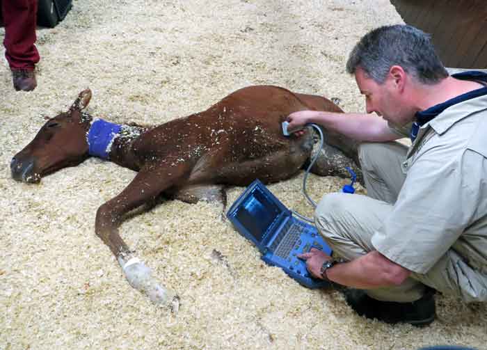
Ultrasonography can provide additional information in the examination of a horse with colic, especially in foals and small horses where rectal examination cannot be performed (Figure 6). Abdominal ultrasound can be performed transcutaneously or per rectum.
Abnormalities commonly detected include peritoneal effusion, adhesions, masses, small intestinal distension, ileus, intussusception, and left dorsal displacements of the large colon.
The following factors should initiate consideration for referral of a horse with colic: