14 Oct 2019
Nicola Menzies-Gow discusses issues that may arise with older horses, including aspects of nutrition and common diseases.
Nicola Menzies-Gow
Job Title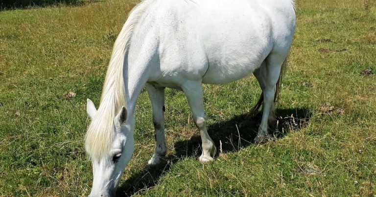
Horses are usually considered to be geriatric when they are either older than 15 or older than 20 years and, in the UK, geriatric horses make up approximately a quarter of the equine population. The number of geriatric horses within the equine population appears to be increasing and these animals may have increased requirements for preventive health care, such as dentistry. In addition, certain diseases may be more prevalent within this subgroup of the population. These factors combined mean a large geriatric equine population in the UK requiring veterinary attention exists.
The term geriatric was first used in human medicine more than 100 years ago to describe the stage of life characterised by a progressive decline in physical condition, organ function and immunity.
The definition of the term geriatric, as it applies to horses, has not yet been firmly established, and different studies have used different ages. However, in most studies horses are considered geriatric when they are either greater than 15 or greater than 20 years of age, and when questioned, owners considered a horse to be old at a mean age of 22 years1.
While it is well documented that population ageing is an important feature of the human population, information relating to the demographics of the equine population is limited. In UK studies, geriatric horses and ponies (greater than or equal to 15 years of age) constituted 25% to 29% of the overall horse population2-4, 11% were aged 20 to 30 years old and 2% were greater than 30 years old2. The British Equine Trade Association’s 2011 National Equine Survey estimated 1.3 million horses were in the UK; this suggests more than 300,000 UK geriatric horses and ponies.
Some evidence exists that more aged horses are being presented for veterinary treatment; referral hospital admissions of horses greater than or equal to 20 years rose from 2.2% to 12.5% within 10 years5. This may be for a number of reasons. Firstly, the role of the horse in UK society appears to be changing towards that of a companion animal, and a strong owner-pet bond often exists, which may contribute to owners becoming increasingly likely to seek and finance veterinary treatment. Longer duration of ownership may enhance this owner-pet bond; for the general equine population, the median duration of ownership has been reported to be 4.9 years, compared to a median of 11.25 years for geriatric horses6.
Many geriatric horses continue to have a useful working life; in one study, 74% were still in ridden work, participating in a similar range of activities reported for the general equine population, with hacking or pleasure riding being the most common use6. Insurance companies are extending the cover provided for older horses, which may make owners more likely to present their older animals for veterinary treatment. Finally, one study found a reduction in the provision of preventive health care measures – such as vaccination, farrier care and routine veterinary checks – with increasing age and retirement7. This reduced frequency of routine health care measures may predispose the older horse or pony to disease.
Therefore, the number of geriatric horses within the equine population appears to be increasing and these animals may have increased requirements for preventive health care, such as dentistry. In addition, certain diseases may be more prevalent within this subgroup of the population. These factors combined mean a large geriatric equine population in the UK requiring veterinary attention exists.
For nutritional purposes, some authors consider that older horses (older than 20 years of age) can generally be categorised as aged-normal – that is, a healthy animal that may be normally overweight or underweight; or geriatric – that is, an older horse with clear signs of senescent changes alone or in the company of concurrent disease, such as pituitary pars intermedia dysfunction (PPID)8. Senescent changes include sarcopenia (loss of skeletal muscle mass and strength associated with ageing), the loss of body mass, and the onset of organ dysfunction with progressive decline of the dental, neural, immunologic and other systems8.
In one UK study, the majority of geriatric animals were found to be in good body condition, greater than 25% were overweight and only 4.5% were in poor condition9. However, in a second UK study, 35% of apparently healthy older animals were in poor or very poor body condition10. The nutritional requirements and advice will depend on the body condition of the individual animal.
If the animal is in ideal body condition, no indication may exist to make specific alterations to nutritional provision8.
In obese animals, controlled weight loss is best accomplished with the animal taken off the pasture. Animals should initially be restricted to 1.25% of body mass (BM) as daily dry matter intake of hay (approximately 1.5% of BM as fresh hay). The hay can be soaked to reduce its carbohydrate content and provision of a forage balancer product is essential. The daily hay rations should be divided and offered as several small meals, ideally using methods to slow consumption, such as doubled, small-gauge hay nets.
Where removal from pasture is not possible, intake can be restricted by strip grazing or with the judicious use of grazing muzzles. Where possible, exercise should be given as an adjunct to dietary restriction. Weight loss should be monitored monthly using a weight bridge/tape or by measuring heart and belly girth, and the diet adjusted according to weight loss achieved.
In the aged-normal older horse that is losing condition, the causes of the weight loss can be multifactorial. Common causes of weight loss include pain, bullying at pasture, chronic gastrointestinal parasitism, dental problems or inadequate nutrition. Identification and correction of these factors should allow a progressive return to normal condition with minimal adjustment to standard nutritional regimes8. Failure to respond to corrective therapy may indicate the onset of senescent changes or disease.
In the geriatric horse, grass remains the forage of choice as grazing promotes gentle exercise, and the higher water content of fresh grass relative to hay/haylage facilitates mastication (assuming dental compromise), swallowing and digestion8.
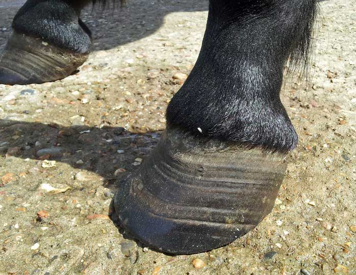
Many severely dentally compromised animals still benefit from turn out to pasture and will obtain a significant proportion of daily intake if the pasture lengths exceed 6cm (Figure 1)8. Soaked, processed, complete feeds are also beneficial in these animals.
Compound feedstuffs should be highly palatable and heavily moistened to aid in the prevention of choke and subsequent digestion. Processed (for example, extruded, ground, pelleted) compound feeds are preferred because the smaller particle sizes are more easily masticated, swallowed and digested, with a reduced risk for impaction colic. Compound feeds containing increased crude protein (12% to 14% compared with 8.5% for younger mature animals) should be chosen8. If increased calories are required, this should be provided as oil (up to 500ml of vegetable, corn or soya oil), which can be introduced over two to three weeks rather than carbohydrate.
Strangulating lipoma and large colon/caecal impaction are two causes of colic that are thought to be more prevalent in older horses5. Among cases referred for colic, geriatric horses aged greater than or equal to 16 years old were twice as likely to have a strangulating small intestinal lesion, compared with non-geriatric adult horses11; however, underlying reason remains unclear.
The increased prevalence of large intestinal impactions is postulated to be related to a decline in the digestive or absorptive processes in the large intestine, possibly secondary tissue damage caused by intestinal parasites, coupled with dental disease that may contribute to a decrease in digestibility and increased length of fibre entering the large colon predisposing to simple obstruction5.
The overall short-term survival of geriatric horses with colic and the survival of geriatric horses treated medically seems to be lower, compared to mature non-geriatric horses11. However, this is most likely due to the owner electing for euthanasia as the horse has a surgical lesion and surgery not being an option.
The survival of surgically managed geriatric horses was not different to that for mature horses and no difference exists in survival between geriatric and mature horses with small or large intestinal strangulating lesions11. Therefore, treatment decisions for geriatric horses with colic should not be based on age alone.
Dental abnormalities are extremely common in geriatric animals; in one UK study, dental abnormalities were identified in 95% of animals, with cheek teeth diastemata, tooth loss, excessive wear/cupped out teeth and focal overgrowths the most frequently identified conditions9. Clinical signs of dental disease vary with the severity of the pathology. Many animals show no clinical signs, despite the presence of multiple dental abnormalities. However, possible clinical signs include quidding, taking longer to masticate, temporary cheek swellings if forage becomes packed between the cheek and the teeth, presence of long fibres in the faeces and weight loss12.
The natural ageing process of equine teeth contributes to the majority of dental disease observed in geriatric horses13. The enamel thickness and enamel infolding decreases apically in the cheek teeth. This means less enamel exists on the occlusal surface as the teeth wear down, resulting in teeth that are less able to resist wear and an increased rate of attrition.
In addition, because the teeth narrow apically, geriatric horses have less cheek tooth surface area and angulation. This means the compression forces in each cheek teeth row are unable to maintain the close interdental contact between teeth, which may in turn result in multiple diastemata developing along a cheek teeth row, termed senile diastemata, with secondary food impaction and periodontal disease14.
An age-related change also exists in the occlusal contact between the cheek teeth, which may contribute to the wear pattern disorders commonly seen in geriatric dental patients, such as wave mouth12. Finally, various management and dietary factors may also contribute to the rate at which teeth are worn and may accelerate age-related changes.
The mainstay of dental treatment in older horses is aimed at ensuring oral comfort and maximising masticatory ability. The short remaining reserve crown limits dental crown reduction treatment in geriatric horses and trying to achieve major changes in the cheek teeth row occlusal profile is more detrimental than more conservative treatment13.
Wave mouth is an uneven wear pattern of the occlusal surfaces with a proportion of teeth overgrown and/or worn within the same cheek teeth row. Treatment of this condition is aimed at reducing the overgrown teeth.
It is important to carefully rasp the overgrown teeth by only a few millimetres, because reduction of these teeth effectively takes them out of occlusion, reducing a horse’s masticatory ability until the continued eruption of worn teeth effects occlusal contact again12. This treatment needs to be repeated approximately three months later. If a reasonably even occlusal surface has been achieved, treatment has to be repeated at six-month intervals to ensure no further deterioration.
When diastemata are due to apical narrowing of the teeth (senile diastemata), treatment is aimed at alleviating the associated gingivitis and periodontal disease. Cleaning out and flushing of periodontal pockets may be indicated. If the diastema is narrower at the occlusal aspect than the gingival aspect, widening with a rotating diastema burr may be beneficial.
Ultimately, in geriatric patients, unlike in younger patients due to the lack of cheek teeth row compression forces, resolution of the diastemata is unlikely to exist. Other adjunct therapies – such as analgesia, antibiotic therapy and dietary management – may be required.
Equine PPID is a slowly progressive neurodegenerative disease with loss of dopaminergic (inhibitory) input to the melanotropes of the pituitary pars intermedia, which appears to be associated with localised oxidative stress and abnormal protein (α-synuclein) accumulation. However, the exact cause remains unknown. The consequent dysfunction of this region results in hyperplasia of this area of the gland and overproduction of pars intermedia-derived hormones; eventually the area undergoes adenomatous change.
The condition is seen in older animals; the average age in retrospective case series ranges from 18 to 23 years15-18. No breed or sex predilection exists, but ponies are more frequently affected than horses in some studies16-18. In one survey, 21% of horses aged older than 15 years of age had endocrine changes consistent with PPID19 and in a second, clinical signs consistent with PPID were documented in nearly 40% of horses older than 30 years of age7. However, it should be noted that the disease is recognised in animals younger than this, with the youngest reported case being in a 7-year-old animal20.
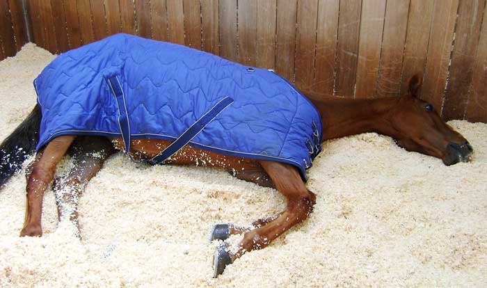
The clinical signs associated with PPID can be roughly divided into those that are seen early in the disease and those that are associated with advanced disease. Early signs include decreased athletic performance, change in attitude/lethargy, delayed haircoat shedding, regional hypertrichosis, change in body conformation, regional adiposity and laminitis. Late signs include lethargy, generalised hypertrichosis (Figure 3), skeletal muscle atrophy, hyperhidrosis, polyuria/polydipsia, recurrent infections, infertility and laminitis.
Diagnosis of PPID is based on the signalment, clinical signs and further diagnostic test results. No ideal further diagnostic test for equine PPID exists; however, plasma basal adrenocorticotropic hormone (ACTH) concentrations and the ACTH response to thyrotropin-releasing hormone are thought to be the most appropriate tests available. In addition, it is recommended tests to assess for insulin dysregulation (ID) are concurrently employed, as ID occurs in a subset of animals with PPID, and appears to be associated with an increased risk of laminitis and a worse prognosis. Appropriate tests for ID include measurement of resting (basal) serum insulin concentration, an oral sugar (OST) or glucose test and the insulin response test (IRT).
Not all owners will elect to treat the disease specifically, depending on which clinical signs are present and how severe they are in an individual animal. The clinical signs can be managed individually – for example, clip excess hair, treat secondary infections, alter the diet to gain or lose weight and treat laminitis. Where medical therapy is employed, the treatment of choice is the dopamine agonist pergolide (initial dose is 2mg/kg by mouth once a day), which is licensed for the treatment of this condition in horses in the UK. It is reportedly effective in 65% to 80% of cases.
The serotonin antagonist cyproheptadine does not appear to be any more effective than non-pharmacological management, but can be used in addition to pergolide in refractory cases. Normalisation or improvement of endocrine tests can be used to monitor the response to therapy starting 30 days after initiation of pergolide treatment; alternatively, improvement in the clinical signs can be used alone or in conjunction with the laboratory response.
Lifelong drug therapy and/or management of the clinical signs is required as all the available drugs will only help control the clinical signs, but not result in a cure. Despite this, many horses continue to have a good quality of life for a number of years.
Musculoskeletal disease is the most prevalent health and welfare issue in aged horses, with OA and chronic laminitis being the most common single disorders21. When 200 geriatric horses were assessed at the walk, 18.6% were lame on at least one limb, while 50.5% were lame in trot9. In addition, the majority of animals (83.5%) had a reduction in range of motion in at least one joint and 80% of animals had hoof abnormalities9.
Treatment of OA in the geriatric horse is basically similar to treatment of performance horses, but aims more at providing optimal comfort rather than at regaining the ability to compete21. Therefore, as well as pain management, there should be a focus on optimising the exercise regimen, improving the living conditions and losing weight if the animal is overweight or obese. Treatment may consist of combinations of local treatment of the affected joint(s); pain management (NSAIDs); and supportive treatments, such as farriery, physiotherapy and exercise management21. Feed additives or nutraceuticals are used on a very large scale worldwide and many of them claim to be beneficial for (chronic) joint disease. Many of these supplements contain glucosamine and/or chondroitin sulfate, with or without addition of a large variety of other compounds. The evidence for their in vivo efficacy remains very thin, but a number of studies have shown a (limited) beneficial effect of some products, such as extract from green-lipped mussels22.
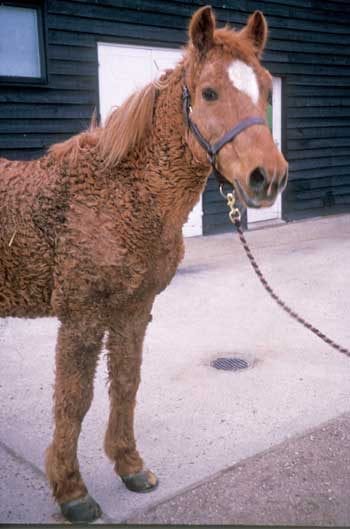
Laminitis (Figure 4) is a painful condition of the equine foot that is now considered to be a syndrome associated with systemic disease (sepsis or systemic inflammatory response syndrome or endocrine disease) or altered weight bearing rather than being a discrete disease entity23. Therefore, laminitis can be divided into three forms: sepsis-associated, endocrinopathic and supporting limb laminitis. Endocrinopathic laminitis is the most common form of the disease, and encompasses laminitis associated with equine metabolic syndrome and PPID. Studies have reported 5% to 7% of geriatric horses suffer from laminitis7 and laminitis in the geriatric horse is most frequently related to PPID. Laminitis in geriatric horses is managed as in the general horse population, with additional benefit from pergolide administration in cases with PPID.
Respiratory disease is common in the geriatric horse: horse owners reported that 14% of geriatric horses coughed7 and veterinary examination of 200 geriatric horses revealed 7.5% had a spontaneous cough during the examination, 18.5% had a nasal discharge, 22% had abnormalities on thoracic auscultation at rest and 31% had thoracic auscultation abnormalities on re-breathing9.
The only respiratory disease that tends to be more prevalent in older animals is severe equine asthma (SEA); in a study of SEA within the UK general equine population, both age and retirement were identified as risk factors for the disease6. Uncomplicated geriatric SEA cases are generally managed in the same way as cases in other age groups.
However, comorbidity is common in the geriatric patient, the concurrent presence of PPID and/or laminitis should be considered before initiating corticosteroid therapy.
Age has been identified as a significant risk factor for certain cardiac diseases in the horse. The prevalence of murmurs associated with left-sided valvular insufficiencies, such as aortic and mitral insufficiency, increased with age24; left-sided valvular regurgitation or murmurs consistent with this are reported to occur in between 13% and 20% of geriatric horses7,24,25.
In the geriatric horse, the aortic valve is the most common site for degenerative valvular pathology, which in turn can be associated with regurgitation. In many horses, aortic regurgitation (AR) is well-tolerated and is often an incidental finding26. When advising on the significance of AR in the older horse, the clinician must consider the horse’s activity and workload27. In geriatric horses that are retired, further investigations may not be required; however, with older horses still in work, echocardiography and ECG are warranted.
The mitral valve is the second most common location for valvular pathology, most often related to degenerative valvular disease. Slowly progressive valvular disease is often well-tolerated until it becomes very severe, but chorda tendinea can rupture spontaneously or after inflammatory or degenerative disease, leading to a rapid change in left heart haemodynamics that is much less well-tolerated27. Mitral regurgitation can also lead to sudden death due to pulmonary rupture secondary to pulmonary hypertension. If the horse is still in work, echocardiography and ECG are warranted.
Age is not reported to be a risk factor for any arrhythmias in horses.
Ophthalmologic abnormalities are common in geriatric horses; in studies involving the UK geriatric horse population, 94% of animals had at least one abnormality identified on veterinary examination7,9. Ophthalmic lesions frequently identified included vitreous degeneration (66%), cataracts (58.5%) and senile retinopathy (33.7%)9.
Vitreous degeneration manifests as clouding or discoloration of the vitreous gel with membranous or cellular debris that appears suspended within the vitreous body, sometimes termed floaters or muscae volantes28. No specific treatment exists.
Typically, early senile cataracts appear as microvesiculation in the posterior suture lines28. This progresses to more dense condensation around the posterior suture, together with perinuclear and cortical (anterior and posterior) cataracts28. Complete and occasionally hypermature cataracts can occur in some cases, with animals experiencing more significant visual compromise28. Cataracts can only be treated by surgical removal of the lens; however, surgery is usually reserved for those horses with a significant visual impairment as a study suggested only 26% of horses remained visual two years after surgery29.
Senile retinopathy affects the retinal pigmented epithelium and photoreceptor layer. It appears as irregular linear hyperpigmentation surrounded by hypopigmented areas, mostly affecting the nontapetal fundus, although the tapetal fundus can also be involved. The pathogenesis of senile retinopathy is not fully understood, but possible causes include oxidative damage or choroidal vasculature disease. Some debate exists as to whether senile retinopathy causes visual deficits or not; some consider it is of no clinical significance, whereas others report vision problems in affected animals, particularly in poor lighting conditions28. No specific treatment exists.
Neoplasia is an important cause of death in the geriatric horse, accounting for almost 20% of deaths in one survey30 in which pituitary adenoma, squamous cell carcinoma, lymphoma and melanoma were the most common malignant neoplasms.
Geriatric horses and ponies represent a significant proportion of the equine population, and are of great importance to their owners. Colic, dental, musculoskeletal, endocrine, respiratory and cardiovascular disorders are highly prevalent in this group of animals. Owners may not be aware of the significance of clinical signs observed in their aged horse and seem to under-recognise some of the most prevalent diseases. Therefore, it is important preventive health care is maintained within this group of animals and owners are educated to ensure clinical signs of disease are not mistaken for benign signs of ageing.