21 Sept 2015
Elisabetta Mancinelli provides a brief overview of the dietary requirements of some species commonly kept as pets, and the need to educate owners and keepers about their needs.
Elisabetta Mancinelli
Job Title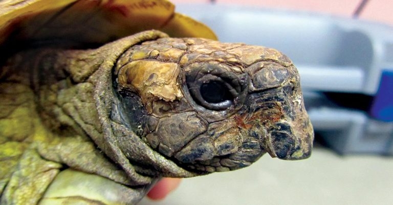
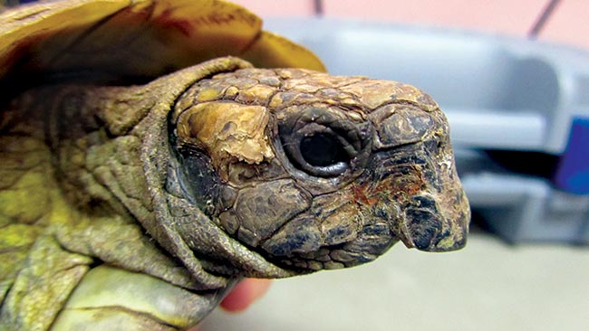
Figure 6. Beak overgrowth can be seen in turtles and tortoises, and can occur in association with inadequate nutrition.
In human medicine, it has been clearly demonstrated how important certain foods are for people’s health and how poor food is linked to various diseases or pathologies.
The constant growth of new information in exotic animal medicine is slowly filling the gap compared to the situation in more common companion species and has the potential to truly impact on the health and well-being of many exotic pets. However, despite increasing knowledge in reptile husbandry and nutrition, nutritional-related disorders are still commonly seen in captive animals.
Lizards, snakes and chelonians make up an extremely large and diverse group of reptiles with thousands of species (www.reptile-database.org). Reptiles are found throughout the world, adapted to a wide variety of climates, habitats and natural diets. Their anatomy and physiology can vary considerably between groups, with each species adapted to its natural environment.
Consequently, different species can have extremely varied captive and nutritional requirements, often difficult to reproduce satisfactorily in captivity. This, coupled with their ability to survive without showing obvious signs of illness, can present a challenge for the veterinary surgeon having to make general recommendations. Anecdotal information, largely coming from experience, is widely – and often too easily – available, but scientific knowledge is becoming more accessible allowing increasing support to husbandry and nutritional requirement recommendations.
eNutritional disorders vary between species and feeding strategies, and can be caused by an imbalance in various nutrients, including protein, vitamins and minerals or by defects or other diseases that prevent proper utilisation of nutrients. For example, snakes fed whole prey are more likely to show generalised starvation in contrast to iguanas and other herbivorous reptiles, which are more likely to suffer from calcium deficiency from imbalanced diets (Donoghue, 2006).
Nutritional secondary hyperparathyroidism (NSHP) is a form of metabolic bone disease and the most common nutritional disorder seen in captive reptiles, especially in lizards, chelonians and herbivorous and insectivorous species. When dietary calcium and/or vitamin D3 (because of insufficient dietary intake in non-herbivorous reptile species or of a lack of adequate exposure to UVB radiation) intake is inappropriate and, occasionally, when diets are high in phosphorus (reversed calcium/phosphorus; Ca:P ratio), NSHP can occur in captive reptiles. Snakes are not affected by NSHP because they feed exclusively on whole prey that contains adequate amounts of calcium and vitamin D3 (Mans and Braun, 2014).
Insufficient dietary calcium intake or inappropriate Ca:P ratio of the diet; insufficient intestinal calcium absorption (Vitamin D3 dependent); insufficient dietary vitamin D intake; or insufficient exposure to UVB radiation are among dietary factors that can lead to NSHP (Calvert, 2004). Provision of UVB to captive reptiles is a controversial topic as in many, but not all, the species examined, exposure led to increased plasma vitamin D levels (Ferguson et al, 2003; Acierno et al, 2006 and 2008; Hedley and Eatwell, 2013). Furthermore, dietary intake of vitamin D3 affects basking behaviour and, therefore, exposure to UVB radiation (Ferguson et al, 2003).
Nevertheless, most forms of artificial UVB radiation provided in captivity are an inadequate alternative to the natural sunlight most reptiles are exposed to in their natural habitat (Mans and Braun, 2014).
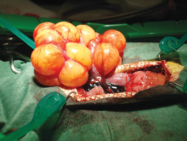
A low calcium level stimulates the release of parathyroid hormone (PTH), which, in turn, induces mobilisation of calcium from bones to maintain normal blood calcium levels. Therefore, clinical signs (Figures 1 to 5) are often related to demineralised bones (deformities, pathological fractures) and eggs (with resulting follicular stasis or dystocia), hypocalcaemia (nerve and muscle dysfunction – twitches, tremors, seizures, tongue dysfunction, crouched stance, cloacal prolapsed and so on; Donoghue, 2006).

Radiographs can reveal reduced bone density, jaw and/or long bone deformities and/or fractures, and can prove invaluable for an accurate diagnosis. Often, total plasma calcium levels have little diagnostic value. Ionised calcium levels should always be obtained whenever possible.
Appropriate treatment must aim at correcting underlying husbandry and management issues, as well as providing adequate supportive care, specific supplementations and management of concurrent disorders, such as dehydration or secondary bacterial or parasitic infections (Donoghue, 2006; Mans and Braun, 2014).
Vitamin A is essential for many biological processes, including vision, growth, reproduction and immune function (Hand and Lewis, 2010). Herbivorous reptiles are able to endogenously synthesise vitamin A from dietary precursors differently from many carnivorous and omnivorous species in which activation of the provitamins into vitamin A does not occur because of the lack of the needed enzymes (Hand and Lewis, 2010).
Clinical signs associated with vitamin A deficiency are commonly seen in aquatic turtles (for example, red-eared sliders), North American box turtles, chameleons and geckos fed inappropriately supplemented diets (Donoghue, 2006; Mans and Braun, 2014).
Anorexia, stunted growth, oedema, inflammation and infections of the eyes, oral cavity and respiratory dysfunction secondary to multifocal squamous metaplasia of epithelia are commonly seen clinical signs, often strongly suggestive of a diagnosis of hypovitaminosis A in reptiles alongside the dietary history.
Confirming this diagnosis can be challenging. Positive response to treatment with vitamin A and other nutrients, as well as the provision of a more balanced diet, is often used to confirm the diagnosis in most captive reptiles.
It is important to remember over-supplementation with vitamin A and resulting clinical signs of iatrogenic intoxication can be as detrimental as its deficiency. Therefore, administration of vitamin A and retinol should be made with caution and provision of dietary carotenoids considered, especially in herbivorous reptiles, which are able to absorb and convert carotenoids into biological active vitamin A (Mans and Braun, 2014).
Thiamine (vitamin B1) is a highly labile vitamin, which undergoes rapid deterioration under normal storage conditions.
Captive fish-eating reptiles (such as garter snakes) and semi-aquatic turtles are most commonly affected if they are fed fish high in thiaminases, or frozen fish thawed slowly over several hours (this process will lead to depletion of thiamine by enzymatic destruction by thiaminase) or inappropriately stored fish. Herbivorous reptiles, such as green iguanas (Iguana iguana), can potentially develop signs of thiamine deficiency if fed frozen then thawed vegetables, which contain phytothiaminases (Calvert, 2004).
Neurologic signs are commonly seen with deficiencies because thiamine is essential to brain function. These clinical signs can be similar to those caused by hypocalcaemia, therefore a detailed dietary history, response to treatment with vitamin B1 and ionised blood calcium level can help identify the primary cause and allow a more specific treatment.
Iodine is an essential trace element, which plays an important metabolic role as it is a component of thyroxine and triiodothryronine. These hormones can influence many physical and mental functions, including growth, brain, nerve tissue and muscle function.
Deficiencies can occur for reasons including insufficient dietary iodine intake or excessive intake of goitrogenic plant material (such as broccoli, Brussels sprouts, cauliflower, cabbage, cress, bok choy, kale, mustard seed, rapeseed and turnips; Donoghue, 2006). These plants should be fed sparingly because their content in goitrogens can lead to interference with the iodine uptake by the thyroid gland and, subsequently, cause nutritional secondary hypothyroidism and goitre unless compensatory dietary iodine is ingested (Mans and Braun, 2014). There have been few reports of clinical signs associated with dietary iodine deficiency (oedema of the neck, head and forelimbs, decreased appetite and lethargy) mainly in herbivorous lizards and chelonians, and, often, the diagnosis is only presumptive (Frye and Dutra, 1974; Calvert, 2004; Mans and Braun; 2014) as, to date, a thyroid stimulation test has not been reported in reptiles and a low thyroxin level may reflect non-thyroid associated diseases or may be due to the influence of environmental factors.
A response to treatment with thyroxin does not help in confirming a diagnosis of hypothyroidism because increased metabolism, appetite and activity are to be expected if thyroxin is supplemented in animals, regardless of whether the thyroid is diseased. A diagnosis of hypothyroidism caused by insufficient iodine intake or excessive intake of goitrogenic plants should be made cautiously and consideration given to the risk of causing intoxication when supplementing iodine.
Abnormal beak growth is often seen in turtles and tortoises and can interfere with feeding (Figure 6). It can occur in association with inadequate nutrition. Treatment consists of burring the excess mouthparts into a more normal shape. The condition usually re-occurs due to the original disorder with the upper and lower beaks growing in uneven positions, and long-term maintenance is required as well as provision of a more “natural” diet.
The consistency of the faecal material passed may vary between reptile species depending on their diet. Soft faeces or diarrhoea may be seen in herbivorous reptiles being fed an inappropriate diet (for example, one low in fibres, too rich in simple carbohydrates and easily fermentable). However, causes of gastrointestinal motility disorders unrelated to the diet – such as environmental factors (for example, low temperature), endoparasites and other infectious causes (for example, bacterial, viral and fungal) – need to be ruled out as well (McArthur et al, 2004; Mans, 2013; Mans and Braun, 2014).
A thorough history including details of husbandry and diet will be extremely important in these cases. Faecal parasitology (wet prep and flotation) may be useful to rule out endoparasites. Further investigations, including diagnostic imaging, such as radiography – with and without contrast – haematobiochemistry and endoscopy may be useful to reach a definitive diagnosis when non-dietary causes are suspected. Treatment will depend on the primary underlying cause and associated disorders. Optimising husbandry and diet is mandatory.
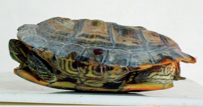
Dehydration is frequently seen in captive reptiles and it can be assumed any anorexic or chronically ill animal is dehydrated. Sunken eyes, lethargy, anorexia, skin tenting or dysecdysis may be seen, but they may also be unreliable clinical signs, not necessarily indicative of dehydration in reptiles (Figure 7; Mans and Braun, 2014).
Dehydration is often secondary to incorrect husbandry (for example, low humidity or inappropriate temperature gradient for the species) and these issues need to be addressed as part of the treatment plan. Fluid loss needs to be replaced and, depending on the severity of dehydration and associated clinical signs, fluid therapy may be started per os or via the subcutaneous, intravenous, intraosseous, epicoelomic or intracoelomic routes. Soaking in shallow warm water is also an effective and non-invasive method of reperfusion in dehydrated reptiles, particularly for cases of mild dehydration of if dehydrated reptiles cannot be hospitalised.
Current recommendations disagree with previous presumptions that diluting commercially balanced electrolyte solutions was necessary to avoid administration of hypertonic solution to reptiles because of their lower intercellular fluid’s osmolality. The belief now is the reptiles’ plasma osmolality is similar or higher to that of mammalian or human plasma, therefore available balanced electrolyte solutions, such as lactated Ringer’s solution, can be used in most cases (Davis and Klingenberg, 2004; Dallwig et al, 2010; Guzman et al, 2011; Orosz, 2013; Mans and Braun, 2014).
When inappropriate diet and lack of exercise allow a positive energy balance, with energy intake exceeding species and individual requirements, obesity may occur. Excessive fat may need to be differentiated from pathological conditions (such as ascites, tympany or over-inflation of lungs/air sacs in certain lizards).
Fat may accumulate in the subcutaneous space, intracoelomically, but infiltration of parenteral organs such as the liver may also occur (for example, hepatic lipidosis).
Unfortunately, little is known about the metabolic needs of many reptile species, especially in relation to their physiologic state in captivity. However, an accurate review of diet and husbandry is important to identify and correct major faults. Alternative preys should be considered, more appropriate and balanced diets should be fed and adequate exercise should be guaranteed in an attempt to avoid obesity in captive reptiles (Barker et al, 1998; Oonincx and Dierenfeld, 2012; Mans and Braun, 2014).
Nutritional disorders are still commonly seen in many captive reptile species (Calvert, 2004; Donoghue, 2006) and this article provides just a short overview of the most common ones.
Many of these problems are often diagnosed late in the disease process when secondary complications have already occurred. Therefore, every attempt should be made to educate reptile owners and keepers about the proper care and, in particular, dietary needs of reptiles under their care because nutritional disorders seen in captive reptiles are often preventable.
Considering the complexity of the subject, the reader is strongly encouraged to refer to the literature provided for more in-depth and comprehensive information.