28 May 2021
Sarah Gough – in the first of a two-part article – looks at the types, causes, treatment and management of infectious diseases.
Sarah Gough
Job Title
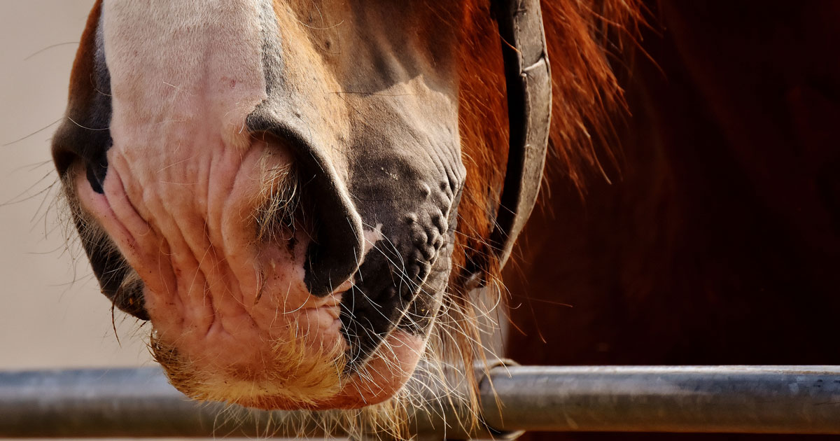
Our equine patients are among the elite of the animal kingdom when it comes to their respiratory capacity.
It is, in part, this large respiratory reserve that enables horses to perform at such high intensities, as seen in sports such as racing, one-day and three-day eventing, and barrel racing, to name a few.
As such, any condition that compromises respiratory function can result in reduced performance in these elite athletes. However, the large respiratory reserve can also work in the opposite way for horses that are not expected to perform at high intensity, as a mildly compromised respiratory function may still provide sufficient oxygen, hence clinical signs may not be immediately evident in these patients.
Many conditions can affect the respiratory tract and result in altered airway dynamics, compromise and varying degrees of systemic effects, and these can broadly be classified into:
However, unfortunately it is not possible to discuss all of these conditions in a single article, so for the purposes of this review the author will focus on the aforementioned infectious causes of respiratory diseases.
While they are by no means an exhaustive list of viral and bacterial causes of respiratory disease in our equine patients, they are arguably the most common conditions practitioners are faced with. A similar review on non-infectious causes of respiratory disease will be provided as part two in an upcoming Vet Times issue.
Strangles is a highly contagious bacterial upper respiratory tract infection that is well known by most equine practitioners. It is caused by the Gram-positive cocci S equi equi and causes disease of variable severity with a high morbidity, but low mortality rate.
The clinical severity varies among patients; however, it is generally characterised by lethargy, fever, lymphadenopathy and abscessation (primarily the retropharyngeal and mandibular lymph nodes, although other lymph nodes can also be affected in atypical cases) and purulent nasal discharge.
Initial febrile episodes can be marked (greater than 39.4°C) although many horses do not display signs of malaise in this early period, so the fever may be missed until further signs develop, typically including more marked malaise, mucopurulent nasal discharge and lymphadenopathy as above.
The severity of clinical signs is often influenced by both age/immunological maturity and previous exposure, with young, old and immunocompromised horses most severely affected. This variable severity can result in misdiagnosis in mild cases, and subsequently disease outbreaks.
Infection with S equi equi typically occurs from ingestion or inhalation of infected mucopurulent discharge (including via fomites), which enables attachment of the bacterium to the epithelium and crypts of lingual and palatine tonsils through the use of the fibronectin binding surface proteins SeM and SEQ2190.
However, once attached the bacterium does not remain on the mucosal surface, but instead has an affinity for the deeper tissues of the mandibular and retropharyngeal lymph nodes1-3.
The incubation period ranges from 3 to 14 days. Bacterial shedding typically occurs within two to three days of the onset of fever, providing a period where patient isolation can be implemented prior to disease shedding; and lasts from two to six weeks or longer if guttural pouch empyema develops1-3. However, as mentioned already, the initial febrile episodes may not be detected in cases that continue to eat and do not demonstrate malaise.
Accurate and timely diagnosis of S equi equi continues to be a challenge for equine veterinarians, particularly in field outbreak situations. As aforementioned, a delay of two to three days exists between the onset of fever and bacterial shedding.
During this period, false negative test results are likely to be obtained as the bacterium does not colonise the epithelial surface (it has an affinity for the deeper tissues), and shedding in nasal discharge has not yet begun, hence nasal, nasopharyngeal and guttural pouch sampling may all fail to identify bacteria.
In addition, these cases are typically too early to show elevation of antibody titres on ELISA. Traditionally, culture was considered the gold standard; however, as this requires live bacteria, potential exists for false negative results due to potentially low bacterial numbers in a sample, poor sample handling and concurrent inhibitory bacteria such as Streptococcus equi subspecies zooepidemicus.
As such, PCR is considered a more sensitive approach, with the triplex PCR the gold standard as it detects three antigens on the SeM gene4. The use of PCR and culture concurrently would be considered the most sensitive diagnostic test protocol1.
So, which samples do we take and when? Much confusion often exists when it comes to selecting the most appropriate sample for identification of S equi equi and this is likely because the most appropriate sample varies depending on the stage of disease, aim of the test (confirming a diagnosis or determining shedding status) and the clinical signs displayed by the animal.
Table 1 provides a summary to guide veterinarians as to which test is likely to be reliable or not in varying scenarios.
| Table 1. Possible sites for sample collection and the most appropriate time to perform each in the clinical course of Streptococcus equi subspecies equi | ||||
|---|---|---|---|---|
| Sample | Asymptomatic/no exposure to disease | Acute clinical signs/exposure to disease | Recovering from disease | Determine carrier status |
| Serology (blood) | Paired serology | Paired serology | Not reliable | Not reliable |
| Nasopharyngeal lavage | Not reliable | Most reliable | Not reliable | Not reliable |
| Nasopharyngeal swab | Not reliable | Not reliable unless marked nasal discharge | Not reliable | Not reliable |
| Guttural pouch wash | Combined with paired serology | May be too acute to detect disease | Most reliable | Most reliable |
| Lymph node aspirate | Not necessary | If lymph nodes are enlarged | If lymph nodes have not yet ruptured | Not reliable |
| Aspirate of purulent discharge from upper respiratory tract | Combined with paired serology | May be reliable | May be reliable | Rule out secondary sites |
Paired serology (two samples 10 to 14 days apart) to determine antibody titres to antigen A (the S equi equi specific portion of SEQ2190) and antigen C (the S equi equi specific N-terminal portion of SeM) is a useful screening tool in some animals3, such as asymptomatic animals or those without known exposure to infected horses (that is, screening prior to moving yards). However, in a disease outbreak it is important to ensure exposure does not occur between the two samples as this would mean the second sample is no longer a true “convalescent” sample.
Both systemic and mucosal immune responses typically occur within two to three weeks of infection and typically signal mucosal clearance2. A positive titre does not confirm disease; however, it does suggest exposure and indicates a need for further bacterial testing, including guttural pouch endoscopy and lavage1. Importantly, antibodies from previous vaccination cannot be differentiated from exposure to disease.
While nasopharyngeal swabs have long been used for bacterial culture and PCR sampling in suspect strangles cases, their use is limited by the small surface area sampled2. This is the least reliable sampling method for S equi equi and the author encourages readers to move away from this technique.
So, where and how should we collect samples for culture and PCR? In the face of acute clinical disease the most appropriate sample is a direct sample from a specific site of infection3; as such, the sample you collect may be different in different cases. Typical sites in the acute phase of clinical disease include an aspirate from an abscessed lymph node, a guttural pouch lavage if empyema is evident endoscopically or nasopharyngeal lavage if no evidence of guttural pouch empyema exists.
Nasopharyngeal lavage has – or should have – replaced nasopharyngeal swabs; it is a more appropriate test and involves “lavaging” the pharynx with 50ml to 60ml warmed saline (an AI catheter works well for this) and collecting the sample in a sterile pot, cup or rectal sleeve as it runs out of the nares.
This samples a larger surface area, providing a more accurate result2, and may be the most accurate or reliable diagnostic test during acute clinical disease, particularly if no evidence exists of purulent material within the guttural pouches.
If the retropharyngeal lymph nodes have not ruptured into the guttural pouch, a guttural pouch lavage will likely yield a false negative result. Conversely, during the chronic stages of disease the bacteria is often sequestered away in sites such as the guttural pouch or sinuses, and in these cases drainage into the nasopharyngeal region is less likely to occur, making nasopharyngeal lavage inaccurate.
Needle aspiration of enlarged or abscessed lymph nodes is a test with high specificity for confirmation of S equi equi, and in appropriate cases (that is, those with lymph node abscessation prior to rupture) may be the most reliable site to sample2,3.
As mentioned, guttural pouch empyema only occurs once the retropharyngeal lymph nodes rupture into the medial compartment, and this does not always occur, or may not have yet occurred, during the acute phase of disease. Hence, reliance on guttural pouch lavage (the “traditional gold standard”) in this instance may result in a false negative.
Likewise, retropharyngeal lymph nodes may rupture externally rather than into the guttural pouch, making external sampling or a direct lymph node aspirate more accurate3. That being said, the guttural pouch lavage remains the gold standard for identification of both carrier status (particularly in animals with chondroids in which the bacteria is often sequestered away and only occasionally shed via the nares) and for confirmation of bacterial clearance.
In the author’s experience it is also important to consider other potential sites of sequestration, such as sinuses, which can also lead to chronic shedding. Historically, it has been advised that three negative samples are required to confirm both clearance of disease and/or rule out disease in an animal.
While this remains true for bacterial culture due to the inherent limitations of culture techniques, a single PCR – assuming sampling has been from an appropriate site and at an appropriate stage of disease – is considered equivalent in sensitivity and specificity to three cultures.
Management of active cases of strangles can be challenging, and in many cases it simply involves providing supportive care while the disease runs its course. Of critical importance is that effective biosecurity is implemented to prevent spread of infection.
NSAIDs can be used to reduce fevers, reduce swelling and reduce discomfort as part of supportive care. However, the temptation is often to provide antibiotic therapy, but this is not always of benefit and may inhibit development of immunity, resulting in reinfection1. Likewise, provision of antibiotics to horses with abscessed lymph nodes is considered contraindicated as it prolongs the maturation of the abscess and hence the overall disease process.
According to the revised consensus statement, antibiotic treatment may be indicated in acutely infected animals with high fever, malaise and no evidence of abscess formation, respiratory distress associated with lymphadenopathy, metastatic abscessation, development of purpura haemorrhagica, and topical use in cases of guttural pouch empyema, with penicillin generally considered the drug of choice2.
However, the risk remains that reoccurrence of disease may occur on cessation of antibiotics2. Conflicting evidence exists as to whether antibiotic use increases the risk of metastatic disease or “bastard strangles”2,3.
Horses that develop chronic guttural pouch empyema and/or chondroids in contrast generally require antibiotic treatment topically once thorough lavage has been performed to remove any remaining empyema. In cases where chondroids are present, their endoscopic retrieval via basket snares or extensive lavage with or without laser fenestration of a salpingopharyngeal fistula may be necessary.
Likewise, with persistent empyema the use of acetylcysteine in lavage solution may be useful. However, this remains controversial and careful dilution to a 20% solution in buffered saline is necessary to reduce the risk of irritation to the many nerves within the guttural pouch2.
Topical treatment involves the use of either a gelatin and aqueous penicillin mixture (see Panel 1 for recipe) to make “penicillin jelly” that is injected via an AI catheter or Foley catheter into the pouch. However, this mixture melts as it warms to body temperature and subsequently drains from the pouch when the horse lowers its head.
Penicillin gel formulation
An arguably more effective approach may be the use of thermodynamic benzylpenicillin5, which can be formulated at 120mg/ml in a 25ml solution by commercial specials manufacturer BOVA. In the author’s experience this product is easy to use, more effective than the gelatin penicillin (likely due to longer retention times within the pouch) and as such can be prescribed via the cascade.
Equine influenza (EI) is a highly contagious respiratory virus in horses that results in varying severity of clinical signs depending on level of immunity (either from prior exposure or vaccination) – from mild to marked fever, serous to mucopurulent nasal discharge and reduced appetite through to complete inappetence, mild lethargy through to obtundation and a cough that may be mild through to harsh and hacking6-9.
This virus is spread via direct contact with respiratory secretions or inhalation of aerosolised respiratory secretions, which can travel up to 2km 8, with the former including contact with fomites (personnel, equipment, environmental objects such as feeders or water buckets)7,9.
EI was initially associated with the H7N7 virus, although this has not been definitively identified as the causative virus since the 1980s. The current strains of EI are sublineages from the H3N8 virus, of which there are European and American lineages.
It is the Florida sublineage of the American lineage that is most commonly associated with EI outbreaks in current times. However, the Florida sublineage has further diverged into two clades – Florida Clade 1 and Florida Clade 2 – with the former responsible for the 2007 epidemics in Japan and Australia, and the 2018-19 epidemics in Europe and the UK. Prior to this, Florida Clade 2 had been most commonly associated with disease in Europe and Asia6.
EI historically resulted in regular epizootics throughout the UK until the implementation of widespread vaccination throughout the country. Consequently, until the outbreak in 2019 there had not been suspension of racing due to EI since 19796.
The contagiousness of EI was highlighted in 2007 when an outbreak occurred in Australia – a country previously free of EI and in which vaccination is not performed. The introduction of a single infected horse resulted in more than 76,000 cases on 10,000 separate premises, with a cost of approximately AU$1 billion (£549.5 million) to control and eradicate the virus10.
Diagnosis of EI requires identification of virus in nasal secretions via reverse transcription-PCR on nasopharyngeal swabs or confirmation of a rising antibody titre via ELISA7,9.
Although nasal swabs have historically also been used, the sensitivity of nasopharyngeal swabs is superior to nasal swabs – as such, the author discourages the use of nasal swabs due to risk of false negative results. However, as discussed previously with strangles, temporal aspects exist that influence which samples are most reliable at which time point.
Nasal shedding of virus is likely to occur within 2 days of infection and often ceases alongside resolution of the fever; although isolation for a minimum of 14 days after the onset of clinical signs is recommended6. As such, the most appropriate times to collect nasopharyngeal swabs are at the time of onset of clinical signs (within two to three days of infection) through to approximately day five, although some nasal shedding can continue through to around day seven8. After this point a nasopharyngeal swab is likely to yield a false negative result, hence swabbing of in-contact horses may be more appropriate.
Nasopharyngeal swabs should be transported in virus transport media so that virus retrieval for typing can be performed if necessary; however, importantly the amount of media should not be more than that required to soak the swab (that is, they should not be submerged in a pool of media).
Serology can also be used to confirm recent exposure, with a baseline sample ideally collected within the first week followed by a convalescent sample 10 to 21 days later with a fourfold increase in titre consistent with exposure9.
Completion of a convalescent sample is important given the potential for widespread prior exposure via vaccination in horses in the UK. This can become more complicated in vaccinated horses, although the ProteqFlu vaccine allows for differentiation of infected and vaccinated animals. Importantly, horses that have been previously vaccinated may develop a milder clinical disease, which complicates diagnosis by both shortening the period of nasal shedding and the potential for subclinical infection with concurrent nasal shedding8,9.
Treatment is largely symptomatic and supportive, including NSAIDs to manage fever, IV fluid therapy, and potentially partial or total parenteral nutrition in severely affected and anorexic patients.
Currently no evidence exists to support the use of antiviral medication in treatment of EI6. Infection with EI may result in damage or complete loss of the ciliated upper airway epithelium and hence the mucociliary escalator8,11. Consequently, accumulation of mucous and debris within the airways may occur, predisposing to secondary bacterial infections. Additional nursing steps, such as feeding horses from the floor, are therefore indicated to help facilitate clearance of debris from the lower airways.
In the event of suspected secondary infection, an aseptically performed tracheal aspirate should be obtained for culture and bacterial susceptibility testing to determine the need for antimicrobial therapy, and in turn guide selection of an appropriate antimicrobial. However, most horses recover from EI without complication, although regeneration of the ciliated epithelium may take several weeks9,11, and it is therefore important that horses are afforded an adequate convalescent period to allow the restitution of the ciliated epithelium.
While it is difficult to determine the exact amount of time necessary, as a guide providing a week of rest for each febrile day during the acute phase of illness, followed by a slow return to exercise, is recommended.
The mainstay of preventing EI in the UK is vaccination and ensuring strict biosecurity in the event of an outbreak. Three main EI vaccines are available in the UK (Table 2) – all of which induce cell-mediated immunity, mucosal immunity and a humoral response, although the number of viral proteins to which immunity is stimulated varies among vaccines.
Importantly, there is more to vaccine efficacy than simply the included clade, and as such, the specific adjuvant/antigen composition, cross-protectivity and vaccine technology all need to be considered when evaluating a vaccine6. With this information in mind, both pros and cons of each vaccination are available that need to be considered; of note, ProteqFlu is currently the only vaccine that fulfils all of the World Organisation for Animal Health recommendations for vaccinations.
While a thorough analysis of the available vaccinations is beyond the scope of this article, Table 2 provides some basic information about some of the available vaccinations in the UK.
| Table 2. Three of the equine influenza vaccines available in the UK and their respective types, included viral proteins and vital strains | |||
|---|---|---|---|
| Vaccine | Vaccine type | Viral proteins | Viral strains |
| ProteqFlu | Live attenuated canarypox | Haemagglutinin proteins and carbomer adjuvant | Florida Clade 1 and Florida Clade 2 |
| Equilis Prequenza | Whole inactivated virus | Haemagglutinin proteins and immune-stimulating complex | Florida Clade 1 and Newmarket |
| Equip F | Subunit vaccine | Virus proteins and immune-stimulating complex | H3N8 European and H3N8 American |
Inevitably discrepancies exist between the vaccination schedules recommended by the vaccine manufacturers, those recommended by sporting jurisdictions and those identified by epidemiological research, and as such the following is a recommendation that incorporates important aspects of current research while still meeting manufacturer and sporting jurisdiction regulations:
Five main equine herpesviruses (EHVs; designated EHV-1, EHV-2, EHV-3, EHV-4 and EHV-5) affect horses, resulting in various syndromes of varying severity and economic impact. EHV-3 causes equine coital exanthema, a venereal disease of the reproductive system, and as such won’t be discussed further in this article.
Both EHV-1 and EHV-4 typically result in rhinopharyngitis and tracheobronchitis, and associated serous nasal discharge that typically progresses to mucopurulent; fever, malaise and ocular discharge in some horses12-14, and EHV-4 is arguably one of the predominant respiratory viruses causing morbidity worldwide12.
However, EHV-1 has a propensity to spread beyond the respiratory tract, resulting in cell-associated viraemia including the endothelium of small blood vessels, hence a widespread vasculitis that is most detrimental when it affects the reproductive tract during pregnancy or neurological system, resulting in abortion and neurological dysfunction (EHV myelitis), respectively15-17.
Although rare, highly virulent endotheliotropic strains of EHV-4 have been associated with abortion also15. Further discussion on the abortogenic and neurological forms of EHV-1 is beyond the scope of this article; however, it is primarily the avoidance of these sequelae that drive vaccination in broodmares.
EHV-2 and EHV-5 are also respiratory viruses with a high prevalence among the horse population, resulting in pharyngitis of varying severity, with concurrent fever, lymphadenopathy and rhinitis. However, in many cases the initial disease is asymptomatic or subclinical, resulting in seropositivity developing in the absence of evidence of clinical disease.
Latency is again a feature of EHV-2 and EHV-5, and the prevalence of these viruses in the population is approximately 50%16. However, of more clinical significance is the development of keratoconjunctivitis associated with EHV-2, and to a lesser extent EHV-5 and equine multinodular pulmonary fibrosis (EMPF) associated with EHV-5.
The equine alphaherpesviruses consist of EHV-1 and EHV-4, both of which are considered respiratory pathogens with infection occurring via inhalation and replication occurring within the upper respiratory tract epithelium7,14. However, once within the respiratory tract these two viruses can have drastically different effects on morbidity.
The respiratory form of EHV-1 and EHV-4 are unable to be differentiated clinically from one another, and both are highly contagious with rapid spread among susceptible in-contact horses. It is common to see outbreaks of disease characterised by high fever that may be biphasic in nature, rhinopharyngitis and pneumonitis among young horses at the time of weaning, and secondary bacterial infection is not uncommon13.
In one study, 37% of horses testing positive to EHV-4 were younger than one year of age and a further 23% were aged from one to five years12; similar to another study in which 5.1% of horses younger than one year of age and 30.8% of horses aged one to five years with respiratory disease and/or acute neurological disease tested positive to EHV-113, further highlighting the frequency of this virus causing disease in young horses.
Once infected, the virus establishes latency in the trigeminal ganglia, resulting in lifelong carriers, and the potential for virus recrudescence and shedding via the respiratory tract during periods of stress7,13,17. Vaccination has been shown to reduce the severity of EHV-1 and EHV-4 respiratory disease, including reducing viral shedding, as well as reducing the risk of EHV-1 induced abortion; however, it is not completely protective and does not prevent recrudescence of latent virus15.
The equine gammaherpesviruses consist of EHV-2 and EHV-5, and both are again transmitted via inhalation of virus, followed by penetration of the respiratory epithelium and B-lymphocytes – the latter of which results in systemic dissemination7,16. The respiratory disease associated with these viruses is typically pharyngitis of varying severity, with concurrent fever, lymphadenopathy and rhinitis. However, in addition, EHV-5 has been associated with EMPF, with increasing evidence to substantiate a causative role of this virus in the development of EMPF18-22.
Typical clinical signs of EMPF include variable hyperthermia, tachypnoea, increased respiratory effort, tachycardia, abnormal thoracic auscultation findings (wheezes and crackles) and often weight loss; all clinical signs that are commonly attributed to severe equine asthma (EA)19. In many cases these horses are initially treated for EA, with minimal response. Likewise, EHV-2 has been associated with the development of keratoconjunctivitis in both foals16 and adults that typically responds to antiviral topical medication23-25.
As most horses are exposed to herpesviruses at a young age, seropositivity is high among the population, making diagnosis using a single serological measurement inappropriate. Accurate diagnosis of EHV-1 and EHV-4 can be achieved through the use of quantitative PCR (qPCR), virus isolation (VI) or through paired serology (samples collected during acute infection followed by a convalescent sample 10 to 14 days later) to show a fourfold rise in titre12,15.
In a study of 3,028 horses in the United States with upper airway infection, 10.8% were positive on qPCR for EHV-4 and 1.4% were positive for EHV-1; this is in comparison to 9.3% being positive on qPCR for EI and 6.7% for S equi equi12.
As with other disease, consideration of temporal factors is critical in accurate diagnosis of the alphaherpesviruses in horses. Identification of virus is most likely to be achievable within five days of the onset of fever; and at this stage, the clinical signs may be limited to a fever only15.
In one study, 61.5% of horses were febrile prior to nasal shedding of virus, supporting the temporal association between fever and nasal shedding26. Nasopharyngeal swabs passed through the nasal passage to the level of the medial canthus can be used for virus identification through qPCR and/or virus isolation15,17, although the latter is less sensitive than qPCR15.
As discussed previously for EI, nasal swabs or the use of short cotton-tip swabs typically used for bacteriology are not appropriate for sampling, as they do not adequately sample the nasopharyngeal mucosa. Sample handling involves placing swabs into virus transport media as described previously for EI and refrigerating for transportation to a laboratory. In addition, qPCR or VI on blood (ethylenediamine tetra-acetic acid sample) may identify virus within the buffy coat cells during systemic viraemia, which typically occurs between days 2 to 11 after infection, although a negative result on blood does not rule out disease – rather, it suggests there is not currently systemic viraemia15,17.
The gold standard for diagnosis of EHV-5 and EMPF involves histopathological evaluation of pulmonary tissue acquired either via transcutaneous pulmonary biopsy or at postmortem examination from horses with clinical signs supportive of EMPF; however, qPCR on bronchoalveolar lavage fluid (BALF) or a combination of nasal secretions and whole blood can also be used to support the diagnosis27.
However, the typical cytological profile of tracheal wash fluid or BALF is a predominance of non-degenerate neutrophils, and reduced lymphocyte and macrophage counts, with or without eosinophilic intra-nuclear inclusion bodies in macrophages19.
A useful test to help differentiate severe EA and EMPF is administration of bronchodilators such as atropine or Buscopan, which should result in improved respiratory effort within half an hour in horses with severe EA, but will have minimal effect on the respiratory rate and effort of a horse with EMPF19.
Additional diagnostic tools such as radiography and ultrasound examination may be useful, with most horses with EMPF showing a diffuse, nodular interstitial pattern that is often severe on radiography – particularly in advanced cases. Early cases may initially present with an interstitial pattern that becomes nodular over time. Likewise ultrasound examination can be useful, with initial findings being fairly non-specific such as diffuse pleural irregularity, although this generally progresses to the development of multifocal superficial nodules of varying size19.
Treatment of the respiratory forms of EHV-1 and EHV-4 involves symptomatic and supportive care that may include administration of NSAIDs, feeding from the ground to encourage drainage of airway secretions, provision of a low-dust environment and IV fluid therapy and/or parenteral nutrition in more severely affected or anorexic animals.
As with other viral disease, infection can result in damage or loss of the mucociliary escalator, as well as impairment of alveolar macrophages, resulting in increased risk of secondary bacterial infection28. In cases where secondary bacterial infection develops, management involves collection of a tracheal aspirate for bacterial culture and susceptibility testing to guide antimicrobial therapy.
Treatment of EMPF has been associated with an approximately 50% response rate, with improved response in cases treated early in the course of disease19.
Treatment protocols vary, although one recommended treatment includes an initial six-week course of combined therapy with corticosteroid (dexamethasone or prednisolone), doxycycline for both antibiotic and anti-inflammatory effects, and acyclovir for antiviral effects19. However, overall the prognosis should be considered guarded in cases of advanced disease.
Treatment of EHV-2 associate keratoconjunctivitis with topical antiviral ophthalmic preparations typically results in resolution within a couple of weeks, although progression of disease despite treatment has also been reported, necessitating enucleation or euthanasia if bilateral16,23-25.
Respiratory diseases are a common cause of morbidity in equine patients, and vary in severity from mild disease to severe compromise and even death. In addition, the implications of disease outbreaks at both a yard and national competition level can be huge – particularly if cases are not accurately diagnosed and isolated in a timely manner.
Timely and accurate diagnosis of respiratory diseases requires accurate evaluation of clinical signs, consideration of patient signalment and history, and correct sampling techniques and sample handling based on the temporal aspects of each individual case.