19 Aug 2025
Roger KW Smith MA, VetMB, PhD, FHEA, DEO, DipECVSMR, DipECVS, FRCVS discusses where the components of the examination should be performed within the diagnostic journey and what it can tell you, while also acknowledging its limitations
Roger KW Smith
Job Title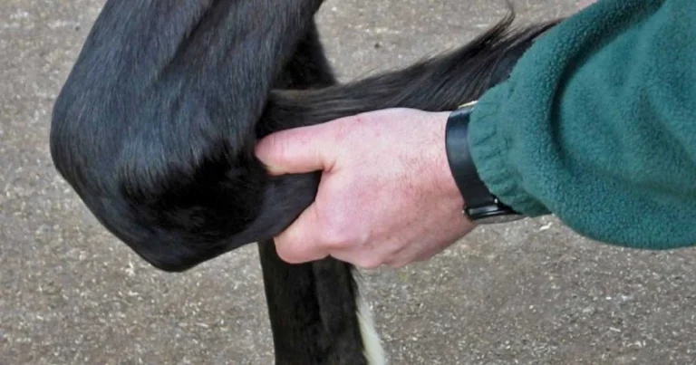
Proximal suspensory ligament palpation on forelimb.
With all the diagnostic technology now available – to the practitioner, as well as the hospital clinician – it is all too tempting to forget about those “human” skills that can make such a difference to the case because it can be quick and (relatively) cheap.
We are talking about the clinical skills of palpation and physical manipulation of the equine limb. The horse’s distal limb (where most causes of lameness are located) is a paradise for the palpating practitioner. The limb has minimal soft tissue cover, so that many structures such as tendons, ligaments, bones and synovial (joint and tendon sheath) cavities can be easily identified, and swelling detectable by comparing weight-bearing limbs and their significance as a cause of pain-related lameness confirmed when pressure is applied to the affected structure – usually best with the limb raised, so the tendons and ligaments are slack.
A protocol for (usually) starting at the distal part of the limb and working proximally without missing out any structure needs to be developed and practised by every vet. This takes time, which is why this author prefers to do the detailed palpation after the lameness examination, so it can be targeted at the affected limb(s).
A convincing and repeated response to palpation can obviate the need for diagnostic analgesia, while diagnostic imaging is usually still needed to make a definitive diagnosis and clarify its severity.
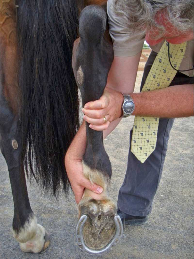
Of course, limitations to the capabilities of palpation exist: swelling and pain may have resolved, or the palpation of the structure is not possible. This is particularly true for structures within the hoof because of the rigidity of the hoof wall and sole. Hoof-testers are, therefore, essential to apply sufficient pressure to the sole to detect sites of pain (such as pus in the foot).
Furthermore, careful attention to where structures extend proximal to the limits of the hoof capsule can still provide clues – such as distension of the dorsal pouch of the distal interphalangeal joint, swelling of the distal interphalangeal joint collateral ligaments, and swelling and pain on pressure over the individual lobes of the deep digital flexor tendon in the distal pastern.
The latter shows sensitivity, with deep digital flexor tendon lesions within the foot in around one-third of cases, either because pressure on the tendon in the distal pastern either causes pressure in the painful area of the tendon inside the foot or, alternatively, because of the high frequency of combined pastern and foot lesions of the deep digital flexor tendon. This means that a positive response can be helpful, but a negative response does not eliminate the diagnosis, as is true for most palpation.
This relatively low “strike rate” should not deter clinicians from performing palpation because it can still be rewarding for a significant number of cases – especially in the acute stages –and its sensitivity and specificity can be improved in the hands of an experienced clinician. In spite of this, subsequent steps of diagnostic analgesia and imaging may still required for definitive diagnosis.
In addition to palpation, the components of the clinical examination of a lame horse includes visual assessment for conformational, evident swellings and muscle atrophy, evaluation of the gait (ideally including an objective assessment), and flexion tests.
Flexion tests are designed to place abnormal stresses on a region of the limb to exacerbate lameness and, thereby, give clues of the location of the site of pain. However, they are relatively insensitive, at least to some joints, and applying excessive stress and/or for excessive time can increase your false-positive rate, such as more “normal” horses showing a response.
Consequently, most people apply moderate pressure for around one minute and apply the same test to both limbs (fore or hind or all four) and also do distal and proximal limb tests, separately.
In reality, it is most discriminatory when the same person does all the flexion to “standardise” the test. The most significant responses are when a difference exists between limbs or between proximal and distal flexion tests, and so, is less helpful with bilateral pain in the same location.
Other variants have been proposed, such as extension tests for the foot (where the foot in placed on a wedge to elevate the toe, and the contralateral limb raised to apply extra weight to the limb with the toe elevated off the ground), or asymmetric loading of the foot with a board to apply asymmetric load to the structures within the foot (and potentially more proximal).
Manipulation of the limb (or other parts of the body, such as the neck) can reveal reduced range of motion, or pain, although these findings are not always relevant to the current site of pain, and so, should be interpreted with caution.
In summary, a thorough physical examination, including careful palpation, flexion and manipulation, is not only an essential, but also a rewarding part of the examination of any lame horse.
Even though the existence of false-positive and false-negative findings must be recognised, it can be a rich source of diagnostic information, which can prompt more specific subsequent diagnostic steps, such as diagnostic analgesia or imaging and, thereby, result in a more accurate and quicker diagnosis with cost savings to the client.
Think about attending the BEVA Congress this year and attending the talk if you want to learn more about the pros and cons of these commonly performed actions.
Roger KW Smith is professor of equine orthopaedics at the RVC, with particular interests in orthopaedic surgery, imaging, lameness and tendon research. Having qualified from the University of Cambridge in 1987, after two years in general practice, he undertook a residency in equine surgery at the RVC and then a PhD on the extracellular matrix of equine tendons. Roger holds the Diploma of Equine Orthopaedics, is diplomate of two European specialist colleges, and is an RCVS specialist in equine surgery. He was awarded an RCVS fellowship in 2016 for meritorious contribution to knowledge and was BEVA president in 2023.