18 Jul 2015
Aimi Duff highlights the vital contribution VNs can make in helping vets to assess horses that present with abnormal gait or stance, including useful techniques.
Aimi Duff
Job Title
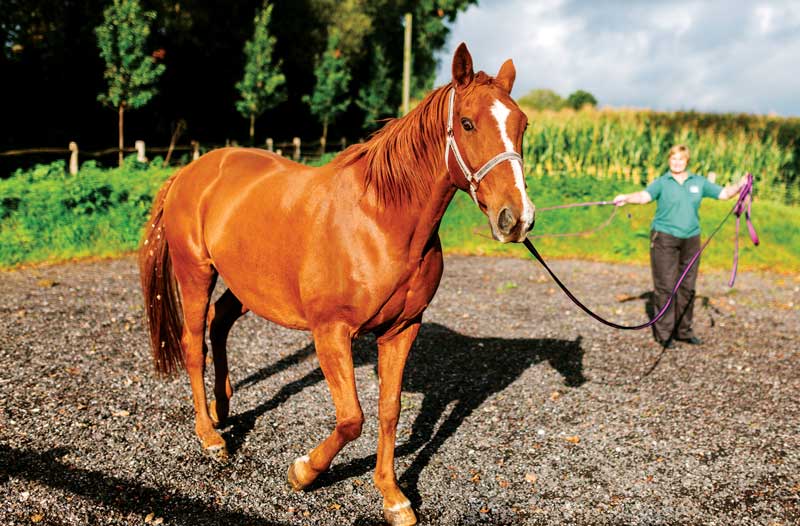
Walking and trotting a horse to assess its gait may be carried out on hard and soft ground as part of the lameness investigation process. IMAGE: ©urbancow/iStock.
Lameness is a common presentation in equine practice and the equine veterinary nurse plays many key roles in the investigative process.
In many practice situations, the veterinary nurse receives the initial communication from the owner and is involved in triaging the client. Veterinary nurses are crucial for assisting the veterinary surgeon in the diagnostic and treatment stages, before discharging patients and providing ongoing client support.
Lameness in horses can be variable in presentation and when the client contacts the practice, the veterinary nurse or receptionist receiving the telephone call must ascertain the urgency with which the patient should be seen.
Lameness emergencies requiring immediate veterinary attention include severe non-weight bearing lameness with a wound near a joint or apparent fracture, and severe lameness causing patient distress.
Clients may telephone to request an initial consultation with the veterinary surgeon or a “lameness work-up”. The latter is a more involved process, generally requiring the patient to be hospitalised where facilities permit while local anaesthetic agents are used to localise the lameness.
By placing local anaesthetic drugs around nerves and into joints, regions of the leg can be sequentially “numbed” to help isolate the painful area. This enables diagnostic imaging of the problem area so the cause for lameness might be identified and treated.
Several important considerations should be explained to the owner before a horse undergoes lameness investigations:
When a horse is lame on multiple limbs the veterinary surgeon may advise the horse be worked up over a couple of days. This may require hospitalisation, so the owner should bring any rugs, feeds and so on with the horse so its routine can be maintained as close to normal as possible.
A veterinary nurse has a key role in presenting a horse for lameness investigation. The veterinary surgeon will need to evaluate the horse’s gait, which involves assessing the horse walk and trot in a straight line and also observing the horse on the lunge.
The horse may be evaluated on hard and soft ground as part of the investigation processes – for example, lameness on a soft surface is more likely to reflect soft tissue injury.
The handler should wear a hard hat, gloves and steel toecap boots for this because horses in pain and in strange surroundings can be unpredictable.
A well-fitted head collar, bridle or chifney may be necessary to control the horse for gait evaluation.
When presenting the horse for gait assessment the handler should walk and trot the horse on a flat and even surface, away from and towards the veterinary surgeon, as requested. The horse should be led as straight as possible and the head allowed to move freely so a head nod indicative of lameness may be appreciated. The horse should ideally be lunged in a purpose-built lunge ring that contains the horse safely.
The veterinary surgeon will advise on whether to walk, trot or canter the horse and in which direction. Working on a circle increases the strain on bones, joints and soft tissue structures, which can increase the severity of lameness.
Because this might exacerbate the degree of pain the horse is suffering, the handler should be cautious of how the patient might react.
The veterinary surgeon might also wish to perform flexion tests on the patient. These involve the veterinary surgeon holding the limb in a flexed position for approximately one minute before the handler trots the horse off on a straight line. This causes an increase in intra-articular and subchondral bone pressure, stresses the joint capsule and causes soft tissue compression, so any lameness associated with these structures can be accentuated.
The normal horse may trot off lame for the initial two to three strides, but if this lameness persists it would suggest a problem with one of the joints flexed.
Following gait evaluation, the veterinary surgeon will examine the patient for indicators of disease and may then embark on perineural anaesthesia to localise the painful area.
Perineural anaesthesia or nerve blocks are best performed without sedation so they can be rapidly evaluated. This may warrant the use of a nose twitch or feed distraction if the patient is particularly fractious. It is up to the veterinary surgeon to decide whether the injection area is clipped before aseptic scrubbing to prevent infection-related complications.
The success of a nerve block can be evaluated by testing for skin sensation within five minutes of injection. If successful, the patient is then evaluated dynamically to detect an improvement in lameness, suggesting the cause of lameness is associated with a structure innervated by the nerve anaesthetised. If there is no improvement in lameness, further nerve blocks must be performed.
Once nerve blocks have localised the region of pain, diagnostic imaging can be used to identify the cause of lameness (radiography, ultrasound, MRI and so on) enabling treatment.
Once the source of lameness has been identified, a treatment plan can be formulated. This may involve strict rest or controlled exercise and various pharmacological therapies, which the veterinary surgeon will discuss with the owner.
On collection, the discharging veterinary nurse should ensure the owner is confident with the treatment plan and prescribed medications and aware of the dose and route of administration. Providing a written discharge form can help affirm recommendations when the owner returns home as, invariably, only a fraction of the information communicated verbally is retained.
This document can state exercise guidelines, medication instructions, feeding recommendations, any special instructions – for example, bandaging or cold hosing – and practice contact details.