23 Jun 2026
Mélanie Perrier MRCVS discusses a common issue in equine practice, including advances on diagnostics.
Mélanie Perrier
Job Title
Image: Valeri Vatel / Adobe Stock
Poor performance is very often a multifactorial issue in horses, but lameness remains one of the most common problems identified in equine veterinary practice.
Lameness refers to any change in movement caused by pain, injury or mechanical dysfunction in the limbs, joints or soft tissue structures. While some cases are straightforward, some will only appear as decreased athletic ability or behavioural changes. Early detection is deemed essential to protect both the horse’s welfare and performance.
One of the earliest recognised signs of lameness is a decrease in performance. This can be seen clinically as a reluctance to jump, reluctance to accept the bit, not taking the contact evenly, difficulty with transitions, getting disunited or reluctance to go forward.
Riders may also notice loss of freedom and elasticity of movement, shortened strides, the horse feeling uneven or an unwillingness to go forward. In many cases, these signs can be mistaken for poor behaviour or lack of training when they are often an early indication of pain prior to a consistent lameness being noticeable. Common causes of lameness include tendon and ligament injuries, joint disease, foot imbalance and bone pathology.
Several diagnostic steps are available to help identify lameness at an early stage.
The process usually begins with a detailed history followed by a thorough physical and gait examination, during which the horse is observed walking, trotting and turning.
Flexion tests may also be performed as an additional diagnostic tool. In poor performance cases, it is important to also perform a ridden examination as some of the clinical signs may only be seen under saddle.
To localise the exact source of pain, diagnostic nerve or joint blocks can then be used. Imaging techniques such as x-rays, ultrasound, and, more recently, magnetic resonance imaging (MRI), computed tomography (CT), nuclear scintigraphy and PET scan are valuable tools for obtaining a complete and accurate diagnosis1.
Common causes of lameness and reduced performance include degenerative joint disease of the hocks, suspensory ligament desmitis, synovitis and foot pain/imbalance. A good knowledge and understanding of the discipline the horse is being used for is also useful to understand the common pathologies that affect a particular type of horses2,3. Several publications outline specific conditions depending on the main use of the horse4,5,6.
For example, dressage horses are predisposed to metacarpophalangeal/metatarsophalangeal joint region injuries (osteoarthritis or suspensory branch injury). Suspensory ligament injuries in particular are a common cause of poor performance and lameness in these horses.
Dressage requires advanced movements that increase loading on the hindlimbs and place repeated strain on the suspensory apparatus. Over time, excessive stress may lead to inflammation, fibre damage or degeneration of the ligament. One of the challenges of suspensory ligament injury in horses is that clinical signs are often subtle at first. Horses may not show obvious lameness, but instead demonstrate reduced performance.
Common signs may include loss of impulsion, difficulty engaging the hindquarters, shortened stride length, resistance to collection, difficult transitions and reduced ability to perform lateral movements. Riders may also notice behavioural changes such as tail swishing, irritability or resistance under saddle7,8.
Diagnosis of suspensory ligament injuries is usually made using a combination of clinical examination, gait assessment (subjective and objective) and diagnostic anaesthesia. Ultrasound is the most common imaging technique used to evaluate ligament fibre damage and enlargement. In some cases, MRI or CT may also be helpful to detect lesions not readily seen on ultrasound. Modern technologies such as AI-assisted gait analysis and motion sensors are also increasingly used to identify small asymmetries associated with suspensory pain.
Overall, suspensory ligament injury is a significant cause of poor performance in dressage horses, and early recognition and accurate diagnosis are essential to protect the horse’s welfare and support a successful return to athletic work.
Other conditions that can be associated with lameness and poor performance and that may not be easily detectable include neck pathology and muscle dysfunction.
Neck abnormalities are becoming an increasing, but sometimes overlooked, cause of poor performance in horses. The equine neck plays a major role in balance, movement, flexibility and communication between the forelimbs, back and hindquarters. Problems affecting the neck can therefore reduce athletic ability, alter gait and cause pain or resistance during exercise. In many cases, horses with neck disorders show subtle signs of reduced performance before obvious lameness develops.
Common conditions include arthritis of the cervical vertebrae, nerve compression, trauma, congenital deformities, cervical vertebral stenotic myelopathy and stiffness caused by poor posture or training methods. Horses with neck abnormalities often show performance-related signs rather than clear clinical lameness. These may include resistance to bending, difficulty collecting, poor head carriage, stiffness in transitions, uneven rein contact, reluctance to jump, reduced impulsion or problems maintaining balance9,10.
Diagnosis is made using a combination of clinical examination and imaging techniques. Physical examination includes palpation of the neck muscles and joints, assessment of range of motion, and observation of the horse moving in hand and under saddle. Additionally, neurological examinations may be performed in cases where spinal cord involvement is suspected. Imaging methods such as radiography (x-rays), ultrasound, scintigraphy, and more recently CT help identify specific conditions.
Traumatic muscle injuries are also and increasingly common causes of recognised poor performance in horses, particularly in athletic and competition animals. These injuries occur when muscle tissue is damaged due to excessive strain, direct trauma, overexertion or accidents, such as slipping or falling. Muscle injuries may affect any part of the body, but they commonly occur in the back, neck, shoulder and hindquarters. Horses involved in racing, jumping, dressage and eventing are especially at risk because of the high physical demands placed on their muscles. Poor conditioning, fatigue, inadequate warm-up and sudden increases in workload can increase the likelihood of injury.
There are several types of traumatic muscle injuries. Muscle strains occur when fibres are overstretched or torn, while contusions result from direct impact or bruising. More severe injuries may involve tearing of larger muscle groups or damage to surrounding tendons and ligaments. In some cases, repeated strain can lead to chronic muscle pain and stiffness.
Signs of muscle injury are often associated with poor performance rather than consistent lameness. Affected horses may show stiffness, shortened stride length, reluctance to move forward, reduced speed, difficulty bending, poor jumping ability or resistance during work. Swelling, heat, pain on palpation and muscle spasms may also be present. Horses with back or hindquarter injuries may struggle with collection, transitions or engagement from behind. Behavioural changes, such as irritability, tail swishing, bucking or unwillingness to work, are also common indicators of discomfort11,12,13.
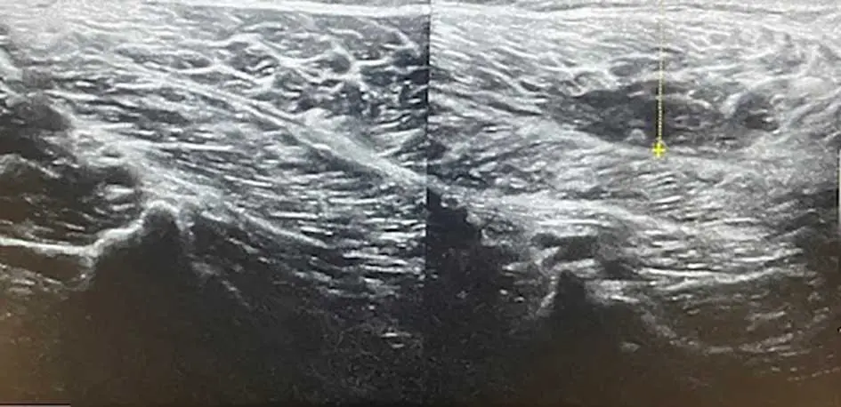
Diagnosis is achieved through clinical examination and diagnostic imaging. Palpation is used to identify painful or swollen areas, while observation of the horse moving helps assess the effect on gait and performance. Ultrasound is commonly used to evaluate muscle fibre damage, fluid accumulation and healing progress (Figure 1). Ultrasonography, scintigraphy, MRI and blood tests measuring muscle enzymes such as creatine kinase (CK) and aspartate aminotransferase (AST) may also assist diagnosis.
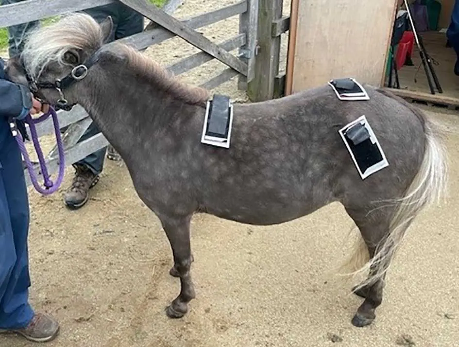
As mentioned, early diagnosis is key in managing any horse presented for lameness/poor performance as it will allow prompt treatment and helps prevent more serious injuries. Advances in equine sports medicine have improved the understanding on how different pathologies affect performance. Researchers increasingly use gait analysis14, pressure sensors and AI-assisted movement tracking to study changes in performance and biomechanics (Figure 2). Similarly, ethograms and behavioural assessments are also being used to identify pain-related behaviours linked to orthopaedic problems.
Artificial intelligence (AI) technology can also be used to improve the accuracy and consistency of diagnosis. Because the analysis is data-based rather than purely subjective, it reduces differences between observers and helps practitioners monitor changes over time. Repeated measurements can show whether a horse is improving, remaining stable or becoming worse, not only during training, but also during the rehabilitation period.
In the past few years, researchers have explored the use of machine learning and predictive models in equine medicine. These systems may eventually predict injury risk before clinical signs appear. Some smart stables already use continuous monitoring devices to track horses daily and alert owners to changes in gait or activity levels.
Some of the limitations of AI-assisted gait analysis include the costs of the required equipment and its difficulty of interpretation. Environmental factors such as uneven surfaces or poor sensor placement may also affect results. Therefore, AI technology should be used to support, but not replace, clinical examination by veterinarians that should remain the gold standard.
Overall, AI-assisted gait analysis is an important advancement in equine sports medicine. By enabling earlier and more accurate detection of lameness, it helps improve horse welfare, prevent injury and support athletic performance.
Predictive injury modelling is an emerging area of equine sports medicine that uses AI, biomechanics and data analysis to predict the likelihood of injuries before obvious clinical signs appear. The goal is to identify horses at risk of developing lameness or musculoskeletal problems early enough for preventive action to be taken. This approach is becoming increasingly important in racing, dressage, show jumping and endurance sports, where injuries can significantly affect both performance and welfare.
Traditional veterinary diagnosis often occurs only after a horse begins to show signs of pain or reduced performance. Predictive injury modelling aims to shift veterinary care from reactive treatment to proactive prevention. By analysing substantial amounts of data collected over time, AI systems can find subtle patterns associated with future injury risk.
These models rely on several types of information. One of the most important sources is gait analysis data collected through wearable motion sensors, inertial measurement units (IMUs), force plates and high-speed video cameras. These tools measure stride length, limb symmetry, joint movement, acceleration and weight distribution. Even very small changes in movement may indicate underlying issues.
Training and workload data are also important. Information such as exercise intensity, frequency of competition, speed, heart rate, recovery time and surface conditions can be analysed to determine whether a horse is being overworked. It is, for example, recognised that sudden increases in training load are strongly associated with tendon injuries and stress fractures.
In addition, predictive models may include veterinary records, imaging findings, shoeing history, genetic factors and environmental conditions. Machine learning algorithms process these complex datasets and compare them with patterns from previously injured horses. Over time, the AI system “learns” which combinations of factors are most strongly linked to injury.
One practical application is the prediction of stress-related bone injuries in racehorses. Research has shown that subtle gait asymmetries and changes in stride patterns may appear before a catastrophic injury occurs. Early warnings allow veterinarians and trainers to reduce workload, modify training or carry out further diagnostic tests before serious damage develops.
Predictive injury modelling has several advantages. It improves horse welfare by reducing preventable injuries, helps extend athletic careers, lowers treatment costs and supports safer training practices. It also provides more objective decision-making for trainers and veterinarians15,16,17.
However, there are still limitations. Accurate predictive systems require access to large datasets and continuous monitoring, which can be expensive and time-consuming. Horses also vary greatly in conformation, discipline and movement style, making prediction complex. AI predictions therefore cannot replace veterinary expertise and clinical judgement14,18.
Despite these challenges, predictive injury modelling is considered one of the most promising developments in modern equine medicine. As AI technology and data collection continue to improve, predictive systems may become a routine part of equine health management, helping veterinarians prevent injuries before they become clinically visible.
An ethogram is a detailed list or catalogue of behaviours displayed by an animal. In horses, ethograms are used to observe and record specific behaviours that may indicate pain, stress, discomfort or disease. In equine veterinary medicine, ethograms have an increased use in identifying lameness and musculoskeletal pain, especially in cases where physical signs are subtle or difficult to detect19.
Traditionally, lameness diagnosis focused mainly on gait analysis and physical examination. However, researchers now recognise that horses may often express pain through changes in behaviour before obvious lameness appears. Behavioural observation using ethograms could therefore provide an additional tool for early detection20.
Common signs to look for in a lameness ethogram include ear pinning, tail swishing, head tossing, reluctance to move forward, visibility of the sclera, resistance during transitions, abnormal posture, stumbling, bucking, or unwillingness to be saddled or mounted. Horses experiencing pain may also show facial expressions linked to discomfort, such as tension around the eyes, nostrils, and mouth. To that effect, a “Ridden Horse Pain Ethogram” (RHpE) was developed to help identify musculoskeletal pain in ridden horses. The RHpE includes a checklist of behaviours observed while the horse is under saddle. Research has shown that horses with higher RHpE scores are more likely to have underlying lameness or back pain21,22.
Ethograms are valuable because they encourage thorough observation and they help owners, riders and veterinarians recognise that behavioural problems may have a physical cause rather than being purely training issues or disobedience.
Modern technology is also improving the use of ethograms. Video analysis and artificial intelligence systems are also being developed to automatically monitor horse behaviour and identify patterns associated with pain or stress. Combining behavioural data with gait analysis and wearable sensors may provide a more complete understanding of equine lameness in the future.
Despite their usefulness, and similarly to other diagnostic tools used in the investigation of poor performance, ethograms do have limitations. Some behaviours may be influenced by temperament, rider skill, environment or training methods rather than pain alone. Therefore, ethograms should not replace a full veterinary examination, but should be used alongside clinical assessment and diagnostic imaging.
In conclusion, a good knowledge of the discipline the horse is used for and regular evaluations using a variety of diagnostic tools including more objective data analysis such as gait analysis, AI systems and integrating an ethogram in serial evaluation of horses may be useful in early detection of lameness as a cause of poor performance in horses. In return, this would help put in place an early comprehensive treatment and rehabilitation plan that would optimise return to athletic function.
Mélanie Perrier graduated in France, then went on to complete a surgical internship, followed by a surgery residency, in the US. She was then a clinical assistant professor on equine surgery at the University of Tennessee, before returning to private practice in the Middle East and France. Mélanie is a lecturer in equine surgery at the RVC.