13 Jul 2021
Ann Derham looks at the most common types of lameness in horses, the importance of an orderly and logical examination and preventive strategies.
Ann Derham
Job Title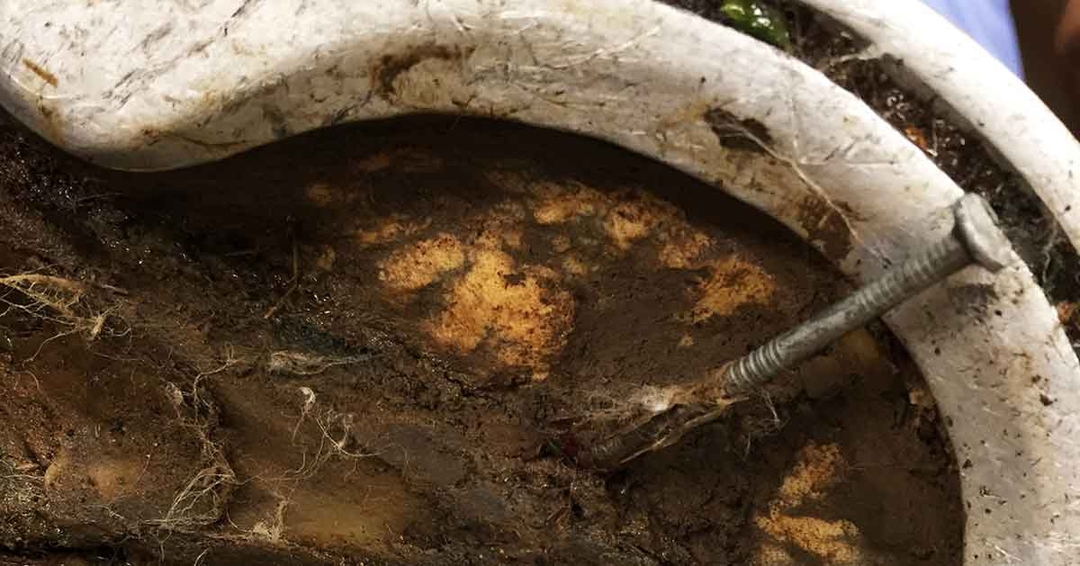
Lameness is a common problem, and can vary from subtle and intermittent to acute non-weight-bearing. Lameness examinations may seem daunting; however, knowledge of the basic anatomy and common diagnostic analgesic techniques will allow the clinician to provide a good standard of lameness examination that both you and your client will be happy with.
The following article covers the most common types and causes of lameness, while highlighting common investigation challenges and factors that predispose horses to lameness.
Equine lameness can be broadly divided into two types:
These may be due to a single traumatic event or represent a previously missed subtle lameness that has suddenly been exacerbated. Horses with a sudden onset of lameness are typically moderately lame to non-weight-bearing. In such cases, it is often best to assume a worst-case scenario (for example, a fracture) until proven otherwise. In the case of acute tendon injuries, septic synovial structures and/or fractures, these should always be considered an emergency, as prompt diagnosis and treatment is critical to preventing further damage – and a worsening prognosis.
These injuries are related to repetitive trauma that is sustained over time (such as desmitis or OA). Lameness is variable and presenting complaints may be vague (for instance, refusing at fences or ill-defined lameness) or very specific (left forelimb lameness worsened when turning on hard ground).
The underlying cause of lameness in the horse may be reasonably straightforward – for example, septic joint or fracture – or more complex, such as palmar heel pain due to chronic conformational issues.
A primary or baseline lameness is one where the gait abnormality is seen when the animal is walked or trotted in a straight line – that is, it doesn’t require flexions to make it lame. A secondary or compensatory lameness is commonly seen in the contralateral limb, or the hindlimb of a primary forelimb lameness (or vice versa), due to increased loading of the unaffected limb.
In most horses, lameness is more commonly found in the forelimb. This is due to the horse’s centre of gravity being positioned closer to the forelimbs, resulting in a forelimb/hindlimb load distribution of 60 per cent to 40 per cent, respectively. Also, during canter and gallop only one forelimb is weight-bearing during the stride cycle. In trotters or pacers that work with a sulky, and in sport horses that are performing collection and propulsion through most of their work, the load is increased in the hindlimbs, which results in a higher proportion of hindlimb lameness. Causes include:
Soft tissue and bone inflammation resulting from a single event (fracture or dislocation), repetitive trauma (arthritis, desmitis, tendonitis or neuropathies), or an immune-mediated event (severe rhabdomyolysis).
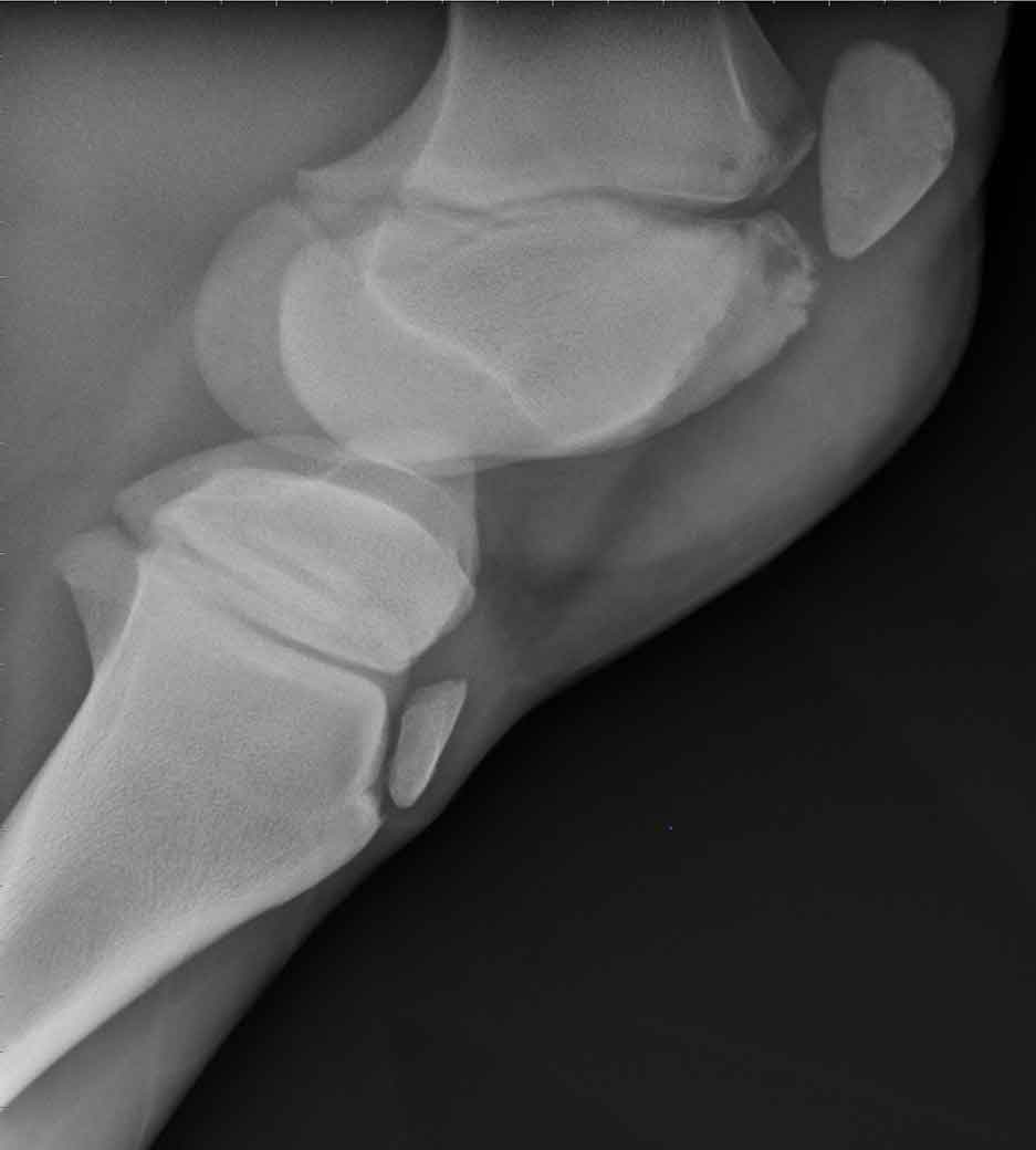
Types include osteomyelitis (Figure 1), septic arthritis/tendinitis/physitis (Staphylococcus aureus, Streptococcus equi or Rhodococcus equi) or lymphangitis.
These can affect the bone, soft tissues, or both. Examples include osteochondrosis (the most common), and flexural and angular limb deformities.
Improperly balanced feet, toed-in/toed-out and broken hoof-pastern axis have been linked to musculoskeletal injuries due to increased stress and overloading of the associated joints and soft tissue structures1-3.
Includes laminitis in at-risk horses suffering from equine metabolic syndrome and pituitary pars intermedia dysfunction and selenium toxicity; bone diseases/developmental disorders due to imbalance in trace minerals (copper, magnesium or zinc); high-protein diets; and excessive/deficient levels of vitamins A and D.
Lameness investigation should always be performed in an orderly and logical manner (thorough history and clinical exam, followed by lameness evaluation, diagnostic analgesia or diagnostic imaging). However, not all examinations will go according to plan, and sometimes the clinician must adapt to the situation. Some common challenges include:
In such cases, depending on the history and degree of lameness, the diagnostic analgesia part of the exam should be avoided – and the animal imaged first to rule out an underlying fracture.
Light sedation may be warranted to prevent injury to the handler, examiner and the horse itself. The use of acepromazine and low-dose xylazine may be required to safely perform the exam. If it is not possible to do flexion tests or diagnostic analgesia safely, referral for advanced imaging – such as nuclear scintigraphy – may be warranted.
Dry, flat and hard surfaces are best for lameness evaluation. Where possible, arrange for the animal to be examined in a suitable, quiet and safe area.
Make sure enough time is set aside for the lameness examination. This is especially true in cases where more than one limb may be involved.
This is one of the more frustrating challenges to deal with. It is best to discuss estimates with the client prior to starting any lameness evaluation. That way you can work out if you have, for example, to rule out certain diagnostic tests or have to be more selective with your nerve/joint blocks.
Withdrawals on local anaesthetic and sedation should always be considered if you are examining a competition horse or racehorse.
Perineural nerve blocks and intra-articular anaesthesia of the equine limbs – and their function in diagnostic evaluations – are well documented4,5. The interpretation of responses to the blocks can be difficult, especially in abolishing and assessing horses with subtle lameness. Subchondral bone pain, incomplete fractures and OA may be inconsistently abolished by intra-articular analgesia, as it does not penetrate the subchondral bone sufficiently. In these cases, peripheral nerve blocks may work better. A similar issue is seen in articular pain, which may arise from more than one source (for example, synovium, periarticular ligaments or subchondral bone) where peripheral nerve blocks may be more appropriate5. False negative responses due to a variance in the neuroanatomy can make interpretation very difficult. It should be stated that intra-articular blocks are not benign procedures and, if these are to be performed, full aseptic technique must be followed to avoid causing a septic arthritis.
The examination is made more difficult when a bilateral lameness is present, with neither limb predominant, and the horse doesn’t appear overtly lame on baseline until one limb has been blocked – for example, racehorses with bilateral third carpal bone disease. Some disorders, such as stringhalt or shivers, can complicate/confuse the diagnosis of a completely separate primary lesion causing lameness.
Good conformation is key to a sound athletic animal, as it plays a vital role in lameness development, with many well-recognised conformational faults leading to lameness problems. Angular and flexural limb deformities (including carpal/fetlock varus/valgus or contracted tendons) should be noted in young animals and managed appropriately – for example, through farriery or surgery. Horses with poorly balanced feet should be corrected, and routine farriery should be performed every four to six weeks to ensure feet are properly trimmed, balanced and shod to meet the horse’s work demands. Allowing conformation issues to progress leads to overloading of the soft tissue structures of joints, increasing wear and tear; predisposing to OA, tendonitis and desmitis; and significantly shortening the performance life of an animal. Evaluation of conformation is, therefore, an essential part of the lameness examination.
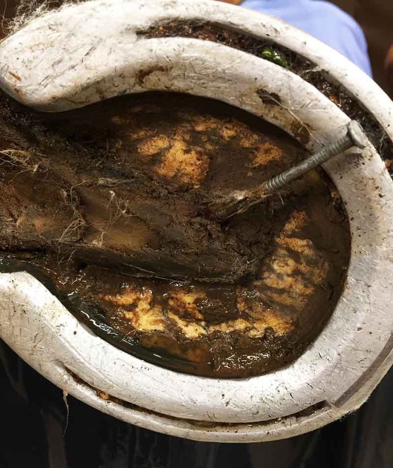
Ensure your clients understand the importance of performing daily checks on their horses and looking for any swellings or focal areas of heat that may warrant further investigation before becoming a serious problem. Daily hoof care is of paramount importance and is often underestimated. Poor hoof hygiene can reduce hoof quality and, coupled with mineral-deficient diets and excessive or uncontrolled exercise, can lead to long-term hoof damage and lameness. Clients should be advised to keep arenas, paddocks and stalls in safe condition to prevent injury (Figure 2). General health practices, such as deworming programmes and vaccinations, will help limit systemic illness that may have knock-on effects on predisposing the horse to injury.
Maintaining a horse in optimal body condition provides energy for the musculoskeletal system, and helps minimise joint and soft tissue injuries. Obesity places excessive force on joints and soft tissue structures, risks insulin resistance that predisposes horses to laminitis, and disrupts endochondral ossification in young growing horses that may play a role in osteochondral (OC) disease development. For youngstock, it is recommended high carbohydrate diets are avoided during periods of rapid growth, as it has been linked to OC disease development and causing angular limb deviation. Always make sure diets are balanced to avoid them being too high or too low in trace elements such as calcium and phosphorous, which are fundamental for bone metabolism.
A basic level of exercise is needed for proper development of the musculoskeletal system, especially for articular cartilage. All exercise regimes should be introduced gradually and consistently on good surfaces, and high peak intensity should be avoided. Consideration should also be given to the animal’s age, type and discipline.
Pacers and trotters will have a higher incidence of hindlimb lameness compared to racing Thoroughbreds because of the added load of the sulky. Similarly, hindlimb issues are more common in dressage and showjumpers versus pleasure horses, due to the collection and propulsion needed in their performances.
Lameness is a common problem in our patients, and while treatments are available for many conditions, prevention of lameness in our patients should always be the goal.
Clinicians should pay particular attention to poor routine management (for example, foot care or diet), abnormal conformation and the type of work or exercise the horse performs or is expected to do, as these can all lead to poor performance and lameness problems in the future.