13 Jul 2022
Lameness may or may not be related to pain, and vets must rule this out in the first instance. Vets leave vet school with lots of knowledge about evaluating and diagnosing pain, but Edele Grey also points out an increase in tools to help clinicians carry out a subjective assessment.

Image © Terri Cage / Adobe Stock

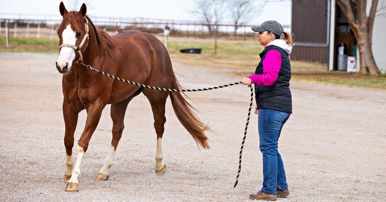
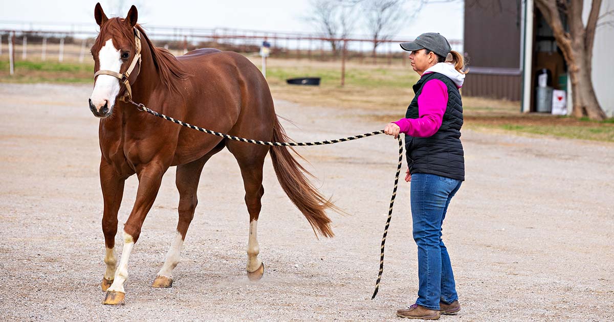
Lameness is generally considered to be an aberration in a horse’s gait and movement. It may or may not be related to pain; however, as veterinarians entrusted with the welfare of our patients, we must responsibly rule out pain as a source in the first instance. Orthopaedic injuries are arguably the most important cause of days lost from training (Dyson et al, 2008; Egenvall et al, 2013).
On graduation from vet school, we’re all equipped with the tools to evaluate and diagnose many causes of lameness in our equine patients. Additionally, in the past decade, the number of commercially available systems providing an objective evaluation to complement a clinician’s subjective assessment has increased.
Most common of these are arguably inertial measurement unit (IMU) systems and, although beneficial, these systems should not be fully relied on – with interpretation of their results being nuanced (Pfau et al, 2016).
In the rapidly changing and extensively researched area of lameness evaluation and diagnosis, it can feel overwhelming when trying to remain up to date. The purpose of this article is to help veterinary surgeons interpret the most clinically relevant results of this research.
Certain causes of lameness can be prevented through management, such as hoof care and farriery, which can aid in preventing thrush and hoof wall defects, while training modification may reduce the incidence of dorsal metacarpal disease, which is also known as “bucked shins” (Verheyen et al, 2005).
Conversely, septic arthritis or fractures due to an unforeseen injury can’t feasibly be prevented (Hinchcliff et al, 2013). Prevention of some stress fractures and proliferative periostitis of small metacarpal and metatarsal bones, or “splints”, may not be as simple as modifying the training programme. These also require early detection using diagnostic imaging and careful monitoring of at-risk athletes (Hinchcliff et al, 2013).
Jackson et al in 2020 reported on the use of parathyroid hormone fragment peptide 1-34 within a hydrogel as a treatment for subchondral cystic lesions. While this use has been described since 2007 (Fuerst et al, 2007), this most recent paper assessed the response at different dosages. This may facilitate commercial use of these gels.
Chronic foot pain can be due to a variety of disorders and a range of management strategies is available to practitioners, from remedial farriery to surgical neurectomy. Dau et al (2020) reported a case series on the use of two per cent ammonium chloride as a neurolytic agent. This case series, while limited to just 10 horses, indicated that 3ml of the solution injected perineurally to the palmar digital nerves resulted in lameness improvement for up to two months in up to 72 per cent of horses. This improvement did appear to inversely correlate to the severity of radiographic lesions (Dau et al, 2020).
Lameness assessment is inherently subjective, with significant disagreement between practitioners. In recent years, objective gait analysis has become more popular with equine practitioners. IMUs are one of the most commonly seen evaluations in private practice. The asymmetry detection precision of the motion sensors is 3mm to 7mm, which is below the threshold visible for practitioners (Pfau et al, 2016). However, in purchasing one of these systems, veterinary surgeons should be aware of the limitations of sensors in that they currently cannot quantify every gait adaptation that’s made by lame horses.
Subtle lameness localisation can be difficult to interpret with multiple vets selecting opposing limbs as the lame one. Even more disagreement exists between vets and the public, with up to 65 per cent of horses in one study deemed sound by the owner, but lame by an examining veterinarian (Dyson and Greve, 2016). Other studies have found lameness in up to 72 per cent of horses deemed “owner sound” (van Weeren et al, 2017).
While much debate surrounds the commercialisation of these objective gait analysis systems (van Weeren et al, 2018), they can play a role in the modern lameness investigation. These analysis systems are meant to complement the clinician’s skills, not replace them.
A ridden horse pain ethogram (RHpE) has recently been described (Dyson, 2021) that has identified 24 behaviours more likely to be seen in lame horses compared with non-lame horses. Verified in horses that perform dressage-type movements (Dyson et al, 2020), this tool shows promise, although further investigation in other disciplines is required. One benefit of the RHpE is its potential use in poor performance situations where no overt lameness is visible to the veterinarian.
Some of the parameters are intuitive and likely considered to potentially be pain-related among a number of equine professionals, including tail swishing, repeatedly disunited in canter, repeated stumbles or gait changes, and hindlimbs not tracking with forelimbs, but deviating to one side.
Others are more subtle and may not be as obvious to the observer, such as exposed sclera or an intense stare for more than five seconds. It was noted that not all lame horses will display a sufficient number of behaviours within the RHpE to fit the classification. In the development of the RHpE, all non-lame horses scored below the threshold cited (Dyson et al, 2020).
In 2019, a critically appraised topic reviewed available research into the use of alpha-2 adrenergic agonists when performing lameness investigations (De Cozar, 2019). To date, studies appear to support the use of alpha-2 agonists without impeding lameness assessment. Young or fit athletes in pain can be highly strung or even fractious, which makes handling during a lameness evaluation difficult and, in some cases, even dangerous for both patient and personnel.
The use of short-acting xylazine in low doses doesn’t appear to mask lameness, but caution should always be used when interpreting mild lameness cases (Rettig et al, 2016; De Cozar, 2019). In 2021, Termansen and Meehan found that while the limited research indicates that the use of alpha-2 agonists (with or without concurrent use of the opioid butorphanol) may be suitable for use in lameness investigations, their use needs more standardised research – and some gait changes are noted on objective measurement assessment.
While it has been understood that diagnostic anaesthesia isn’t as specific as once thought due to diffusion of local anaesthetic, an interpretation bias by investigating clinicians has been noted (Pilsworth and Dyson, 2015). This tends to occur when the veterinarian expects an improvement to a particular block, possibly due to preconceptions about the clinical signs and athletic discipline. Partial resolution may indicate multiple sites of pain (Pilsworth and Dyson, 2015), not just that the nerve block needs longer to work.
The traditional distal to proximal diagnostic anaesthesia approach may not always be most suitable, due to the diffusion of medication, and some may advocate intra-articular anaesthesia in the first instance if localising signs are seen (Gómez Álvarez, 2019). A caveat to this relates to diagnostic intra-articular anaesthesia of the stifle. Radtke et al (2020) indicated this might improve foot lameness in up to 33 per cent of horses within just 30 minutes.
Laminitis remains a persistent concern for horse owners, with equine obesity persisting in the leisure populations. An initial three-part study with a short observation period (five days) of experimentally induced laminitis indicated the possibility of a surgical option to prevent rotation of the distal phalanx (P3; Carmalt et al, 2019).
The short sample size and observation period are major limitations to this study. However, promising results do show that a single 5.5mm diameter cortical bone screw placed through the dorsal hoof wall may withstand the cyclical loading of P3 in living horses to prevent rotation during an episode of laminitis.
This early research may provide hope for laminitis patients in the future. However, the degree of P3 rotation isn’t a reliable prognostic indicator compared with distal displacement (sinking) of P3 (Orsini et al, 2010). However, many vets use this in conjunction with the patient’s comfort and integrity of the sole to assess the prognosis for each horse.
A recent study described the use of intralesional plasmid DNA that encoded species-specific growth factors to treat naturally occurring tendinitis and desmitis (Kovac et al, 2018). This research shows promise, though much further investigation is needed before it may become viable for routine clinical use. The promise held in this may lie in the targeted management of angiogenesis to promote soft tissue regeneration in tendons and ligaments, which are notoriously poorly vascularised.
Some interesting research is emerging in the field of lameness investigation, including objective assessment of gaits that may assist veterinarians in complex lameness cases –although limitations exist.
New determinations on the use of diagnostic anaesthesia/analgesia, and how inadvertent desensitisation of areas distal to the site of injection may impact our interpretation of nerve blocks, have also been made. Novel gene therapies and the potential for commercialisation of parathyroid hormone fragment peptide within a fibrin hydrogel provide exciting opportunities for alleviating lameness in our equine patients.
A word of warning, though – other regenerative methods, such as autologous bone marrow and platelet-rich plasma, held promising results in their experimental models, although they have poorly translated to clinical treatment of severe tendonitis and desmitis in our equine patients.
It is also the author’s opinion that practitioners shouldn’t be overly concerned with the use of xylazine in low doses when performing lameness examinations on fractious patients.
Recent research indicating that intra-articular anaesthesia of the stifle can actually improve lameness in the foot should give veterinarians pause before performing nerve blocks, to ensure they are conscious of further limitations of diagnostic anaesthesia/analgesia than once thought.