
04 August 2026
In this article from Hallmarq, vets are asked if it is time to review how they select their standing MRI cases.

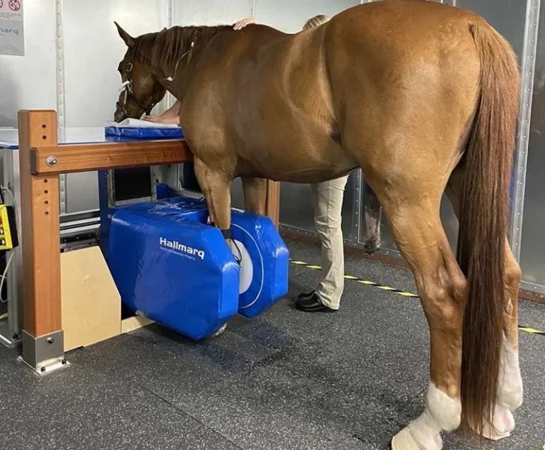
Image: Dr Natasha Werpy
Standing MRI (sMRI) has long been the gold standard for imaging the equine foot in the standing horse. That label is well earned and it’s not going away. But there’s a question worth asking in 2026: is it time to review my standing MRI case selection?
If sMRI is (and always has been) the gold standard for the foot, then what lies ahead for the rest of the limb?
The answer, with two significant advances now live on Hallmarq’s standing MRI systems, is: considerably more than it used to be.
This piece is for the vets who already use standing MRI and the colleagues who refer to them. It’s about what iNAV and AI-enhanced imaging (pAIce) have changed – not in theory, but in practice – and why now is the moment to review the cases you might previously have approached differently.
When standing equine MRI was first introduced, scanning above the coronary band was a genuine challenge. The further up the limb, the more movement there was and movement is the enemy of diagnostic image quality. For years, the foot remained the primary focus of sMRI not because MRI wasn’t valuable higher up, but because consistent diagnostic outcomes were harder to guarantee.
That limitation shaped referral habits. Vets learned, reasonably, to reach for sMRI when localisation pointed to the foot and to consider other options when it didn’t.
The technology has moved on significantly. The referral criteria probably haven’t kept pace.
iNAV is Hallmarq’s advanced motion correction software. Built specifically for veterinary applications, it tracks the position of the horse’s limb three times per second during image acquisition. iNAV is a step change in sampling frequency compared to what came before – and corrects for motion as the image is processed.
The result is immediately visible in the scans. Where proximal limb images once carried the telltale blur of patient sway, iNAV-corrects images to reduce patient artefact. It shows the kind of clarity that was previously associated only with foot scans: the proximal suspensory, the carpus, the tarsus, the fetlock: all areas where iNAV makes a material difference to diagnostic confidence.
As Dr Natasha Werpy, DVM, DACVR, puts it:
“The introduction of iNAV has dramatically advanced magnetic resonance imaging of the standing horse and strengthened our confidence in diagnosing clinically significant injuries in the proximal limb.”
For existing Hallmarq customers, iNAV is already on your system. It’s integrated into scanning protocols to solve the problem of motion impact on image quality. And, as with all Hallmarq software upgrades, this comes at no additional cost.
What AI brings next: pAIce
If iNAV addresses the motion problem, then AI-enhanced imaging (using Hallmarq’s pAIce technology) addresses the time problem.
Using advanced AI denoising algorithms, pAIce removes noise from MRI images while keeping resolution and contrast fully intact. The result is scan time reductions of between 50-75%. That’s not a marginal efficiency gain. Such a significant reduction in scan times changes the clinical calculation in meaningful ways:
Simon Yeung, machine learning engineer, Hallmarq Veterinary Imaging, says:
“We have developed an AI model that can denoise images, providing customers with reduced scan times and – often as a result of less patient movement – improved image quality. It’s an exciting project to work on and we’re anticipating great things as it enhances overall efficiency.”
Why NOW is the time to review your case selection!
Together, iNAV and pAIce fundamentally change the risk/benefit assessment of scanning above the foot in the standing horse. There implementation paves the way for a much wider range of cases that may previously have been overlooked as unsuitable for sMRI.
The historic objections – uncertain image quality in the proximal limb, long scan times demanding extended sedation, unpredictable diagnostic yield – are each materially reduced. What remains is what has always been true, that:
MRI provides excellent soft tissue evaluation while also enabling detection of early osseous changes, particularly bone oedema-like signal, with standing MRI currently representing the only modality capable of detecting these changes in the standing horse, and can identify pathology earlier than radiography or ultrasonography in a significant proportion of cases.
For referring and referral vets alike, reviewing your case selection is worth working through systematically.
Hallmarq’s case study library includes a growing collection of iNAV and pAIce-supported cases across proximal limb anatomy, including long medial collateral ligament injury of the fetlock joint and abnormal sclerosis of the central tarsal bone, among others.
These cases are worth reviewing. Not just for the clinical detail but for what the images themselves communicate: the quality that iNAV delivers in regions that would have been technically difficult to scan to diagnostic standard only a few years ago.
If you’re referring cases to a Hallmarq sMRI centre, the message is straightforward: the technology behind the system that your colleagues are scanning with has changed, and the outcomes you can expect from proximal limb referrals have changed with it.
iNAV and pAIce are already live. Both are built into the platform your patients will be scanned with. When you refer a horse with suspected proximal suspensory injury, pathology involving the carpus, tarsus, fetlock, metacarpus/metatarsus,pathology, you can do so with the same confidence that has always applied to foot referrals.
The diagnostic outcome is there. The scan times are shorter. The case for early referral – before compensation patterns become established and treatment options narrow – is stronger than it has ever been.
Hallmarq’s standing equine MRI earned the gold standard designation for the foot through clinical outcomes, replicated across thousands of cases over two decades. That standard is not a fixed point, it’s a direction of travel.
iNAV and pAIce are part of that ongoing journey. The foot remains the foundation. But with the proximal limb now genuinely within reach of consistent diagnostic imaging in the standing horse, the ceiling for what sMRI can offer has moved.
With iNAV to address motion, and pAIce to address speed, you’re in the perfect position to get more from your standing MRI than ever before. So why not AIm higher!