4 Sept 2017
Sonya Miles reports the case and treatment method of a rabbit that presented with symptoms of this condition.
Sonya Miles
Job Title
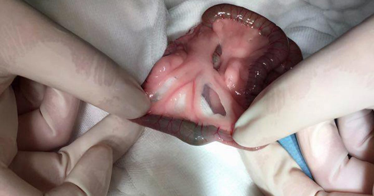
Figure 2. The intestinal obstructions (hair pellet).
One of the most common reasons domestic rabbits (Oryctolagus cuniculus) are presented to veterinary practices is due to gastrointestinal disease (Harrenstien, 1999; Huynh and Pignon, 2013). Gastrointestinal disease in rabbits has a number of aetiological agents, including genetic factors, lack of fibre in the diet causing dental disease, other sources of pain and dehydration (Harrenstien, 1999). A multitude of symptoms were demonstrated by a rabbit with gastrointestinal disease, including a hunched posture, bruxism, lack of faeces, anorexia, bloating of the stomach and collapse (Harrenstien, 1999; Huynh and Pignon, 2013).
In cases of acute anorexia, bloating of the stomach is common and often caused by an intestinal obstruction, which prevents the outflow of the constantly produced saliva from the stomach (Harcourt-Brown, 2007b). Most cases of gastric distension caused by intestinal obstruction need urgent stabilisation and surgical intervention (Harcourt-Brown, 2007a; b).
A four-year-old, neutered, male domestic rabbit – weighing 1.9kg – presented with a 12-hour history of anorexia and lack of faecal production.
The rabbit was found in a hunched position; bruxism and pain were noted when picked up by the owner. The rabbit was lethargic and the owners had not seen it drink anything in a 24-hour period. The companion rabbit was showing no clinical signs, but was brought in to keep its bonded companion company. On initial clinical examination, obvious gastric dilation and pain were noted on palpation; no gut sounds were heard on auscultation. The rabbit was tachycardic with a heart rate of 340 beats per minute; tachypnoeic with a respiratory rate of 250 breaths per minute, as well as hypothermic (36°C) and hyperglycaemic (28.4mmol/L).
Warmed, IV fluids therapy (IVFT) was initiated via the marginal auricular vein, with an initial bolus of 10ml/kg Hartmann’s IV buprenorphine (0.05mg/kg) also administered. The rabbit was monitored closely, while its temperature was actively raised using a heat pad in a dark, quiet kennel.
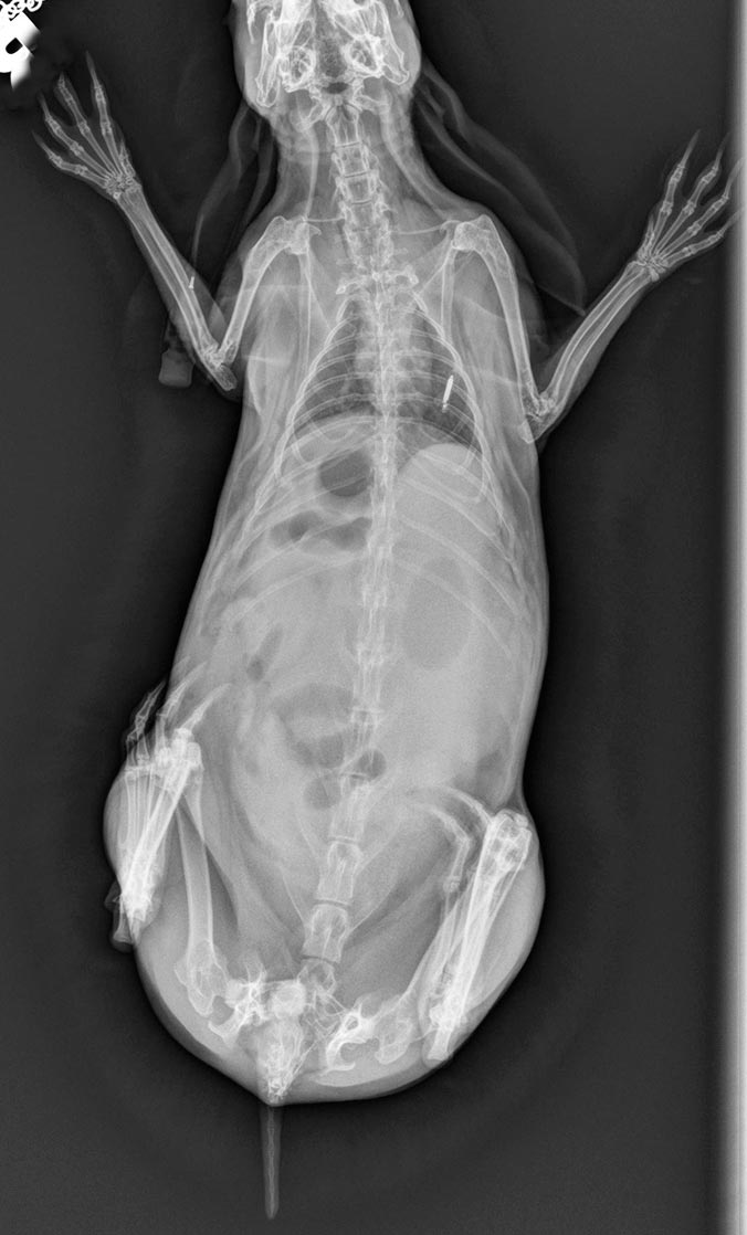
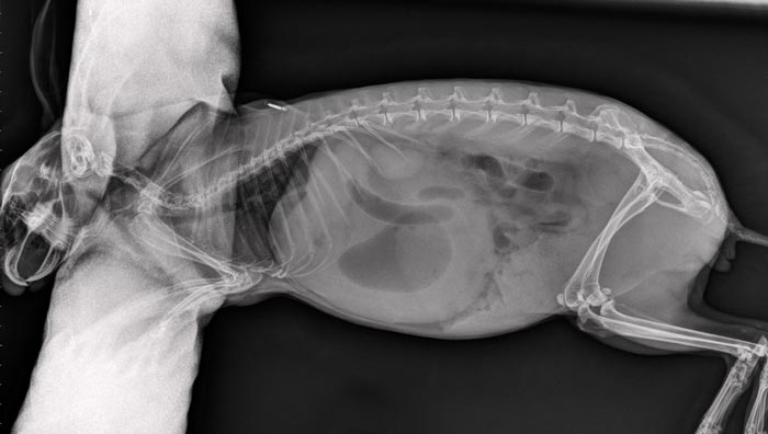
After 60 minutes, the rabbit was normothermic, but still tachycardic, tachypnoeic, painful on palpation and hyperglycaemic. The stomach was more dilated on palpation. Conscious left lateral and dorsoventral radiographs centring on the cranial abdomen revealed a distended stomach filled with a material of soft tissue density, as well as an obvious gas cap. The intestines were distended with gas (Figure 1). Flow by oxygenation was provided throughout the procedure.
In light of the findings on clinical examination, the persistent hyperglycaemia, and radiographic images, an intestinal obstruction causing the gastric dilation was suspected; therefore, an emergency exploratory laparotomy was performed. The rabbit was given a premedication of medetomidine (0.1mg/kg) IM. The buprenorphine IV was still in effect.
After 30 minutes the rabbit was induced with IV ketamine, a 10mg/kg dose was drawn up, diluted to 1ml with sterile water and given slowly to effect. The rabbit was then intubated via direct visualisation and maintained on 3% to 4% sevoflurane, in 3.5L/minute of oxygen; 10ml/kg IV of Hartmann’s boluses were continued hourly throughout surgery. Increases in volume and frequency were based on non-invasive blood pressure readings.
The rabbit was placed on a thermostatically controlled heat pad and a probe was used to monitor rectal temperature. Heart rate, respiratory rate and end tidal CO2 were monitored with a capnograph, and by an experienced exotic nurse throughout. Non-invasive blood pressure readings were taken at regular intervals throughout the surgery.
After aseptically preparing the surgical site with chlorhexidine, the abdomen was entered via a midline approach. The gastrointestinal tract was examined with as little handling as possible with gloved hands and swabs. An intestinal obstruction (hair pellet) in the distal small intestine was discovered and subsequently milked into the caecum (Figure 2). A gastrotomy was performed, allowing the evacuation of a large amount of food, fluid and hair from the stomach. A complete obstruction of the pylorus with a large mat of hair and fibrous food was present.
The gastrotomy site was closed with 4-0 poliglecaprone 25, followed by an abdominal lavage with 1L of warmed saline. The abdominal muscles were closed with a simple interrupted patter of 3-0 polydioxanone and the skin with intradermal 4-0 poliglecaprone 25.
The rabbit was closely monitored throughout recovery, which was uneventful. Once awake enough, the rabbit was started on BID PO ranitidine (5mg/kg), once daily SID PO meloxicam (0.4mg/kg) and PO syringe feeding with critical care food 10ml/kg to 15ml/kg every six hours. The rabbit’s IVFT and buprenorphine IV was to be continued six-hourly.
The rabbit resumed normal eating and defecating 48 hours after surgery. At this stage the rabbit’s buprenorphine and fluid therapy were discontinued, and the rabbit was sent home with a further five days of the aforementioned medications. The rabbit made a full recovery and had no further symptoms.
The gastrointestinal tract of rabbits is specialised for a high-fibre diet and even a small diet change or the normal gastrointestinal process can cause significant gastrointestinal disease (DeCubellis and Graham, 2013).
Normal gastrointestinal activity can be altered in a number of ways, including reduced dietary fibre, pain, stress and dehydration (DeCubellis and Graham, 2013; Harrenstien, 1999), all of which, however, can cause a multitude of symptoms, including anorexia, lack of faecal production, painful postures and bruxism (Harrenstien, 1999; Huynh and Pignon, 2013).
Once gastrointestinal hypomotility has been initiated, colonic transit will be reduced. This decreases faecal output, which in turn will increase dehydration of the intestinal contents and disrupt the enteric microflora of the digestive tract, creating a worsening cycle of anorexia and hypomotility (DeCubellis and Graham, 2013). In severe cases, obstructions can be formed, causing life-threatening bloat (DeCubellis and Graham, 2013; Harcourt-Brown, 2007a; b). Diagnosis must be swift and often involves radiographic studies (DeCubellis and Graham, 2013). In cases of gastrointestinal bloat, emergency surgical intervention is often needed (Harcourt-Brown, 2007a; b).
This case reports the successful surgical treatment of a domestic rabbit with gastric distension, caused by intestinal obstruction. Hyperglycaemic levels in rabbits more than 25mmol/L are an indicator of intestinal obstruction and, therefore, routinely monitoring the blood glucose of rabbits with symptoms of gastrointestinal disease is suggested.
The swift diagnosis and intervention of this case was key to the rabbit’s survival, as many rabbits with this condition pass away following vascular compromise or, sometimes, even rupture of the stomach or intestines (Harcourt-Brown, 2007a).