19 Feb 2018
Sonya Miles details the examination, surgery and treatment of a five-year-old corn snake with multiple desiccated follicles and retained infertile eggs.
Sonya Miles
Job Title
Image © 127071 / Pixabay
A five-year-old female corn snake was referred to Highcroft Veterinary Referrals with a history of anorexia, coelomic swelling and submandibular oedema.
The snake’s temperament had also changed from placid to aggressive. It had been owned by the owners its entire life and had never been housed with other snakes. On questioning, the environmental care of the snake and its diet were ideal.
On clinical examination, the snake had a good body score and weighed 0.76kg. Its submandibular region was swollen, with obvious distention of the middle and caudal third of its coelomic cavity.
On palpation, multiple, non-painful, ovoid masses were present inside its coelomic cavity. Its skin colour was dull and its spectacles clouded, so it was likely approaching shedding. The rest of its clinical examination was within normal limits. In light of its imminent shed and given the fact it was clinically stable, surgery was postponed until it had completed its shed.
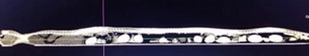
The patient was eventually admitted for sedation to permit a CT scan, using 10mg/kg of IV alfaxalone. The CT scan confirmed a significant amount of fluid present in its coelomic cavity, and multiple desiccated follicles and retained infertile eggs (Figure 1). Blood was taken for biochemical and haematological profiles. For completeness, a faecal parasitology was also performed.
The only changes noted were consistent with reproductive activity and a moderate lymphocytosis. Its faecal parasitology test indicated no parasitic infestations. Based on these results, and after an in-depth discussion with the client, it was decided an exploratory coeliotomy should be performed.
The snake received preoperative analgesia in the form of 1mg/kg IM morphine and 0.5mg/kg IM meloxicam, as well as antibiosis in the form of 20mg/kg IM ceftazidime. Anaesthesia was induced using 10mg/kg IV alfaxalone. It was intubated then ventilated with sevoflurane throughout the surgery.
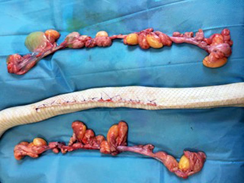
An approximately 25cm incision was made between its first and second row of lateral scales, preserving its ventral surface. Ovarian tissue and oviductal tissue was isolated, exteriorised then removed using an electrothermal bipolar tissue sealing system to aid haemostasis. The oviductal tissue was removed as, on gross inspection, it was abnormal. Its coelomic cavity muscular layer was closed using 5-0 suture in a simple continuous pattern. Its skin was closed using an interrupted everting pattern of 3-0 polydioxanone suture (Figure 2).
Postoperative analgesia and antibiosis were continued in the form of 0.5mg/kg meloxicam given orally and 20mg/kg IM ceftazidime. It was recommended when the snake shed it was to be brought back to remove the previous skin from around the stitches.
Five weeks after surgery, the snake’s sutures were removed, the wound had healed very well, and it had returned to its normal temperament and feeding regime.