25 May 2015
Elisabetta Mancinelli
Job Title
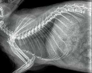
Figure 6. Sagittal MRI of the same chinchilla showing no evidence of structural abnormalities.
A one-year-old, neutered male chinchilla (Chinchilla lanigera), weighing 555g, was presented with a six to eight week history of alleged seizure activity.

One to two episodes per month were described, during which thrashing noises were heard in the cage. The animal was then found in one corner, lethargic and unresponsive for a few minutes. It did not seem to have ever lost consciousness during any of the episodes. The recovery time was variable, but up to several minutes in some cases, after which the chinchilla appeared to be behaving and eating normally. However, the owner had noticed frequent head shaking and teeth grinding after some of these episodes.
The last suspected seizure was reported to have occurred 1.5 weeks prior to presentation. Otherwise there was no change in the animal’s activity level, and appetite and thirst were normal. The chinchilla was kept in an indoor cage with another neutered male chinchilla and fed a commercial pelleted diet for chinchillas, hay and shredded wheat. No obvious exposure to toxins was reported and there was no history of previous medical problems or trauma.
The physical examination was unremarkable. Auscultation of the thorax did not reveal any heart murmurs or arrhythmias. A full neurological assessment was attempted, including cranial nerve assessment, testing of postural reactions and spinal reflexes, but findings were extremely difficult to interpret due to the nature of the patient. Further investigations were therefore scheduled.
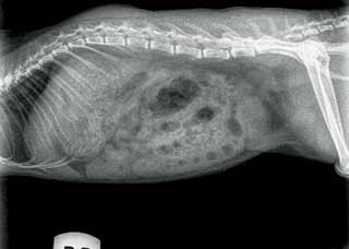
A urine sample was collected for complete urinalysis. The following day general anaesthesia was induced in an induction chamber with 8% sevoflurane in 1L/min oxygen flow. Anaesthesia was then maintained with 2.5% to 3% sevoflurane in oxygen via a face mask attached to an Ayre’s T-piece. A blood sample was collected from the cranial vena cava for routine haemato-biochemistry and blood lead levels. A full set of skull, thoracic and abdominal radiographs was taken (Figures 1 to 3). A CT scan was then performed (Figure 4). The recovery from anaesthesia was uneventful and the chinchilla was discharged the following day with no medical treatment.
One month later, the chinchilla was brought in for a recheck. Physical examination findings were unremarkable, but the owner reported two further episodes two weeks prior to presentation. An MRI scan was therefore scheduled under general anaesthesia. No medical treatment was advised at this stage unless episodes were becoming more frequent or intense. One year later, a telephonic update was given by the owner. The chinchilla had had no further episodes and it was doing fine.

Epilepsy is a neurological condition that can arise from brain injury or genetic susceptibility to cortical hyper-excitability due to a single gene mutation or arising from a complex of polygenic trait (Veres et al, 2013). It is considered a debilitating disease characterised by recurring seizures and is a substantial health problem in humans worldwide (Veres et al, 2013). Animals, especially rodents, are often used as models of the human disease. Cases of spontaneous epilepsy in chinchillas have been anecdotally reported in the literature (Hollamby, 2009). A case of a spontaneous human herpes virus type one infection causing ocular and neurological signs including seizures has also been reported in a one-year-old chinchilla (Wohlsein et al, 2002).
Heat stress, head trauma, lead toxicosis (Hoefer, 1995; Morgan et al, 1991), Frenkelia and toxoplasmosis infection (Dubey et al, 2000), bacterial encephalitis, calcium/phosphorus imbalances, lymphocytic choriomeningitis virus (LCMV) infection (Hollamby, 2009; Saunders, 2009), neoplasia and cardiovascular disease, are all possible differential diagnoses that should be considered when a chinchilla is presented with a history of compatible neurological signs (Hollamby, 2009). In gerbils, a deficiency of glutamine synthetase, the enzyme that catabolises glutamate, is the cause of seizures, which are sometimes triggered by handling or stress (Scotti et al, 1998). Whether there may be a similar reason in chinchillas is not known.
An analytical approach to rule out the major causes for seizures is necessary to identify a possible underlying cause. In this case the physical examination findings were unremarkable. Heart murmurs are often (23%) found incidentally on routine auscultation of chinchillas and echocardiography is recommended to detect structural cardiac abnormalities (Pignon et al, 2012).
An ultrasonographic examination was not considered necessary in this case in view of the clinical history and examination findings.

The haemato-biochemistry results and urinalysis parameters were within the reported reference ranges for this species and failed to identify any signs of infection or electrolyte imbalances. Blood lead level was 2μg/dl, which was considered normal. No abnormalities were detected on the radiographic study. The CT findings were also unremarkable (Figure 5) and no structural explanation for the seizure activity could be identified on MRI (Figure 6). Normal cerebrum, cerebellum and brainstem were seen. Ventricles and CSF spaces were normal for the species. Midline structures, bases crania, tympanic bullae and membranous labyrinth were examined and considered normal.
Furthermore, the clinical condition of the animal did not appear to deteriorate over time, therefore more serious bacterial or viral infections causing encephalitis were considered unlikely.
Epilepsy has been extensively studied and many rodents have been used over the years as animal models in an attempt to understand the genetic and environmental causes as well as the triggering factors of this complex neurological disease (Frankel, 2009). In a 2009 study, Frankel reported a seizure episode may be triggered by specific sensory stimuli or might appear randomly. There are also idiopathic forms of epilepsy that do not have any obvious organic cause or identifiable lesion and may be of genetic origin (Frankel, 2009). There are no genetic studies using chinchillas as animal models and no information could be found in the literature (to this author’s knowledge) regarding this disorder in laboratory or pet chinchillas. Furthermore, seizure disorders, whether spontaneous or experimentally induced, are very complex, multifactorial and can vary greatly in severity, incidence and form.

It is very difficult, in absence of structurally evident abnormalities that could have explained the described clinical signs, to speculate on a possible genetic or other cause and therefore prescribe a specific treatment. CT and MRI are extremely useful diagnostic imaging techniques. The first permits evaluation of the brain, vertebral column and calcified structures while the latter can better identify soft tissue abnormalities such as soft tissue masses, oedematous or inflammatory soft tissue changes (Keeble, 2006). MRI, in particular, has been extensively used in rodent models of different brain disorders and it is considered an extremely useful diagnostic modality capable of giving detailed information without causing harm to live tissues (Denic et al, 2011). In this case, both CT and MRI failed to identify any structural abnormality and the cause for the seizure disorder of this chinchilla remained unknown.
In other species (Keeble, 2006), diazepam or midazolam is suggested for symptomatic relief of seizures while addressing the underlying cause. Medical treatment with phenobarbital (1mg/kg to 2mg/kg daily) is recommended even in absence of a final diagnosis, but the prognosis is considered poor. In gerbils, seizures usually do not cause permanent damage and treatment is seldom required (Scotti et al, 1998). Considering the very low frequency of the episodes and the good quality of life of this chinchilla otherwise, medical treatment was not considered necessary.
Elisabetta Mancinelli
Job Title