11 Feb 2019
Sonya Miles describes her approach to removing a large abdominal mass from a lethargic, poorly cared for lizard.
Sonya Miles
Job Title
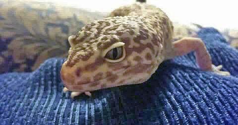
Figure 1. The lizard was in adequate body condition.
A four-year-old male leopard gecko presented with a four-week history of anorexia and increasing levels of lethargy.
It had been kept in substandard conditions with inadequate heating and ultraviolet light provision. It had also been kept on a sand substrate with poor calcium supplementation, and was provided with an inadequate diet of non-gut-loaded mealworms.
Despite the poor care, on clinical examination it was noted the lizard was in adequate body condition (Figure 1). It did, however, have a large mass taking up the vast majority of its coelomic cavity. When palpated, this mass was mobile, but firm, and elicited the reptile to attempt to bite – so it was, I assumed, therefore, painful. The rest of the clinical examination was within normal limits.
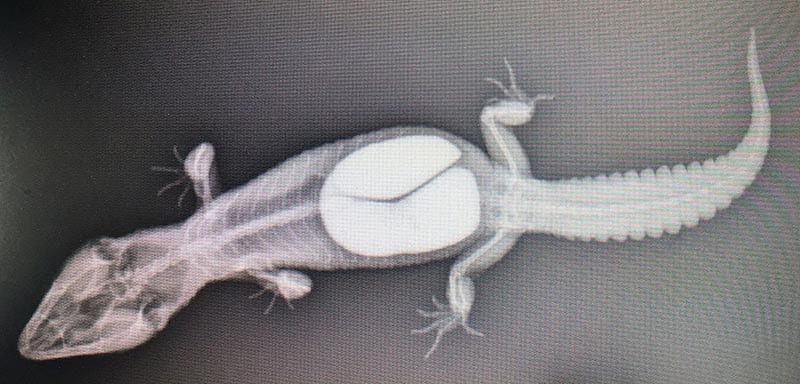
A conscious dorsoventral radiograph was performed to ascertain the extent and density of the coelomic mass (Figure 2). Ideally, a contralateral view would have also been taken; however, funds were limited. The owner was given two options: either to euthanise the reptile – as medical management for an impaction of this size would be unlikely to work – or opt for an exploratory coeliotomy. The latter option was undertaken.
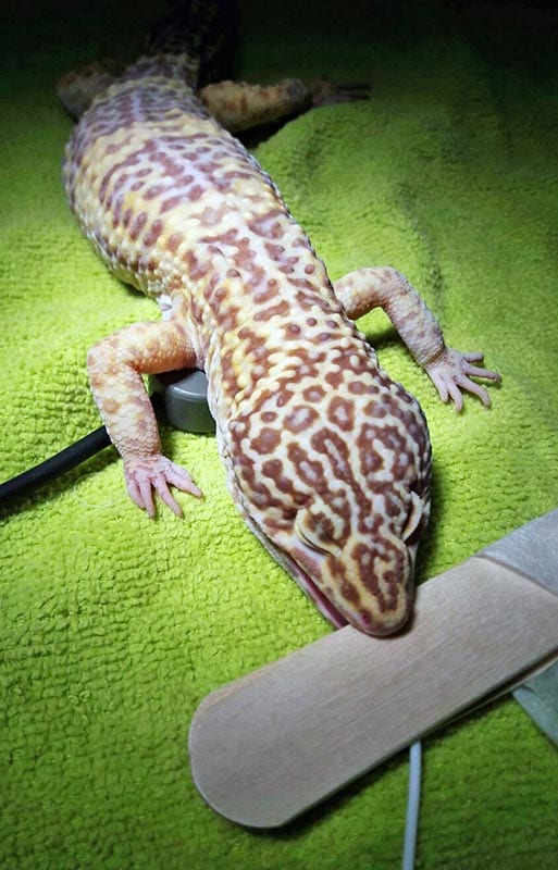
The patient was anaesthetised by placing it inside a small knock down box and inducing unconsciousness with sevoflurane. Once unconscious, morphine was given 1mg/kg IM. The leopard gecko was then intubated with a 1.5mm endotracheal tube and prepared for surgery. Tongue depressors were used to sandwich the tube to prevent the patient from biting through it (Figure 3).
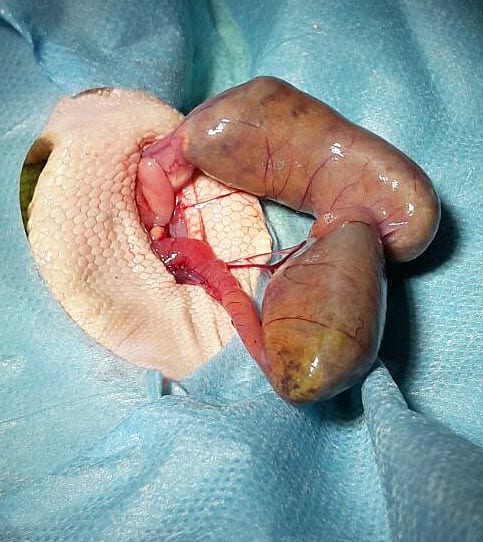
A para-median approach was performed to avoid the ventral abdominal vein, and the intestines containing the obstruction were exteriorised and placed on sterile saline-soaked swabs (Figure 4).
An incision was made distal to the obstruction and the contents – consisting of digested material and copious amounts of sand – were milked from the incision (Figures 5 and 6). The intestinal incision was flushed with sterile saline to remove any particulate matter from the external surface of the intestine. It was then closed with a simple continuous pattern of 5/0 poliglycaprone 25 in a single layer.
The now closed intestinal incision was leak tested by instilling 1ml of sterile saline into the lumen of the intestine via a hypodermic injection, which was then put under digital pressure (Figure 7).
The intestines were replaced, allowing a full examination of the rest of the coelomic cavity. Once deemed free of any other pathologies, the muscle layer was closed using a monofilament simple interrupted pattern (Figure 8).
The skin was then closed using a simple everting pattern. In recovery, the patient was given meloxicam 0.5mg/kg IM, and it was started on a course of 20mg/kg ceftazidime, consisting of five doses to be administered IM.
The leopard gecko had an uneventful recovery and was sent home the following day. The correct care for the species – including the provision of ultraviolet light, a thermostatically controlled heat source, suitable supplementation, and a varied gut-loaded diet – was discussed at length.
At its seven-day postoperative check, the owners reported a return to normal feeding habits. The sutures were removed five weeks post-surgery, at which stage the lizard had gained sufficient weight and returned to normal activity levels.