3 Jul 2017
Sonya Miles describes the case of an adult Syrian hamster that presented with anorexia, alopecia and extreme skin scaling.
Sonya Miles
Job Title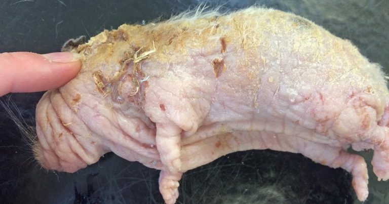
The extremity of clinical signs exhibited by the hamster. The pictures were taken postmortem, after an increase in severity of its condition.
Hamsters are a commonly owned exotic pet, with a range of species available. As one of the larger and more docile species, Syrian hamsters are commonly presented for a variety of reasons – skin conditions being one.
Skin disease in these animals can be the result of multiple causes, from endoparasites, endocrine diseases to cutaneous neoplasia. Skin tumours in hamsters, however, are not common (Harvey, 1995), but, when they do occur, epitheliotropic lymphoma is the second most common cutaneous neoplasm (Harvey et al, 1992) – most often occurring in older hamsters (Paterson, 2006; Hoppmann and Barron, 2007). A multitude of clinical signs can be noted by the client, from anorexia, lethargy, alopecia, pruritis and infected encrustations (Paterson, 2006; Hoppmann and Barron, 2007).
This article will discuss the presentation, diagnosis and outcome of an adult Syrian hamster that presented with anorexia, alopecia and severe dermal scaling.
A two-year-old male Syrian hamster (Mesocricetus auratus) presented with a two-month history of progressively worsening alopecia, pruritus, erythema and severe epithelial scaling over the majority of its body.
During the seven days prior to presentation the owner had reported anorexia and an increase in lethargy in the hamster. The owner also reported vocalisation when being handled and the hamster had bitten on numerous occasions, which was unusual. When discussing the clinical history, it was noted the hamster was fed a good, balanced diet and housed well.
Other hamsters in the household did not show any clinical symptoms that matched the presenting one and, although they did not have direct contact with each other, they did share exercise space.
On clinical examination the animal weighed 120g (body condition score 3 out of 5). Severe scaling over the dorsum was seen, with only minimal hair being present on the hamster in general. Multiple excoriations were present and the hamster resented being touched, often vocalising when the more severe lesions were examined. Its heart rate was within normal limits, but the respiratory rate was elevated. The patient was pyrexic and abdominal palpation was within normal limits.
A frank discussion was held with the client, where the severity of the signs, the apparently poor quality of life and guarded prognosis was discussed at length. The client declined immediate euthanasia in favour of further investigation.
Due to the inability of rodents to vomit, no pre-anaesthetic fasting was required (Longley, 2008), therefore, also minimising the risk of hypoglycaemia, which is of concern in hamsters due to their small size (Capello, 2011). Fluids were administered (5ml of sterile Hartmann’s solution) SC prior to, and during, surgery.
The hamster was given a pre-medication of 0.05mg/kg buprenorphine IV. Thirty minutes later, the hamster was placed in an induction chamber and preoxygenation was performed for five minutes, before the patient was anaesthetised with 8% sevoflurane and 3L/min of oxygen. Once the hamster had lost its righting reflex, a Darvall breathing circuit was used to allow sevoflurane maintenance at 4%. The patient’s mouth was checked and cleared of food to minimise the risk of aspiration.
The hamster was placed in dorsal recumbency and 1ml of blood was taken for biochemical and haematological analysis from the cranial vena cava, after cleaning the skin in this area. Skin scrapes, tape strips and a full-thickness skin biopsy were also performed under anaesthetic.
While the hamster was recovering from anaesthesia, meloxicam at 0.2mg/kg SC was administered. The hamster was maintained at its optimum environmental temperature and monitored closely throughout recovery, which was uneventful.
Haematological analysis demonstrated a marked leukocytosis with a lymphocytosis. Skin scrapes and tape strips were negative for ectoparasites, but did demonstrate a superficial infection. The skin biopsy yielded a diffuse presence of neoplastic cells invading the epidermis and follicular infundibula of all samples provided.
Neoplastic cells had a round shape and pale cytoplasm, and showed moderate to marked anisocytosis and anisokaryosis. Scattered mitoses were observed and diffuse epidermal hyperplasia was evident, with marked laminated to compact orthokeratotic hyperkeratosis and a large serocellular crust. A mild interstitial infiltrate of mononuclear cells was observed in the superficial dermis extending to the panniculus in samples from truncal skin.
On these results, and owing to the fact the hamster’s clinical signs were significantly worse by the time the results had returned, euthanasia was discussed at length and finally permitted. A full postmortem was, however, declined.
In general, skin tumours in hamsters are not common (Harvey, 1995); however, when they do occur, epitheliotropic lymphoma – which resembles mycosis fungoides seen in humans – is the second most common cutaneous neoplasm (Harvey et al, 1992). Cutaneous lymphoma most often occurs in older hamsters and can cause a variety of clinical symptoms, from anorexia, lethargy, alopecia, pruritus and encrustations, which are often secondary infected (Paterson, 2006; Hoppmann and Barron, 2007).
A full clinical history and clinical examination can allow for a tentative diagnosis (Paterson, 2006), with impression smears or scrapings from ulcerated lesions often demonstrating signs of lymphocytic infiltrate with cells showing signs of malignancy (Paterson, 2006). A definitive diagnosis is made by biopsy only (Paterson, 2006).
Treatment is rarely successful and euthanasia is most often recommended under welfare grounds – as in this case – with the clinical signs progressing quickly in a mean of 10 weeks (Paterson, 2006). Anecdotal reports of chemotherapy exist (Hoppmann and Barron, 2007).
This case describes the diagnosis and eventual euthanasia of a Syrian hamster with cutaneous lymphoma, diagnosed by full-thickness biopsies. Cutaneous lymphomas progress quickly, affecting the animal both topically and systemically (Paterson, 2006).
Palliative treatment quickly becomes refractory and, ultimately, euthanasia is the recommended course of action.