13 Nov 2017
Elisabetta Mancinelli presents the third part of a her series on spontaneous mammary tumours, this time focusing on hamsters and hedgehogs.
Elisabetta Mancinelli
Job Title
A Djungarian hamster. IMAGE: Fotolia/Piotrma.
Spontaneous mammary tumours are one of the most important diseases among females of all species, including humans.
These tumours are described in many domestic and laboratory species, but their morphology and biological behaviour vary between each of them, and interspecies comparative studies are considered important as they may contribute significantly to the understanding of human breast cancer.
Furthermore, as the standards of small mammal pet care improve and their age increases, many of these studies also represent an invaluable tool for the vet seeing them in daily practice as they may improve knowledge and provide clinically applicable information.
Following parts one and two, the final part of this series of articles presents latest research on hamsters and hedgehogs. Readers are advised to refer to the literature for more detailed information.
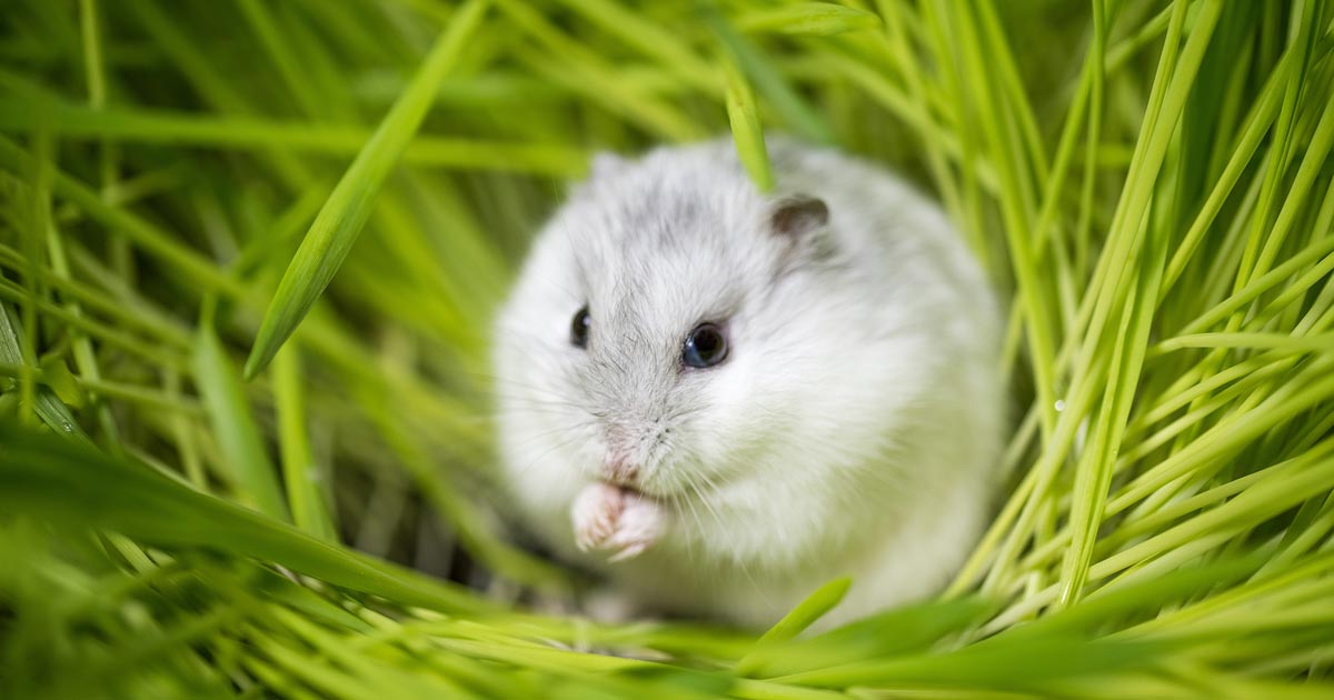
The prevalence of mammary tumours in Djungarian (Siberian or Russian) hamsters (Phodopus sungorus) is higher than in Syrian hamsters, with a reported incidence of 57.1% (12 out of 21) in females kept as pets in Japan (Fernandez et al, 1996; Kondo et al, 2009).
In a 2015 study, Yoshimura et al aimed to characterise histological and immunohistochemical features of spontaneous mammary tumours encountered in this species.
A total of 45 cases of mammary tumours (47 biopsy specimens; 2 of which were obtained from recurrent tumours), surgically resected from Djungarian hamsters kept as pets in various regions in Japan and submitted to the laboratory, were re-examined. In all 45 animals, mammary tumours were solitary and classified into seven histological types: adenoma (14 cases), adenocarcinoma (18 cases), lipid-rich carcinoma (1 case), adenoacanthoma (2 cases), malignant adenomyoepithelioma (2 cases), benign mixed tumour (1 case), and balloon cell carcinosarcoma (7 cases).
In Djungarian hamsters, it has been reported mammary tumours tend to develop at a younger age, compared with other tumour types (Kondo et al, 2009). In the Yoshimura et al (2015) study, authors observed the mean ages of Djungarian hamsters with benign types of mammary tumours (adenoma and benign mixed tumour) were lower than those of malignant types (adenocarcinoma, lipid-rich carcinoma, adenoacanthoma, malignant adenomyoepithelioma and balloon cell carcinosarcoma). In addition, the size of tumour masses among benign histological types also tended to be smaller than those of malignant histological types.
Kondo et al (2009) reported apocrine secretion in all 12 cases of a series of Djungarian hamster mammary tumours, and a relationship with the expression of androgen receptor was suggested. Apocrine secretion was also noted in most glandular components of the mammary tumours of the Yoshimura et al (2015) study, including adenomas and adenocarcinomas. Unusual histological types were also reported, including two adenoacanthoma, which exhibited not only glandular growth, but also extensive areas of epidermoid structure (the squamous epithelial areas accounted for approximately 50% of the whole tumour).
Furthermore, two cases of malignant adenomyoepithelioma (characterised by a biphasic proliferation of luminal epithelial and myoepithelial components) and seven cases (15.6% of cases) of carcinosarcoma (composed of an admixture of malignant glandular and polygonal-spindle cell components) were diagnosed. This last finding is surprising as carcinosarcomas are considered rare in humans, dogs and cats. Different theories have been proposed to explain the coexistence of the carcinomatous and sarcomatous components in carcinosarcomas (Meyer, 1920).
The majority of researchers agree most carcinosarcomas are monoclonal neoplasms that derive from a single epithelial cell (or stem cell), as these tumours exhibit a transition from carcinomatous to sarcomatous components, which is supported by morphological and immunohistochemical evidence (Wang et al, 1992; Wick 1993; Zarbo et al, 1986).
In the seven Djungarian hamster cases in the reported study, authors recognised histological and immunohistochemical features suggestive of the fact a glandular tumour developed initially, then a portion subsequently transitioned to polygonal cells, and subsequently to sarcomatous spindle cells, as observed previously in human and mouse carcinosarcomas. Since these features are suggestive of epithelial-mesenchymal transition (EMT), EMT-phenotype tumour may be a more appropriate diagnostic term, as it is also used for mammary carcinosarcomas in mice.
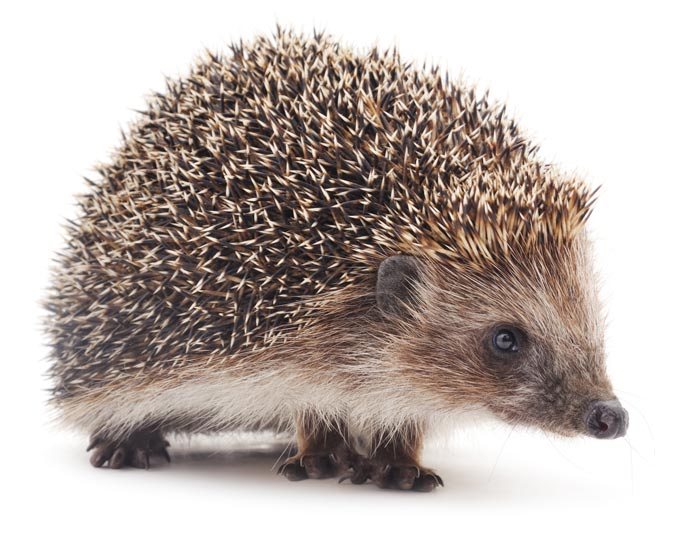
Spontaneous neoplasms are more frequently recorded in African hedgehogs (Atelerix albiventris) compared to European ones (Erinaceus europeus). In a study, Raymond and Garner (2000) noted mammary tumours occurred as single and multiple nodules in African hedgehogs, and consisted of benign and malignant variants. Papillary adenomas, or solid, tubular and papillary carcinomas, or unclassified adenocarcinomas, were identified in this species.
Malignant neoplasms often were locally infiltrative and occasionally showed vascular invasion. Some of the affected animals concurrently showed additional neoplasias (Raymond and Garner, 2000; Wellehan et al, 2003). In their study, DÖpke et al (2007) described the morphological and immunohistochemical features of spontaneous mammary tumours in European hedgehogs. A total of 48 surgical tissue samples submitted for histopathological analysis and 404 necropsies (including 200 males, 188 females and 16 of unknown gender) were reviewed.
Mammary tumours were found in 11 of the surgical samples and in 5 of the necropsied animals. Specific details were recorded and tumours classified according to the World Health Organization (WHO) histological classification of mammary tumours of the dog and cat (Misdorp et al, 1999). Similarly to what was previously reported in African hedgehogs in which mammary tumours were predominantly malignant (Raymond and Garner 2000, 2001; Wellehan et al, 2003), the mammary tumours described in the Raymond and Garner (2000) study in European hedgehogs were all malignant.
Neoplasms grossly appeared as firm, circumscribed nodules on the ventral aspect of the thorax or abdomen, round to ovoid in shape and with a grey-white to yellow-brown cut surface. In all animals, neoplasms were classified as simple tubulopapillary carcinomas of the mammary gland composed of proliferating epithelial cells, similarly to what Raymond and Garner (2000) and Wellehan et al (2003) had described in African hedgehogs.
According to the WHO classification of mammary tumours in dogs and cats, simple carcinomas are divided into tubulopapillary, solid and anaplastic – reflecting increasing malignancy (Misdorp et al, 1999).
Whether this can be directly extrapolated and applied to hedgehogs cannot be determined at this stage due to the lack of clinical data and low number of cases collected. Cystic cavities, necrosis, ulcerated skin, calcification, focal haemorrhages and suppurative mastitis were seen associated in some of the cases.
Focal infiltration of the surrounding fibrous connective tissue, invasion of lymphatic metastases and blood vessels were seen in 13, 1 and 6 cases respectively. No distant (for example, in the lung) metastases were recorded in any of the animals studied. In African hedgehogs, vascular invasion was detected in three out of seven cases (Raymond and Garner, 2000). In dogs and cats, simple carcinomas have a high tendency (up to 50%) to infiltrate surrounding tissues and vessels (Misdorp, 2002).
The infiltrative behaviour of mammary carcinomas in African and European hedgehogs seems similar to that of dogs and cats, although the low number of cases examined does not allow conclusions to be drawn on the metastatic potential of mammary tumours in hedgehogs.
When immunohistochemistry analysis was performed, cytokeratin (CK) 20 was clearly demonstrated in normal and neoplastic mammary epithelial cells of European hedgehogs, while mammary myoepithelial cells were consistently negative. In humans, CK20 expression is mainly seen in epithelial cells and tumours originating from the gastrointestinal tract, urothelium and Merkel cells.
In dogs and cats, mammary tumours do not demonstrate CK20 expression, which is instead seen in carcinomas of the thyroid gland, intestine, lung and kidney in cats and carcinomas of the gastrointestinal tract and ovary in dogs.
The mammary gland of hedgehogs in the Raymond and Garner (2000) study was consistently negative for CK7, which is instead expressed in human, feline and canine mammary epithelial cells and their tumours. These findings represented a significant difference from the CK profile shown by mammary epithelial cells of other mammalian species, including dogs and cats. CK expression in epithelial cells may, therefore, vary significantly between different mammalian species.
Only one animal in the Raymond and Garner (2000) study presented another tumour concurrently with a mammary neoplasia. Occurrence of multiple neoplasm was also described in previous studies (Raymond and Garner, 2000; Wellehan et al, 2003) in four African hedgehogs. The cause of this finding is unknown, as is the cause of mammary neoplasia in hedgehogs. Retrovirus infections have been associated with such tumours in mice and cats (Weijer et al, 1974; Percy and Barthold, 1993). Retroviral particles have been detected in a parosteal sarcoma, osteosarcoma and an intestinal lymphosarcoma in African hedgehogs (Peauroi et al, 1994; Raymond et al, 1998).
A viral cause for mammary carcinoma of European hedgehogs cannot be either confirmed or ruled out at this stage and other possible factors (including hormonal stimuli, food, genetic factors, irradiation and environmental factors) should be taken into consideration.