28 May 2021
Sonya Miles recounts the case of this reptile that presented with anorexia and was reported to have passed gritty urates.
Sonya Miles
Job Title
Figure 1. The female African spurred tortoise (Centrochelys sulcata).
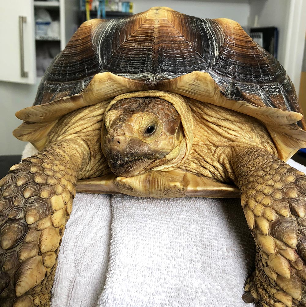
A six-year-old female African spurred tortoise (Centrochelys sulcata) presented with a 48-hour history of anorexia. Her owner also reported that she had passed a large volume of gritty urates along with brown-coloured liquid (Figure 1).
Before this, the tortoise was reported to be very active and eating well. She was fed a diet consisting of 75% grass and hay, and 25% freshly picked edible plants. She was soaked regularly and had access to fresh water at all times. On clinical examination she weighed 7kg, was of good body condition and was very strong, limiting a complete, conscious clinical examination.
Conscious radiographs were performed demonstrating a 5.6mm × 5.5mm calcified structure in the region of the bladder; a large bladder stone was suspected (Figure 2). Bloods were taken to assess biochemical and haematological values. These were all within their respective reference ranges.
As a result of the combination of the clinical history and radiographic findings, it was decided that the tortoise should be taken to surgery.
A pre-femoral approach (Figure 3) – instead of a plastromy approach – was undertaken to ensure a faster return to normal behaviours, and because the long-term medications often associated with the plastromy approach would have been difficult in this species.
The tortoise was premedicated with 1mg/kg morphine IM and a course of eight injections of 20mg/kg IM ceftazidime was initiated; 10mg/kg IV alfaxalone was given to sedate the tortoise enough to allow intubation and then ventilation using sevoflurane.
The tortoise was positioned in dorsal recumbency, with each leg extended and tied in a position that allowed for easy access of the pre-femoral windows (Figure 4). The surgical site was sterilely prepared and the skin, muscle and coelomic membrane incised, providing a vertical opening to the coelomic cavity; a Lone Star retractor was used to aid visual inspection of the coelomic cavity (Figure 5).
Once the tortoise’s coelomic cavity was open, her reproductive tract became immediately obvious (Figure 6), obscuring the surgical field. To visualise the bladder and remove the bladder stone, the reproductive tract was removed. This was performed on both sides, through each of the pre-femoral fossae.
Once there was a clear surgical field, an assistant put pressure on the bladder through the hole in one of the pre-femoral fossae, pushing the stone and the bladder closer to the other side. This allowed partial exteriorisation of the bladder.
The surrounding coelomic cavity was packed with sterile, saline-soaked swabs; stay sutures were placed to hold the bladder open once a cystotomy had been performed (Figure 7).
The stone was broken up inside the bladder and the stone removed in a piecemeal fashion (Figure 8). The bladder was then flushed and closed in a double inverting patter with monofilament suture material.
The coelomic cavity was flushed with warmed, sterile saline, then the coelomic membrane, muscle and skin were closed separately, again with monofilament suture material, with the external layer being closed in an interrupted everting pattern (Figure 9).
In the recovery period the tortoise was given 0.5mg/kg IM meloxicam, which was continued for 14 days post-surgery in oral form. A complete course of ceftazidime was given and 1mg/kg IM morphine was given daily for 72 hours post-surgery.
The tortoise returned to normal eating habits 12 hours after surgery and has made an amazing recovery.