5 Oct 2015
Fiona Froehlich and Neil Forbes explain the causes of severe skin-related conditions in parrots, the treatments and why regular assessment is vital.

Figure 1. Severe ulcerated inguinal dermatitis.
Avian skin essentially consists of the same structures as mammalian skin, namely epidermis and dermis.
The epidermis contains germinative, intermediate and corneous layers and the dermis has blood vessels, fat deposits, feather follicles, nerves, neuroreceptors and muscles.
Avian skin is significantly thinner and does not contain sweat glands or sebaceous glands, except for the ear canal and the preen gland.
The epidermis produces neutral fats and phospholipids, which make the skin strongly lipogenic (Stettenheim, 2000). This mechanism is thought to support waterproofing and inhibits growth of bacteria and fungi.
A significant subcutis layer is only present in the ventral midline and axilla.
The skin is attached to the skeleton at the skull, wing tips and sternum, but lies otherwise on the muscular layer underneath.
A thick cornified epidermis, footpads and other adaptations are found on the extremities of different bird species according to their lifestyle.
Some species, in particular cockatoos and African grey parrots, have specialised powder-down feathers. The bird chews them to produce a dry powder of keratin debris to assist with preening and maintaining feather condition.
The shedding of keratin from the shafts of new developing feathers can be prolonged in sick animals and may lead to itching and discomfort (Cooper and Harrison, 1994).
Parrots are often presented for skin-related conditions such as bite wounds, burns and parasitic, bacterial or viral disease. However, the most common presentation is the plucking bird.
A detailed history must be obtained to assess diet and husbandry, including details about other birds in the collection.
Systemic liver, kidney, intestinal, pancreatic, toxic, nutritional, developmental or bone marrow disorders can manifest as skin conditions and affect plumage, moult, skin and wound healing.
A dermatological examination should include a thorough inspection of the skin and plumage, focusing on inflammation or redness, wounds, feather development abnormalities, stress lines, powder down and parasites.
Further diagnostics such as skin scrapes, tape exam, feather pulp cytology, bacteriology and histology may be indicated.
Superficial chronic ulcerative dermatitis (SCUD) or chronic ulcerative dermatitis (CUD) is often encountered in lovebirds and medium-sized parrots, such as African grey parrots and cockatoos.
Many factors are thought to contribute to this condition including infections with polyomavirus or circovirus (Cornelissen et al, 2001), Mycobacteria species or Giardia species infections, skin cancer, xanthoma and hypovitaminosis E (Jepson, 2009).
Methicillin-resistant Staphylococcus aureus (MRSA) was isolated from three parrots suffering from chronic pyoderma (Forbes and Smith, 2007).
Affected areas include the underwing, groin, side of the neck, back or tail head (Figure 1).
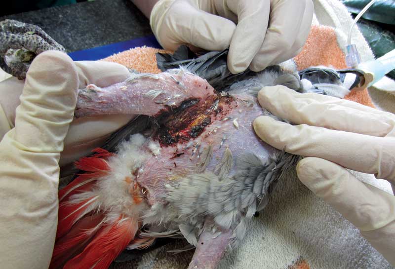
Skin lesions typically demonstrate long-term thickening, often with ulceration and/or a wet discharge, and patients are often presented with pain and severe itching.
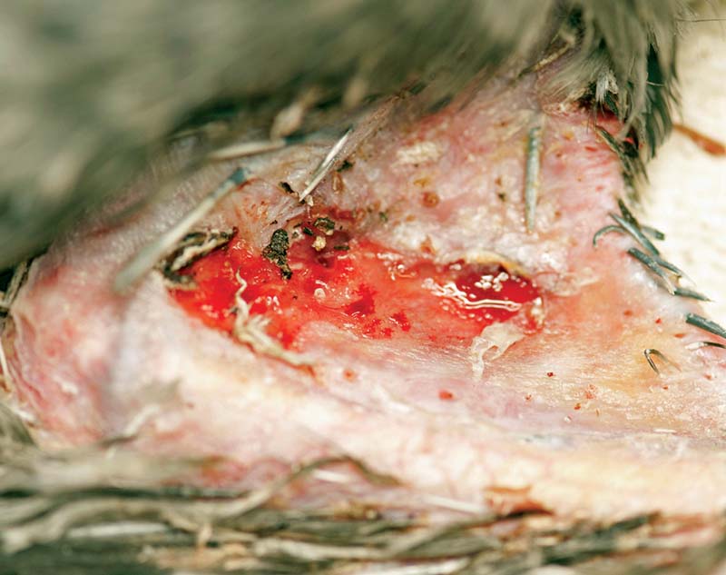
The underwing and “armpit” are especially prone to severe long-term dermatitis due to constant movement, which impairs healing, and a tendency to be a sweaty, poorly ventilated area resulting in moist conditions (Figure 2).
Scar tissue can develop and lead to pain, restricted movement and long-term self-mutilation.
This causes severe skin damage and presents an extra challenge in managing dermatological disease.
Regular assessment will help formulate a plan for pain relief, antibiosis and supportive treatment.
The inflammatory phase of wound healing occurs within five days of injury and is characterised by signs of inflammation (redness, swelling, heat and pain).
The reduced pH during this period pre-exposes the skin to secondary bacterial and fungal infections.
During the second proliferative phase of wound healing, neovascularisation, epithelialisation and wound contraction take place, followed by the maturation and remodelling phase (Hildreth, 2014).
Samples for culture and sensitivity testing must be obtained from the wound before local or systemic antibiotic treatment.
Staphylococcus species and environmental Gram-negative bacteria are often isolated (Chitty, 2005).
Skin biopsies may be required to confirm neoplastic or fungal elements on histopathology. In cases of chronic skin disease, a full diagnostic work-up including haematology, biochemistry, serology, full-body radiographs and faecal examination are recommended.
There are many products for wound lavage and treatment.
Chlorhexidine (1:40 dilution with sterile water or saline) and povidone-iodine have effective antimicrobial properties. If used excessively, however, both products may have negative effects on fibroblasts and granulation tissue formation.
F10 antiseptic solution may also be used in a 1:250 dilution, which is “gentle” on tissue and will control bacterial, fungal and viral pathogens.
Wound lavage should be performed with a sufficient sized syringe and needle creating adequate pressure.
Surgical debridement of necrotic or desiccated tissue is necessary to allow granulation tissue development.
Severe SCUD cases often require repeated debridement and regular wound assessment under general anaesthetic. Wound healing is achieved by second intention healing.
Systemic and local antibiosis must be given according to culture and sensitivity results. Local treatments are aiming to control infection and aid wound healing.
Ointments should be avoided in avian patients because they can cause feather damage.
Silver sulfadiazine is effective against most Gram-positive and Gram-negative bacteria and most fungi. Mupirocin can be used to prevent antibiotic-resistant bacteria developing, but only use with caution after sensitivity testing.
Manuka honey and aloe vera gels aid healing and have been used successfully on different wound types.
A hydrogel dressing can be used very effectively to provide a moist environment and optimal conditions for healing once infection is under control.
The skin defect and applied medication should be covered with a hydrocolloidal and vapour membrane dressing to avoid desiccation.
Wound dressings can be stitched to the skin providing moisture, protection and limiting tension across the wound.
Excessive wing movement must be avoided, as this may cause further damage to the fragile granulation tissue.
Protective support – for example, radiograph film material – combined with hydrocolloidal dressing can be sewn above and below the propatagium to stop the wing stretching while it heals (Figure 3).
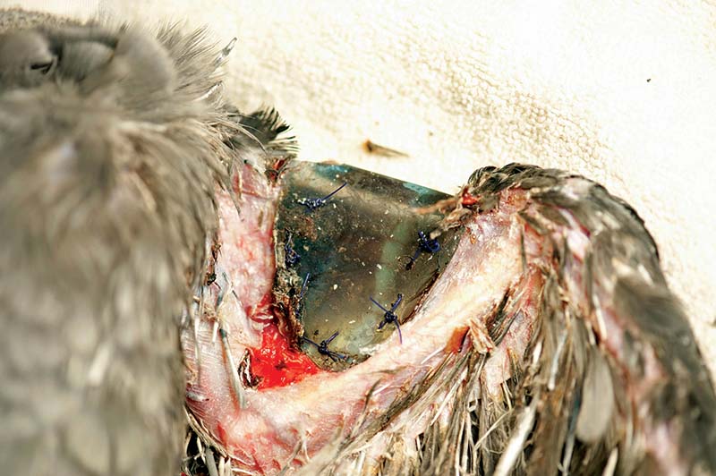
Clinicians must take great care not to injure the tendon of the propatagial muscle, which extends from the shoulder to the wrist joint, when placing dressings surgically.
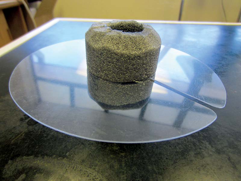
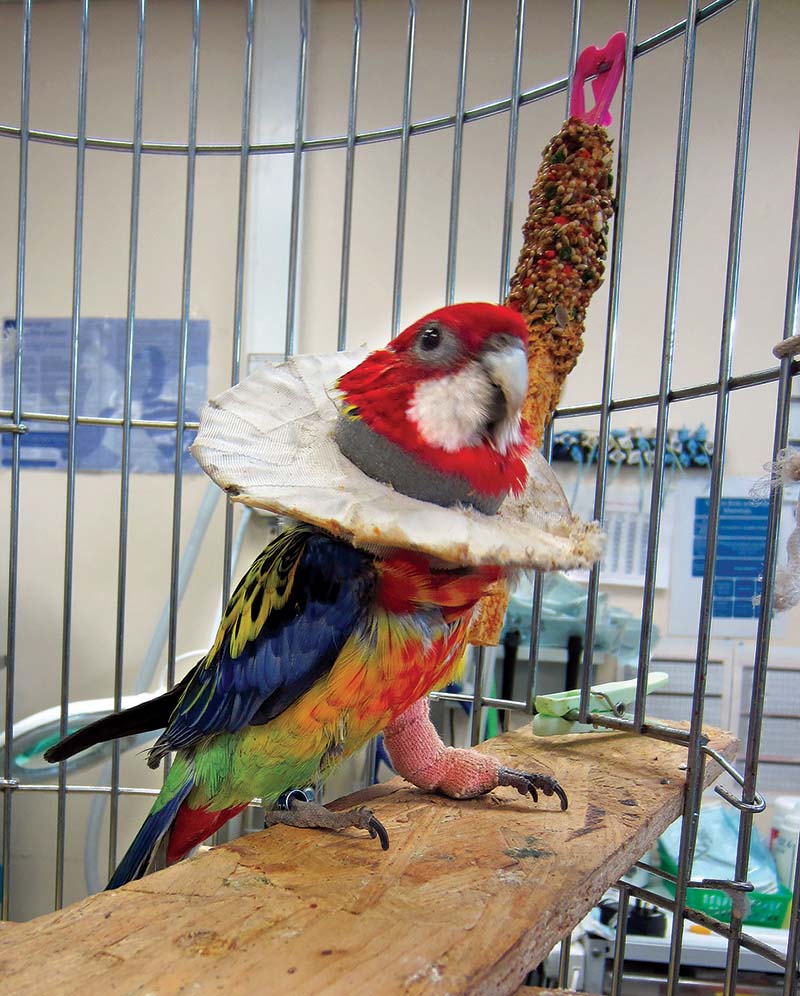
Neck restraint collars, to prevent ongoing self-trauma, should be used to allow unhindered healing. The best results are achieved by using a foam neck support (commercially available as cold water pipe insulation material), with an additional horizontal plastic Elizabethan collar, dependent on the site of the wound (Figures 4 and 5).
Additional integral therapy may include treatment of underlying conditions, optimised diet and omega-3 fatty acid supplementation.
In cases of severe pruritus and agitation, antihistamines and/or benzodiazepine can be used short-term (Schmidt and Lightfoot, 2006).