4 Apr 2022
Urinalysis in chinchillas
From the <em>Vet Times</em> archives – Elisabetta Mancinelli explores research that has come off the back of increasing awareness of this species.

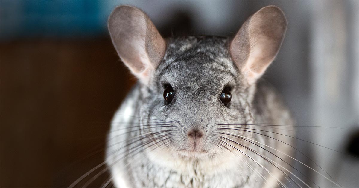
Image © Patryk / Adobe Stock
- This is an extract from “Studies into pet rodent species”, originally published in Vet Times 47.28 (17 July 2017).

Many rodent species, often used as animal models in research, are commonly kept as pets.
The increasing awareness of veterinary professionals towards these species has resulted in an increase in research, which is helping, more often, achieve a definitive diagnosis and establish a more targeted treatment, ultimately improving standards of care.
As practising vets, we should look with interest into these studies, as results may be of clinical interest and applicable in our everyday job.
Renal disease
Urinalysis (including determination of urine specific gravity; USG), chemical analytes (by use of a dipstick and sediment microscopic examination) can be helpful in diagnosing both urinary tract and systemic disorders. Penile fur rings, urinary tract or urogenital diseases are uncommon in chinchillas (Mans and Donnelly, 2012).
In a retrospective review of necropsy results for 202 chinchillas, only 5 had renal disease (Lucena et al, 2012).
Chinchillas can also develop urinary calculi, which are more commonly seen in males, with haematuria being the most frequently observed clinical sign (Mans and Donnelly, 2012).
Within the order caviomorpha, urinalysis results for pet rodents have only been reported in guinea pigs (Bishop et al, 2010; Rueloekke et al, 2016). A study was therefore undertaken, including 41 clinically normal chinchillas (29 females and 12 males) exposed during a breeder exposition, to collect urinalysis data on voided urine samples including USG (measured before and after centrifugation with a handheld veterinary refractometer), urine dipstick analysis, microscopic sediment examination, urine sulfosalicylic acid (SSA) precipitation test and quantitative protein analysis (Doss et al, 2016). The urine varied greatly in colour.
Pigments in the diet have been associated with urine colour in chinchillas, as in rabbits (Banks et al, 2010; Melillo, 2007). Mostly, the samples were clear. The turbid samples had a large number of crystals.
Unlike guinea pigs and rabbits, which excrete a large amount of calcium in their urine, chinchillas excrete calcium primarily through faeces and do not rely on urinary secretion of calcium to maintain calcium homeostasis (Hagen et al, 2014).
In the study, urine samples that were dark in colour generally also had a higher USG than lighter-coloured samples. The USG measured had a wide range (1.010 to more than 1.060), although a large proportion (17 out of 41; 41%) of the chinchillas had a USG less than 1.050. The USG before centrifugation did not differ significantly from that after centrifugation, even in those samples with visible sediment; therefore, centrifugation of chinchilla urine samples prior to measurement of USG with a refractometer is not considered necessary.
Protein detection
Protein was detected in all urine samples on dipstick analysis. Several exotic mammals – including hamsters, mice, rats, gerbils and rabbits – are reported to have small amounts of protein in their urine (Fisher, 2006). However, unexpectedly, 98% of the animals had more than trace amounts of dipstick protein contents, which may be a function of the fairly high USGs for this population.
This prompted further tests, including the SSA precipitation test (a turbidimetric screening test for proteinuria, commonly used to verify positive dipstick protein results) and quantification of urine protein concentration on the samples for which sufficient volume was available (37 and 18 samples, respectively).
The results suggested the dipstick protein and the SSA test results were not accurate. The urine dipsticks used in the study are marketed for use in human urine samples, and values of 1+, 2+, 3+ and 4+ are approximately equivalent to urine protein concentrations of 30mg/dL, 100mg/dL, 300mg/dL and greater than or equal to 2,000mg/dL, respectively. For the 18 urine samples that underwent quantitative protein analysis, the maximum urine protein concentration was 87mg/dL, which suggested the maximum dipstick protein result should have been 1+. In the study, USG was positively correlated with the dipstick protein results, which might have contributed to the unreliability of the dipstick protein results.
In small animals, a high USG and alkaline urine are associated with false-positive urine dipstick protein results (Grauer, 2007). The recorded pH for all samples was 8.5, which was the upper limit of detection for the reagent strip. False-negative SSA test results can occur because grading of turbidity is subjective and variation between individual readers is likely (Grauer, 2007). This was considered unlikely to have occurred in this study.
High alkaline urine may also lead to false-negatives, which may have affected the SSA test results in this study. In Sprague-Dawley rats, it is reported the SSA precipitation test should not be used to verify urine dipstick protein results (Reagan et al, 2007). Glucose or ketones, but not both, were detected in 5 and 6 samples, respectively. In chinchillas, ketoacidosis can develop as a sequela to anorexia. It is possible stress associated with transport and exhibition might have resulted in reduced food intake with subsequent ketones production and urinary excretion. Stress could have also caused hyperglycaemia, which subsequently led to glucosuria, although an association between stress and hyperglycaemia has not been clearly established for chinchillas.
Ketonuria
Ketonuria, in conjunction with glucosuria, has been reported in chinchillas with hepatic lipidosis (Mans and Donnelly, 2012). In this study, subclinical disease could not be ruled out, which represents a limitation. Crystals were observed in 28 of 41 (68%) samples; 27 of those samples contained amorphous crystals, but, to date, their significance is unknown in this species.
Rare epithelial cells, red blood cells and white blood cells were also seen in many samples, and likely represented non-pathological findings.
References
- Banks RE, Sharp JM, Doss SD et al (2010). Exotic Small Mammal Care and Husbandry. Wiley-Blackwell, Hoboken: 125-136.
- Bishop CR, Fischer J, Brossoit A et al (2010). Standardization of renal physiology parameters in guinea pigs via urinalysis, Proceedings of the 31st Annual Association of Avian Veterinarians (AAV) Conference and Expo, AAV, San Diego: 49-52.
- Doss GA, Mans C, Houseright RA et al (2016). Urinalysis in chinchillas (Chinchilla lanigera), J Am Vet Med Assoc 248(8): 901-907.
- Fisher PG (2006). Exotic mammal renal disease: diagnosis and treatment, Vet Clin North Am Exot Anim Pract 9(1): 69-96.
- Grauer GF (2007). Measurement, interpretation, and implications of proteinuria and albuminuria, Vet Clin North Am Small Anim Pract 37(2): 283-295.
- Hagen K, Clauss M and Hatt JM (2014). Drinking preferences in chinchillas (Chinchilla laniger), degus (Octodon degu) and guinea pigs (Cavia porcellus), J Anim Physiol Anim Nutr (Berl) 98(5): 942-947.
- Lucena RB, Giaretta PR, Tessele B et al (2012). Diseases of chinchilla (Chinchilla lanigera), Pesq Vet Bras 32(6): 529-535.
- Mans C and Donnelly TM (2012). Disease problems of chinchillas. In Quesenberry K and Carpenter J (eds), Ferrets, Rabbits, and Rodents. Clinical Medicine and Surgery (3rd edn), Elsevier, St Louis: 311-332.
- Melillo A (2007). Rabbit clinical pathology, J Exot Pet Med 16(3): 135-145.
- Reagan WJ, VanderLind B, Shearer A et al (2007). Influence of urine pH on accurate urinary protein determination in Sprague-Dawley rats, Vet Clin Pathol 36(1): 73-78.
- Rueloekke ML, Spangsberg R and Koch J (2016). Physiologic serial changes in the visual appearance of urine samples from individual pet guinea pigs (Cavia porcellus), BSAVA Congress Proceedings, BSAVA, Birmingham: 553-554.
