12 Sept 2016
Elisabetta Mancinelli looks at the causes, preventive measures and methods of treatment for urinary stones found in this species.
Elisabetta Mancinelli
Job Title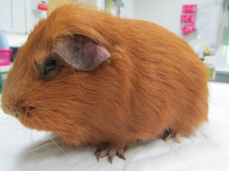
Figure 2. Hunched posture.
Urolithiasis is a significant condition in many small mammals, particularly guinea pigs.

It most commonly affects middle-aged to older guinea pigs and stones may be found anywhere along the urinary tract (bladder, urethra, ureter and kidneys) and sometimes in the seminal vesicles of males (Hoefer and Latney, 2009; Hawkins and Bishop, 2012).
A study found an equal distribution of urinary calculi between males and females older than two years of age, but those located in the ureters seemed to predominate in males (Hawkins et al, 2009). Some authors have considered (but never confirmed) Peruvian guinea pigs to have a predisposition to urolithiasis (Hawkins et al, 2009) and many reports of this condition exist (Spink, 1978; Stuppy et al, 1979; Okewole et al, 1991; Fehr and Rappold, 1997; Gaschen et al, 1998; Stieger et al, 2003; Eshar et al, 2013).
The condition’s aetiopathogenesis remains unclear, although the alkaline pH and the high mineral content of normal guinea pig urine may favour crystal formation and precipitation (Hawkins and Bishop, 2012). Risk factors contributing to urolithiasis include inadequate water intake, urine retention, inadequate cage hygiene, obesity, lack of exercise, food items with high mineral content, renal disease and vitamin or mineral supplementation (Hawkins, 2011). Diet seems to play a role, pointing at calcium and oxalate as main risk factors in stone formation.
In a study that looked into risk factors associated with its development, affected guinea pigs were more likely to be fed a diet high in pellets, low in hay, and with a lesser variety of vegetables and fruits (Hawkins et al, 2008).

Calcium-containing stones, such as calcium oxalate (historically) and calcium carbonate (calcite), are most commonly reported. More recent data seems to suggest calcium carbonate calculi are overwhelmingly prevalent (Hawkins et al, 2008; 2009).
Infections and mechanical factors may also predispose to stone formation. Several bacterial species (for example, Escherichia coli, Streptococcus pyogenes, Staphylococcus, Corynebacterium renale, Streptococcus (bovis/equi group), Streptococcus viridans, Proteus mirabilis and Enterococcus species) have often been isolated from urine of affected guinea pigs and considered responsible for urinary tract infections in association with urolithiasis in this species. However, a direct association has not been confirmed (Hawkins and Bishop, 2012).
Symptoms generally depend on the size and location of the stone(s). Haematuria (Figure 1), stranguria and dysuria may be seen with cystic or urethral calculi. More commonly, clinical signs may reflect abdominal pain, such as a hunched posture (Figure 2) or teeth grinding – or non-specific signs of disease, such as anorexia, lethargy and weight loss.
These symptoms may be seen more frequently when calculi are higher in the urinary tract. Although presentation may be acute, it is common for signs to progress over days to weeks in affected rodents.
A detailed history, including dietary history, is extremely important in association with a complete physical examination. Urolithiasis should be suspected in any rodent with changes in its urinary output or with non-specific signs of illness.
A blood and urine sample should be collected for complete haematobiochemistry and urinalysis to rule out kidney involvement and the presence of infection. Laboratory findings may reflect a high calcium diet (hypercalcaemia) or evidence of cystitis.
The mean specific gravity of 44 guinea pigs with urinary calculi was found to be 1.015 +/− 0.008 (range 1.004 to 1.046) and the urine pH was 8.4 +/− 0.5 (range 6.5 to >9; Hawkins et al, 2009). Haematuria, mucus and lipid droplets were seen on sediment examination in the same study.
Crystal types more commonly seen on sediment examination were calcium carbonate, oxalate and struvite. However, as in other species, this may not be predictive of the urinary calculi composition (Hawkins et al, 2009).
Culture of the urine collected in a sterile way (prior to antibiotic use) or of the bladder wall can direct the antibiotic choice when the sediment examination reveals the presence of bacteria and red and/or white blood cells.
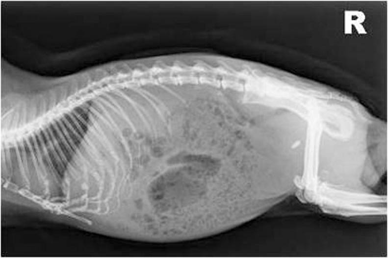
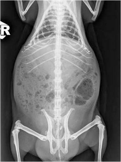
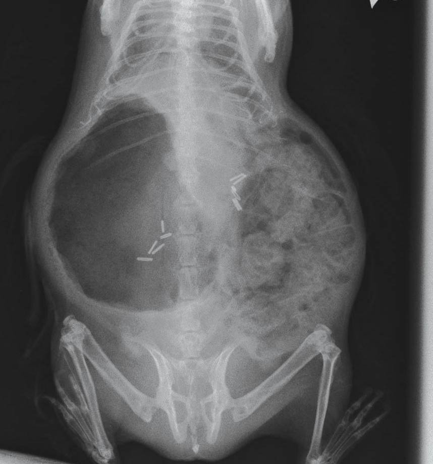
Abdominal radiographs are also valuable as urinary calculi in guinea pigs are generally radiopaque (Figures 3, 4 and 5). However, if a large amount of gas is present within the gastrointestinal tract, anatomical localisation of the calculi may be difficult.
Abdominal ultrasound can help confirm a presumptive diagnosis made using radiographs or it may be necessary to localise stones in the urethra, ureter or kidneys (Figure 6).
Ultrasonography also allows evaluation of changes within the kidneys, ureter or bladder (for example, hydronephrosis, hydroureter and mucosal inflammation), as well as other abdominal (non-urinary) pathologies.
An excretory intravenous pyelogram may permit further functional evaluation of the urinary tract. Alternatively, detailed images can be obtained with a CT scan and contrast agents can be used to elucidate functional abnormalities.
Mineral evaluation of the calculi removed can be performed, taking care the lab chosen for such analysis is able to use techniques capable of differentiating between calcium carbonate and calcium oxalate (Hawkins and Bishop, 2012).
Cystoscopy may also be a useful diagnostic tool for direct visualisation of the mucosal surface of the urethra and urinary bladder, and allows biopsies of mucosal lesions to be obtained (Wenger and Hatt, 2015).
Because the exact mechanism of stone formation is not known in guinea pigs, clear directions for prevention and dissolution of calculi cannot be given in this species.
Surgical removal of the urinary calculi is considered the treatment of choice as medical treatment of urolithiasis is often unrewarding and ineffective (Hawkins and Bishop, 2012). Recurrence of the condition once the calculi have been surgically removed is high and owners should be advised accordingly. The following recommendations are suggested for treatment and prevention of urolithiasis.

Water intake should be increased and dietary calcium and oxalate intake reduced.
According to one study, dietary modification aimed at including more hay and a wider variety of fruits and vegetables seem to decrease the risk of urolith development in pet guinea pigs (Hawkins et al, 2008). No effective stone dissolution diets exist in guinea pigs.
However, reducing, but not eliminating, dietary calcium and oxalate levels (for example, switching to grass, Timothy hay or oat hay, with a lower overall percentage of pellets, rather than using alfalfa-based diets) may help, due to the lower calcium level and higher fibre content.
Food containing high levels of oxalate (for example, spinach, kale, celery, parsley and strawberries) should be limited, especially in conjunction with a low calcium diet.
Mineral blocks or supplements should be removed from the diet.
It is important to remember severe dietary calcium restriction can be dangerous in guinea pigs and cases of metabolic bone disease have been reported following drastic dietary measures for prevention of urinary calculi (Hawkins and Bishop, 2012).
Vitamin C is extremely important in guinea pigs, as they cannot synthesise their own.
When supplementation is limited to 25mg/kg to 100mg/kg per day, it is unlikely to result in excess oxalate levels (vitamin C has high oxalate levels).
Fresh cabbage, kale and oranges are good natural sources of vitamin C.
It can be added to drinking water (200mg/L to 400mg/L daily), but the vitamin is unstable and inactivated quickly with light.
Tablets can also be crushed and sprinkled over vegetables and fruit (Hoefer and Latney, 2009).

Supportive and symptomatic treatment should be provided, including appropriate hydration, adequate pain control, assisted feeding and antibacterial treatment, when indicated and following appropriate testing.
Consider culture/sensitivity as this can be extremely helpful for a more specific medical therapy.
The use of urinary acidifiers, such as potassium citrate (30mg/kg to 75mg/kg twice daily orally), is controversial as guinea pigs normally have alkaline urine, but some clinicians find it helpful in reducing stone formation after surgical removal (Hoefer and Latney, 2009).
The female guinea pig may be catheterised to flush the bladder under anaesthesia to attempt removal of the calculi, but the small size of the male urethra makes this procedure more dangerous (Hawkins and Bishop, 2012). However, catheterisation of the male guinea pig can be performed, taking care to avoid trauma to the urethra, to allow retrograde flushing of the bladder, which may help relieve urethral obstructions (Brown and Mans, 2007).

Cystoscopic urolith removal may also be a more rapid and less invasive alternative to surgical removal (Pizzi, 2009; Wenger and Hatt, 2015). If this is not possible and the guinea pig is not obstructed, supportive care – including fluids and assisted feeding – should be provided and the surgery for removal of the stone scheduled appropriately, depending on the clinical conditions of the patient.
Cystotomy is performed as routine (Figure 7). Surgery is considered the preferred procedure for relief of ureteral obstructions (Figure 8).
Ureteral stones should be milked down into the bladder, if possible. Urethrotomy has been described in guinea pigs (Gaschen et al, 1998; Stieger et al, 2003; Bennett, 2012; Mancinelli, 2012).
The complications reported in dogs and cats following this procedure are significant, including leakage and ureteral stricture. It may also be expected in guinea pigs, especially given the challenges of their relatively smaller size. Given these risks, many cases presented with partial obstruction are often left untreated.
In cats and dogs, urethrotomy can be avoided with placement of ureteral stents, therefore allowing to bypass the obstructed section of the ureter.
The palliative clinical management of a complicated ureteral obstruction in guinea pigs has been described using an ultrasound-guided percutaneous antegrade nephrocentesis and hydropropulsion (Eshar et al, 2013).
Elisabetta Mancinelli
Job Title