21 Sept 2015
Madonna Livingstone considers the treatment options – and the lengthy recovery time in some cases – for tortoises that have suffered serious injuries to their shells.
Madonna Livingstone
Job Title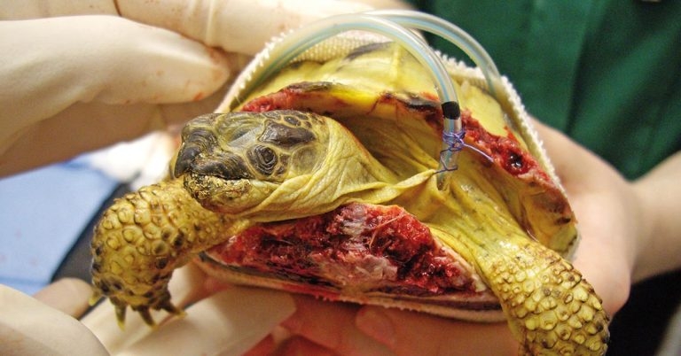
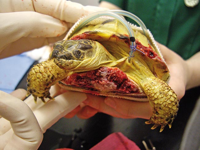
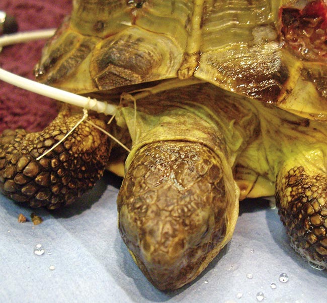
Figure 11. The first case treated by the author.
Shell injuries are relatively common in tortoises and terrapins. This can be intimidating to vets who are unfamiliar dealing with these species, and may lead to many being unnecessarily euthanised or receiving inappropriate treatment.
Hopefully, this article will provide a guide to assessment and treatment of injuries in these species with some of the treatment options available. It highlights how long these animals can take to heal, but also how well they can recover even from severe injuries. Medical and surgical treatments and dressings are discussed. The requirement for cascade forms is also covered, especially in this age of litigatation.
Dog bite injuries to tortoises are relatively common, and it is important not to panic. Many of these animals will make a full recovery with the appropriate treatment, even if they have suffered severe trauma.
So, what to do? Refer on or treat it yourself; it is immensely satisfying to see these amazing creatures heal.
Assessment of the injuries is the first step. A basic overview of tortoise anatomy will help with this (Figure 1).
The shell should be firm. If it is generally soft it could indicate pre-existing metabolic bone disease and the prognosis would be grave.
Tortoises do not have a diaphragm; they use the movement of their legs to inflate their lungs. They can cope with a surprisingly huge amount of damage to their carapace, including full thickness bone loss and lung exposure, and still breathe normally and eat as if nothing is wrong.
Damage to the vertebral scutes could cause damage to the spinal cord, which must be borne in mind as some tortoises can still walk with relatively severe damage. Therefore, bladder and bowel function should be monitored.
Damage to the plastron can be more challenging to assess. Scute loss and some bone loss are healable. Puncture wounds should be assessed to see if they have entered the coelomic cavity. Radiography can help with this.
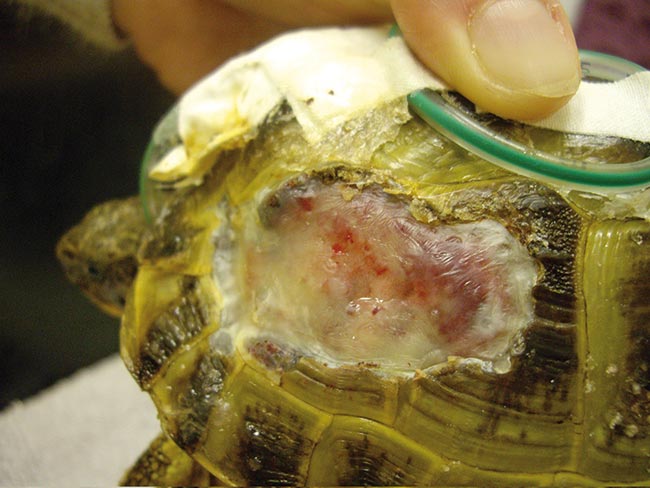
The significance of puncture wounds in the plastron is dependent on position and depth, as internal organs can be severely damaged and may make euthanasia a more viable option (Figure 2).
If the owner is willing to try treatment I always advise it is a long process. It can take a year, it is likely to be expensive and, as with any species, there is never a guarantee of success.
Assuming you are happy to proceed with treatment and the owner is aware of the possible length of treatment and cost, the next thing is to get a cascade form signed. Very few drugs are licensed in reptiles and you will definitely require them.
Analgesia is important because the tortoise will be in pain although reptiles do an amazing job of hiding it (pain scoring in reptiles is very difficult to perform).
Reptiles don’t have many mu receptors (mostly kappa and delta) so butorphanol (0.4mg/kg to 1mg/kg SC, IM q4hr to 12hr) is a much better analgesic than buprenorphine.
However, depending which text you read, this can be controversial. Although they do not have many mu receptors, some experiments have shown morphine has an analgesic effect and is associated with respiratory depression. The efficacy of the drugs also varies between species.
In my experience, butorphanol has worked well with minimal to no respiratory depression. Many reptile vets are now using tramadol (5mg/kg to 10mg/kg PO q48hr to 72hr), which reportedly has good efficacy and comes in liquid form.
Meloxicam should also be given at a dose rate of 0.5mg/kg q24hr to 48hr. It should be continued until the tortoise is eating well on its own.
In an ideal world, swabs would be taken to be sent for bacteriology and culture, but many uninsured owners are keen to limit the costs.
I usually start the patient on enrofloxacin 10mg/kg sid, plus or minus ceftazidime 20mg/kg by injection q48hr to 72hr, depending on the levels of contamination and the time before presentation. Once the ceftazidime is made up it needs to be used within 24 hours or discarded; however, it is possible to fill syringes with the required dose and freeze them.
I will also give a calcium supplement. A liquid form is available that contains the active form of Vitamin D, D3. Many tortoises may have a less than ideal diet for the species and may not have had adequate supplementation. We are trying to repair bone so calcium supplementation needs to be good, but the powdered versions can occasionally put some tortoises off their food.
Most of the time I will lavage the wounds with sterile saline once the analgesia has been given. Sometimes the wounds will be impacted with substrate (for example, soil/sand mixture) or even maggots (Figure 3).
If the tortoise is bright, and hopefully still eating, I will dress the wounds with a hydrocolloid, moisture-retentive wound dressing and cover with vet wrap, initially to help stabilise the tortoise before further treatment (Figure 4).
If the tortoise is dull, I will give a bolus of warmed intravenous fluids into the subcarapacial sinus (saline 10ml/kg to 30ml/kg q24hr split over three doses), as well as the analgesia and antibiotics.
Hydration status can be hard to assess, but generally the eyes will appear sunken. Blood tests should also be considered in case there is an underlying condition – for example, liver or kidney disease – either taken from the jugular or subcarapacial sinus. Fluids can also be given orally or via intracoelomic injection (Figure 5).
Initially, most patients will require hospitalisation so heat provision (from above) and UVB is important.
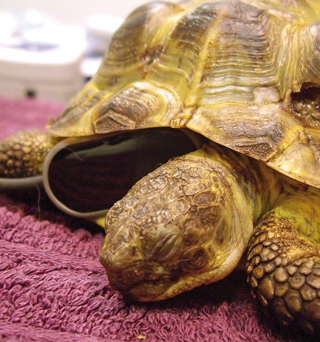
Some species are bolder than others and will tolerate oral medication, but in shy species, such as Horsfield’s (also referred to as Russian or four-toed tortoise), or in anorexic individuals, placement of an oesophagostomy feeding tube is a must.
It is a simple procedure and many of the tortoises will require sedation/general anaesthetic for debridement of their wounds so you can place a tube at the same time (Figure 6).
These tubes can remain in place for a long period – up to six to eight weeks – allowing feeding and medicating in a stress-free manner the owner can do at home with a little training.
Most terrestrial tortoises we see are herbivores (exceptions being red-foot and yellow-foot tortoises that are omnivores), so I use a critical care feed.
About 10 per cent of the tortoise’s bodyweight can be given by volume at each feed, but take into account the 2ml of water used to flush the tube before and after feeding. If there is any resistance to more food then reduce the volume.
The tube can be left in place until the infection has resolved and the tortoise is eating well; it can eat past the tube. No sedation is required – simply cut the suture and remove. The wound heals on its own.
My drug of choice for sedation or general anaesthesia drugs is propofol given IV subcarapacially (3mg/kg to 5mg/kg IV sedation, 5mg/kg to 10mg/kg IV general anaesthesia). This will give enough time, in most cases, to allow thorough debridement of the wounds and feeding tube placement. Ventilation can be performed by moving patients’ legs in and out.
If you feel it is likely to be a longer procedure then intubation can be performed, as the glottis is caudal to the large fleshy tongue (tortoise mucosa is normally a slightly paler pink than our own). In reptiles the normal resting position of the glottis is closed.
A Doppler probe can be positioned in the pre-humeral fossa to monitor heart rate (Figure 7).
The wounds can be cleaned using dilute chlorhexidine or dilute pevidine. I prefer to use gauze swabs rather than cotton wool (better debridement and lower risk of bits being left behind) and use sterile toothbrushes on the bone (most supermarket ones can be put through the autoclave).
If there is significant entry into the coelom, copious flushing of warmed sterile saline and sterile toothbrushing on the edges may be a safer option.
If there is bone die back I use a rongeur to nibble back to healthy bone – for example, until it is oozing normal looking blood (as you would debride tissue in more familiar species Figure 8). Even if the tortoise is fly struck, it is worth trying.
I saw a case where a dog had chewed a tortoise and the owner had been advised to cover all the wounds with manuka honey. No antibiotic or analgesics were given. The tortoise lived in the garden and became fly struck.
The owner (who turned out to be a human pain specialist) sought my opinion. After a long general anaesthetic, and removing many maggots from its lungs (Figure 3), the tortoise made a full recovery.
The dressing used varies depending on the type of wound, contamination, infection and if there is internal organ exposure.
For example, if I had a case with a large amount of lung exposure I would use a wound dressing held on with vet wrap. These dressings should be changed every 48 hours, but this can be expensive. A thin membrane will form covering the lungs, usually within a week or two.
I may then change to a gel dressing to ensure the healing bone edges do not dry out, but sometimes this is not necessary.
Dressing changes can mostly be performed on the conscious patient; however, if the wounds are badly infected then wet to dry dressings may need to be used and would require sedation for removal.
The wounds will require flushing with either dilute chlorhexidine or dilute pevidine then sterile saline at each dressing change.
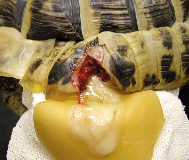
Tortoise shells are examples of membranous ossification – there is no cartilage stage. A thick fibrous membrane will form, which could take up to three months (Figure 9).
At this point dry dressings will suffice and the new bone will finish forming in around a year. I warn owners the fibrous plate may fall off when the bone heals.
Although in the literature epoxy resin is used to close defects, in most cases this is primary closure of a surgical site. In the cases we see there is, more often than not, a lot of contamination, and epoxy resin and fibreglass should never be applied to even a potentially infected wound as it seals the area, causing an abscess. Reptile pus is hard, unlike in dogs and cats (Figure 10). Unless it is a surgical wound, do not consider fibreglass and resin as an option.
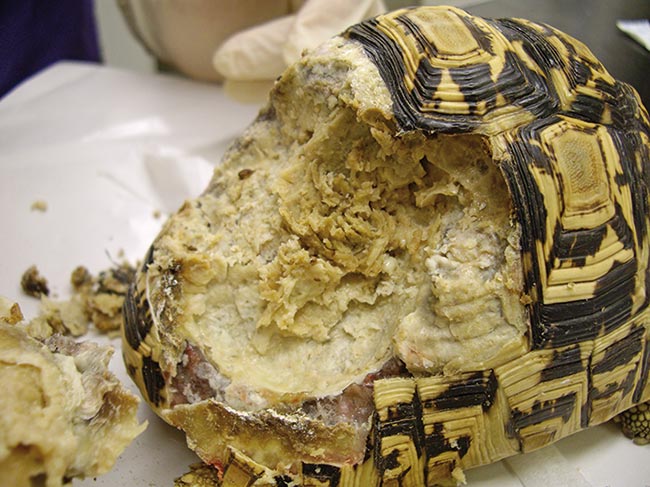
Figure 11 was the first case I treated and a locum who was on duty was surprised I did not just euthanise the tortoise due to its injuries. The locum was astounded at the healing.
Tortoises have an amazing healing ability, given the chance and the appropriate treatment. It never ceases to amaze me how much trauma they can recover from.
It may take months, sometimes more than a year, but given what their lifespan should be, and the dedication of many owners, we have a responsibility to try our best.
If you try, you may be as amazed as I am.