21 May 2018
Ashley Marshall explains the symptoms of this condition, and discusses some of the various treatments and prevention methods.
Ashley Marshall
Job Title
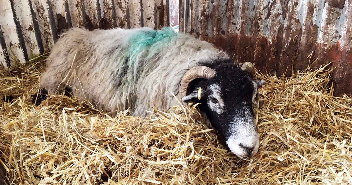
Figure 1. A Swaledale ewe with recumbency and depression. This ewe was treated for ketosis and hypocalcaemia.
Ketosis is a common metabolic condition seen in ewes in late gestation where blood glucose levels are low and ketone body levels are high. Diagnosis is usually made based on clinical signs aided by laboratory diagnostics; however, vigilance on the part of the farmer is important in identifying ewes before they are obviously ketotic.
Treatment is often unsuccessful; however, with prompt intervention the prognosis is improved. Multiple options are available, ranging from medical treatment to induction of parturition or elective caesarean, but these need to be considered on a case-by-case basis. It needs to be communicated to the farmer that ketosis in ewes can lead to other periparturient problems, namely mastitis, and prevention is much more achievable than cure.
The key to controlling and preventing ketosis is to manage and tailor ewe nutrition carefully, and to use all the services available to aid this, such as forage and ration analysis, pregnancy scanning and serological assessment pre-lambing.
Ketosis in sheep, otherwise known as pregnancy toxaemia or ”twin lamb disease”, is a metabolic disorder seen in ewes in late gestation. It is characterised by low blood glucose levels and high ketone body levels.
The high levels of ketone bodies in the blood inhibit hepatic gluconeogenesis, further reducing glucose levels in the blood and worsening the condition.
Ketosis is caused by unmet energy requirements at a time when fetal glucose demand is at its highest. The condition is often associated with insufficient energy provided in the ration, limited rumen capacity due to a uterus full of lambs, or a combination of both. It can also be seen when an acute reduction in feed intake is seen most commonly associated with stress, such as in extremes of weather or after moving location.
Treatment success is variable and highly dependent on early detection of cases. Without prompt intervention, cases can be unrewarding, with high mortality and culling rates.
Risk factors include too low or high body condition score (BCS). Thin ewes have reduced fat reserves for mobilisation, resulting in a negative energy balance, often exacerbated or caused by concurrent issues, such as high helminth burdens (especially liver fluke), lameness or poor dentition. In overweight or obese sheep, lipid mobilisation can easily overwhelm the liver’s capacity, resulting in hepatic lipidosis. Ewes carrying multiple lambs (”twin lamb”) are at higher risk due to increased fetal glucose demands paired with a fuller abdomen; however, it can occur in single lamb pregnancies – especially in thin ewes.
Some ewes, either individually or as a breed-dependent susceptibility, have a degree of insulin resistance. This ineffective use of insulin to facilitate glucose uptake in to cells and regulate blood glucose levels results in impaired glucose supply to maternal tissues, as well as increased lipolysis and ketone body formation (Duehlmeier et al, 2013). These ewes are more likely to shift from a subclinical to a clinical ketosis when feed intake is reduced suddenly.
As vets, we are not often called to examine these cases, but our advice and recommendations for treatment, control and prevention can be invaluable.
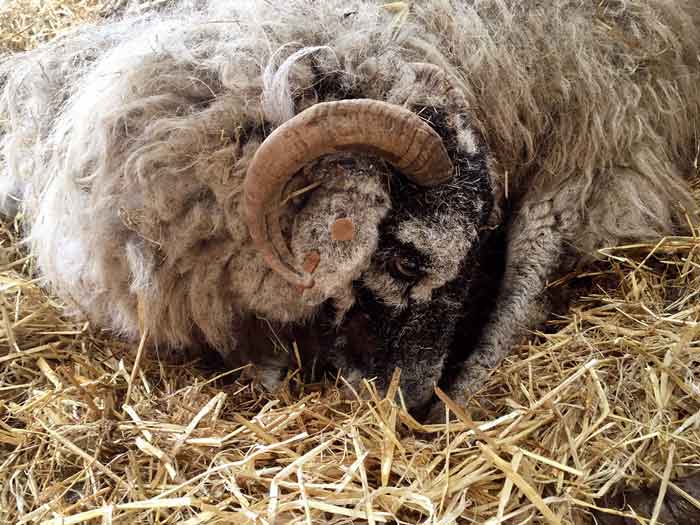
Clinical cases start with anorexia, constipation and mental dullness, progressing to neurological signs and recumbency (Figures 1 and 2). Separation from the flock is an indicator before overt clinical signs; however, this is not always noticed – especially in large flocks.
Opisthotonus, blindness, severe ataxia and tremors can be seen, with teeth grinding a common feature of the condition. Abdominal splaying can be observed in many ewes when abdominal muscle tone is lost.
Diagnosis is made based on clinical signs and can be aided by a handheld ketone meter (normal is less than 0.8mmol/L, subclinical ketosis is greater than or equal to 0.8mmol/L, and clinical disease is greater than 3mmol/L; Olfati et al, 2013). Ketone levels can also be measured in the urine using a dip stick, although obtaining a sample can be difficult. Serum biochemistry will often show signs of dehydration, along with evidence of renal or hepatic damage, as well as increased beta-hydroxybutyrate (β-HB) levels.
Postmortem findings are not pathognomonic, but will often present with evidence of hepatic lipidosis, dead fetuses in varying states of decomposition, and poor or excessive fat stores. In some cases, the adrenal glands are enlarged with haemorrhagic cortices. Diagnosis can be aided by sampling the aqueous humour or CSF, which can be analysed for β-HB levels. Fetal brain can be submitted for histopathology to demonstrate hypoglycaemic encephalopathy.
In first opinion practice, treatment is usually with oral propylene glycol twice daily or another oral solution with readily available energy. Dose rates vary by practitioner, but care should be taken to be conservative due to the diarrhoea associated with overdose.
Oral fluid and electrolytes should be given to correct any dehydration and the ewe penned individually and encouraged to eat palatable food. IV glucose can be useful, but may not be possible if the farmer does not want repeat visits from the vet. Some practitioners use an IV fluid and dextrose combination with varying results. Injectable calcium borogluconate is commonly given to treat concurrent hypocalcaemia, which is present in around 20% of ketotic cases (Andrews, 1997).
Anti-inflammatory drugs are recommended – especially in cases where the ewe is recumbent or has sustained any traumatic injury. However, these are likely to be off licence and, as such, appropriate dosage and withdrawal should be used. The rest of the group should be assessed for subclinical ketosis.
In severe cases, a transient improvement may be seen, but these cases tend to deteriorate and eventually die due to the degree of hepatic or renal damage. In this situation, a judgement call needs to be made as to whether pursuing treatment is appropriate.
In cases of treatment failure, either euthanasia or induction could be offered on a case-by-case basis. The farmer must be made aware death can still occur in the ewe after it has given birth due to renal and hepatic damage, as well as the likelihood of peri-parturient problems, such as retained fetal membranes, poor milk production and increased risk of mastitis (Barbagianni et al, 2015).
Ingoldby and Jackson (2001) evaluated methods of induction and concluded corticosteroids were the least costly and most commonly used drug, with birth occurring around 48 hours after administration. Corticosteroid administration may be ineffective due to high circulating cortisol levels in the ewes, resulting in failure of induction. Elective caesarean may be attempted; however, this would be dictated by closeness to due date and whether the ewe would be considered a suitable surgical candidate. Animal welfare must be evaluated at all times and, if the ewe is unlikely to survive the birth and recovery, euthanasia is the best option.
In farms where a problem has been identified, a plan must be formulated to avoid the same occurring the following year.
Close attention must be paid to the nutritional and energy requirements of ewes throughout pregnancy. This can be aided by pregnancy scanning so ewes can be separated into groups by lamb numbers to facilitate a more tailored diet. Separate groups can also be made using BCS.
A nutritional investigation is always useful – especially when accompanied by forage analysis to ascertain energy levels within feed. This is particularly helpful for homegrown forages where many farmers guess at the nutritional quality or based on the previous year’s analysis.
Meeting nutritional requirements around mating and early pregnancy is essential to optimise cyclicity, ovulation and implantation. A pre-mating increase in energy-dense feed can be given to ewes (flushing), which should be carried on into the first four to six weeks of mating/pregnancy. This is also beneficial for thin ewes to increase BCS and improve cycling and lambing rate (Fthenakis et al, 2012).
During the first 100 days of pregnancy, feeding needs to be maintained so ewes do not put down excessive fat deposits or lose too much weight. Adequate nutrition needs to be provided for growth of the placenta and mammary glands. Correct management of this time period will drastically reduce the risk of ketosis in later gestation.
Fetal growth is rapid in the last month of pregnancy, with 75% to 80% of its birth weight being acquired. This results in a huge increase in energy and protein demand on the ewe, which, when coupled with reduced rumen capacity and feed intake, can result in ketosis. Using a feed that is energy dense and of suitable quality is essential.
Maintaining ewes at optimal BCS throughout pregnancy is advised and scoring should be done at regular intervals, starting at pre-mating. Feed should never be withheld, but can be adjusted gradually to requirement. Rapid weight loss or weight gain needs to be avoided to prevent ewes falling to negative energy balance. Identifying thin ewes may be of benefit in separating out for extra feeding, while fat ewes can be encouraged to exercise by walking between fields to get feed. Adequate trough space must be provided to ensure all ewes have access to sufficient feed. Phillips et al (2014) have provided a thorough overview of feeding the ewe in later gestation.
Blood sampling of late gestation ewes may be helpful to look at nutritional status by evaluating β-HB and protein levels. This can aid in identifying groups at risk of negative energy balance and ketosis, as well as assessing whether the ration supplied is providing adequate energy and whether feeding needs to be adjusted. This may also encourage the farmer to be more vigilant when looking for ewes showing early signs of ketosis.
While ketosis in ewes does not generally carry a good prognosis, early detection is paramount to successful treatment and a full recovery. This condition is more easily prevented than cured, and nutritional management before and during pregnancy is paramount to reducing the risk and incidence.