9 Apr 2018
Nicola Gladden discusses problems and conditions facing dairy cows following calving.
Nicola Gladden
Job Title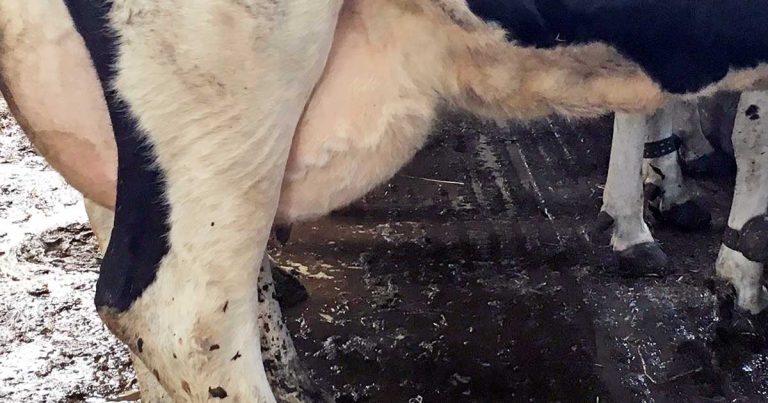
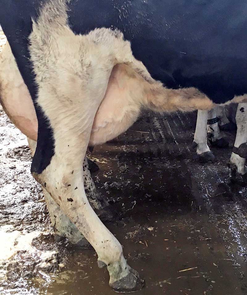
Figure 1. Flexed hock and knuckled fetlock typical of sciatic nerve injury.
Calving is a stressful and potentially dangerous event, and postpartum problems are commonly seen in dairy herds. Problems that occur in the postparturient period can adversely affect the health and welfare of the cow in the immediate term, but can also have longer-term adverse effects on production.
Prompt, appropriate treatment is required to treat the cow in the immediate term, but investigation into management around calving, including dry period management, is also needed to prevent future problems occurring in other animals in the herd. This part reviews some of the evidence available regarding the care of the postpartum dairy cow and management of postpartum complications that may arise.
Following part one of this article, the management of calving injuries and postpartum metabolic disease are reviewed.
Injuries that occur at calving are most commonly soft tissue injuries or neurological injury. Calving injury is most often associated with dystocia and can also occur as a result of inappropriate calving assistance, in particular if excessive or inappropriate traction is used.
Perineal lacerations can occur as a result of feto-maternal disproportion or the use of excessive/inappropriate traction when delivering the calf. Perineal lacerations are classified in accordance with the location and extent of the injury (Table 1; Fubini and Ducharme, 2004).
| Table 1. Perineal laceration classification (Fubini and Ducharme, 2004) | ||
|---|---|---|
| Classification | Definition | Treatment |
| First-degree laceration | The skin and mucosa of the vagina/vestibule are affected only. | Most first-degree tears do not require surgical repair. |
| Second-degree laceration | Extension of the laceration into the fibromuscular tissues. | Emergency treatment is not required; however, surgical correction may be required at a later stage to correct vestibular/vulva conformation abnormalities that can develop secondary to the tear. |
| Third-degree laceration | Complete disruption of the rectovestibular shelf +/− rectovestibular fistula formation. | Faecal contamination of the vestibular/vagina often occurs, which can affect fertility and surgical correction will be of benefit if the animal is to be bred again. |
Third-degree perineal lacerations and rectovaginal fistulae can result in production losses (Dreyfuss et al, 1990; Farhoodi et al, 2000). More minor tears will heal by secondary intention; cows with more severe tears can benefit from surgical repair, in particular if a rectovaginal fistula is present.
Unless the laceration is accompanied by severe haemorrhage that requires emergency repair, it is best to postpone surgical repair until any associated contusions and oedema have resolved (Dreyfuss et al, 1990; Fubini and Ducharme, 2004). Techniques for surgical repair of perineal laceration can be found in large animal surgical textbooks.
Treatment of severe postpartum haemorrhage and perineal lacerations in cattle is not always straightforward or successful and, in some cases, euthanasia may be the most appropriate option. When assisting parturition in cows, measures to prevent calving trauma, such as the use of adequate amounts of lubrication and careful calving technique, should be undertaken. This is of particular benefit in animals shown to be of high risk of developing perineal tears; for example, animals experiencing dystocia and primiparous heifers (Farhoodi et al, 2000).
Obturator nerve injury commonly occurs following a hip-locked calf and, in combination with injury to the L6 nerve root (sciatic), results in adductor paralysis. Cows experiencing obturator nerve paralysis are commonly able to stand on surfaces where they can achieve good purchase, but unable to stand on more slippery surfaces (Divers, 2004).
Calving injury to the sciatic nerve commonly affects the peroneal branch, although the tibial branch can also be affected. The peroneal branch and tibial branches of the sciatic nerve innervate the extensors (peroneal) and flexors (tibial) of the fetlock. Injury to either branch most commonly presents as knuckling of the hoof on the affected hindlimb, as well as flexion of the hock (Figure 1). Unilateral injury is most common; however, bilateral injury can occur.
Treatment of neurological injury is limited to good nursing care of the cow; for example, ensuring she is in a deep-bedded pen with easy access to food and water. Cows with obturator nerve paralysis should be hobbled (above the fetlock) to prevent abduction of the hindlimbs (“the splits”; Divers, 2004; Hartnack, 2017). Many cases will resolve if given time and nursed well, but this can take several weeks. Therefore, it is important to manage farmer expectations with regard to expected speed of recovery.
NSAIDs or corticosteroids can be considered to reduce inflammation, although limited evidence is available to support a beneficial effect with regard to periparturient paresis. One NSAID available in the UK has a licence for the treatment of periparturient paresis. Corticosteroids are not specifically licensed for this use, albeit most corticosteroids in the UK are licensed for general “inflammatory conditions” (Orr et al, 2014).
Postpartum metabolic diseases rarely occur as isolated events and a holistic approach to investigation is recommended. The transition period is widely accepted in the literature to be the period from three weeks before calving to three weeks after calving (Mulligan and Doherty, 2008), although a suggestion has been made to redefine the transition period as the period from 90 days before calving to 30 days after calving (Elanco, 2016).
Metabolic disease postpartum is usually related to prepartum management of the cow and can have longer-term production effects (Mulligan and Doherty, 2008; Vergara et al, 2014). Consequently, management and investigation of postpartum metabolic disease should aim to go beyond the initial treatment summarised in this article.
Postparturient hypocalcaemia is a common occurrence in dairy cows and the risk of developing milk fever post-calving increases with age. The postpartum period sees a sudden increase in demand for calcium and if the cow does not adapt quickly enough to this increase, hypocalcaemia will occur (Goff, 2008; Martín-Tereso and Martens, 2014).
Hypocalcaemia can be classified as either clinical or subclinical. Subclinical hypocalcaemia has been shown to be associated with an increased risk of developing other postpartum diseases, as well as having an adverse effect on immune function (Goff, 2014). Typical signs of clinical hypocalcaemia are well-recognised by both farmers and vets, and include recumbency, constipation and bradycardia.
Treatment of clinical hypocalcaemia is aimed at restoring serum calcium concentration above the commonly accepted minimum concentration of 2mg/dL (Martín-Tereso and Martens, 2014). IV calcium borogluconate is commonly used in practice and is the fastest way of restoring serum calcium concentration to a level above the minimum threshold. Care must be taken not to give IV calcium too quickly, as potentially fatal cardiac arrhythmias can result.
Calcium borogluconate can also be given SC and will maintain serum calcium concentration above the minimum threshold required for longer than IV administration. However, it takes longer to reach an adequate serum concentration and is, therefore, not suitable for emergency treatment. Oral administration of calcium (for example, as a bolus) is best suited for prophylactic treatment or for the treatment of hypocalcaemia in the early stages, before the cow becomes recumbent (Oetzel, 2013).
Prevention of subclinical hypocalcaemia usually aims to improve the adaptation to increased demand for calcium in early lactation. This can be achieved through diet manipulation in the close-up dry period. One example of this is to modify dietary cation-anion difference (DCAD; Martín-Tereso and Martens, 2014).
Hypophosphataemia is commonly identified in recumbent periparturient cattle, although it is also a common finding in clinically healthy periparturient cattle, and the relationship between hypophosphataemia and periparturient recumbency is uncertain (Grünberg, 2014).
One study demonstrated cows with low serum phosphorus concentrations were more likely to become recumbent post-calving (Ménard and Thompson, 2007), but more work is needed to further investigate this relationship. Treatment of acute hypophosphataemia consists of parenteral administration of phosphorus.
Normal bovine plasma concentration of magnesium is 0.75mmol/L to 1mmol/L. If plasma concentration of magnesium falls below 0.5mmol/L, neurological signs start to be seen. As the disease progresses, recumbency and convulsions are seen as cerebrospinal fluid magnesium concentration reduces (Goff, 1999); animals presenting at this stage are emergencies and death can occur.
Treatment consists of IV administration of lower concentration magnesium (available in combination with calcium borogluconate) in addition to SC administration of magnesium sulphate. Treatment of a convulsing animal can be aided by administering the SC treatment before the IV treatment. Alternatively, sedation can be used to alleviate convulsions and assist administration of treatment.
Clinical hypomagnesaemia is rarely seen in housed dairy cows adequately supplemented with magnesium, but is still seen in dairy cows farmed in pasture-based systems (Martín-Tereso and Martens, 2014).
Ketosis commonly occurs soon after parturition when the cow experiences a sudden increase in energy demand for milk production. This is often coupled with a decrease in feed intake, which usually starts in the dry period. Recovery of feed intake postpartum does not keep up with the increase in energy demand, which leads to a period of negative energy balance (Gordon et al, 2013).
Ketosis can be clinical or subclinical, and all cows are at risk after parturition, although older cows appear to be predisposed. Subclinical ketosis is characterised by an increase in ketone bodies in the absence of clinical signs. Clinical signs associated with clinical ketosis include inappetence, weight loss, depressed milk yield, constipation, ataxia and behavioural abnormalities, such as pica and self-trauma.
Clinical ketosis can often be diagnosed from clinical signs and history, but should be confirmed by the demonstration of ketone bodies in urine, blood or milk. Subclinical ketosis requires diagnosis through urine, blood or milk testing. Cow-side tests are available for immediate diagnosis – but detailed discussion of these is outside the scope of this article.
The commonly accepted threshold to indicate the presence of subclinical ketosis in the postpartum period is a serum BHB concentration of greater than or equal to 1.2mmol/L (McArt et al, 2012), although some authors have suggested a threshold of 1.4mmol/L (Duffield et al, 2009).
Treatment of ketosis aims to restore normoglycaemia and reduce the concentration of circulating serum ketone bodies. A number of treatment options are available and a review to determine the most effective treatment recommended 300ml propylene glycol orally once daily for five days, based on available evidence (Gordon et al, 2013).
Other commonly used treatments in the UK are IV dextrose and glucocorticoids. Insulin has been suggested as an adjunct treatment option for cases of clinical ketosis, but does not appear on the allowed substances list and is not licensed for use in farm animals in the UK. Therefore, its use cannot be recommended. A summary of UK-licensed treatment options can be found in Table 2.
| Table 2. Treatment options for subclinical and clinical ketosis (Gordon et al, 2013) | |||||
|---|---|---|---|---|---|
| Treatment | Route of administration | Recommended dose | Mode of action | Licensed in UK? | Comments |
| Propylene glycol | Orally | 300ml once daily for five days | Either it is absorbed directly from the rumen and enters the tricarboxylic acid (krebs) cycle to stimulate gluconeogenesis or it is converted to propionate, which is also used for gluconeogenesis and stimulates insulin release. | AVM-GSL licence in UK. Zero milk and meat withdrawal periods. | Efficacy is well documented. Large overdose can result in CNS depression. |
| Glucocorticoids | IM or IV injection | Dependent on product used | Induces hyperglycaemia and impedes the actions of insulin, allowing catabolism of fat and protein stores. | POM-V licence. Many different licensed products available. Withdrawal period is dependent on the product used. | Commonly used, but few studies and an associated lack of evidence exist to support the efficacy of glucocorticoids in the treatment of ketosis. May cause immunosuppression; therefore, caution should be exercised if concurrent infection is also present (Thanasak et al, 2004). |
| Dextrose | IV injection | 1ml/kg | Rapid reversal of hypoglycaemia. | A 40% solution is licensed for cattle and sheep. Zero meat and milk withdrawal period. | Used to treat clinical ketosis only. Recommended to be used as an adjunct to other treatments. Perivascular administration is very irritant. |
| Vitamin B12/phosphorus | IM, SC or IV (phosphorus only) injection | Dependent on product used | Vitamin B12 and phosphorus are both involved in gluconeogenesis. | Separate products are available and licensed in the UK with zero withdrawal periods. A combined product is not available. | Very few studies are available and evidence is lacking for the efficacy of these products in the treatment of ketosis. Additionally, only use of a combined product has been studied. Some benefit exists to using this as an adjunct to other therapies. |
The definition of retained fetal membranes (RFM) is much debated, although most studies use a definition of failure of detachment of the placenta within 12 to 24 hours postpartum (van Werven et al, 1992; Sheldon et al, 2008). Approximately two thirds of cows will pass the placenta within 6 hours and more than 80 per cent of cows will pass the placenta within 12 hours of calving (van Werven et al, 1992; Sheldon et al, 2008).
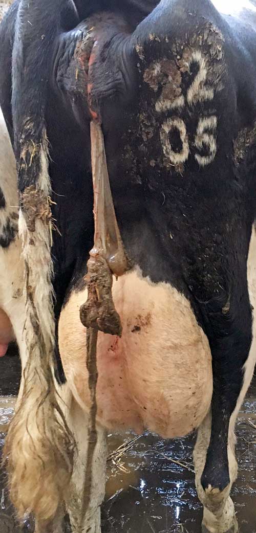
Diagnosis of RFM is straightforward as it can usually be seen protruding from the vulva (Figure 2), although sometimes vaginal examination is required.
A number of treatment options are available, although evidence for these is limited. Manual removal is commonly practised, although some studies have suggested it can cause endometrial damage.
The use of oxytocin or prostaglandin (PGF-2α) has been suggested due to their role in uterine contraction. They may be of benefit in cases of uterine atony, although a number of studies have failed to find evidence to support generalised use of either oxytocin or PGF-2α in treatment of RFM (Beagley et al, 2010).
Evidence exists that the use of PGF-2α following caesarean section may be of benefit in reducing the risk of RFM (Stocker and Waelchli, 1993). The author’s preference is for manual removal only when the placenta can be removed easily without the need for a high degree of traction.
Metritis is a bacterial uterine infection occurring within 21 days of calving and can be classified as either puerperal metritis or clinical metritis.
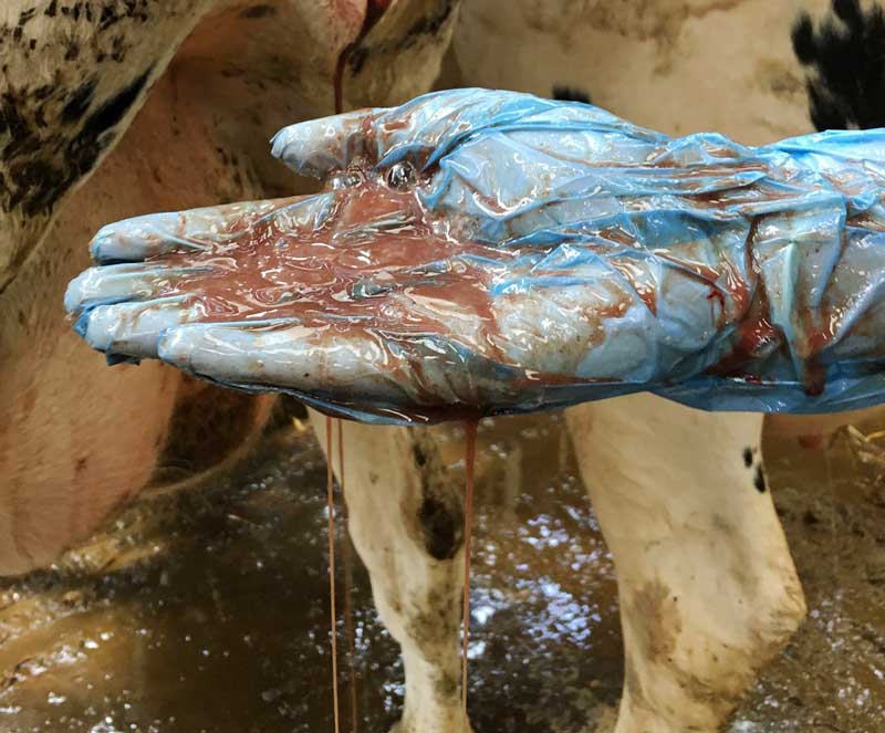
Puerperal metritis is characterised by an enlarged uterus, foetid vaginal discharge (Figure 3), clinical signs of systemic illness and pyrexia (T>39.5°C).
Animals with an enlarged uterus and purulent vaginal discharge in the absence of systemic illness are considered to have clinical metritis (Sheldon et al, 2008).
Metritis has commonly shown to be associated with Escherichia coli, Trueperella pyogenes (formerly Arcanobacterium pyogenes), Fusobacterium necrophorum and Prevotella species (Földi et al, 2006; Sheldon et al, 2008) and antibacterial therapy is indicated when metritis of either classification is diagnosed. Antibiotic choice for the treatment of metritis remains at the discretion of the individual practitioner, but a broad-spectrum antibiotic effective against the commonly implicated bacteria mentioned previously is recommended.
Although cephalosporins are commonly used to treat metritis and have been well-researched (Haimerl and Heuwieser, 2014; Reppert, 2015), ampicillin has been shown to have equivalent efficacy in treating metritis (Lima et al, 2014).
In accordance with recommendations regarding antimicrobial resistance, fluoroquinolones and third and fourth generation cephalosporins should be avoided, if at all possible, when treating metritis.
In addition to antibiotic therapy, analgesia should be considered, as evidence suggests metritis is a painful condition in some cows (Stojkov et al, 2015). The use of NSAIDs in the treatment of metritis may be of particular benefit in cases with concurrent pyrexia.
Left displaced abomasum can occur at any time in the lactation, but the first four weeks postpartum is the highest risk period. Diagnosis is usually made by history and clinical examination (auscultation of a “ping” on the left-hand side) and a number of corrective methods are described.
A summary of corrective options can be found in Table 3, adapted from an article published in In Practice (Mueller, 2011); the reader is advised to refer to this article or surgical textbooks for details of techniques.
| Table 3. Corrective methods described for left displaced abomasum (Mueller, 2011) | ||
|---|---|---|
| Method | Advantages | Disadvantages |
| Rolling (conservative) | Non-invasive and economical. Easily performed. |
Lower success rate compared to other techniques and high risk of recurrence as the abomasum is not fixed in position. Health and safety risks of rolling (to people as well as the cow) need to be considered. Risk of regurgitation when in dorsal recumbency. |
| Grymer/Sterner toggle technique (“toggling”) | Minimally invasive and anchors the abomasum in place. | Blind nature of the technique means the toggle does not always fix the abomasum. Health and safety risks of rolling (to people as well as the cow) need to be considered. Risk of regurgitation when in dorsal recumbency. |
| Left-sided laparotomy with ventral abomasopexy/omentopexy (Utrecht method) | Relatively straightforward technique requiring only one surgeon. Abomasum can be partially visualised. Cow remains standing. | Increased risk of contamination and peritonitis compared to “closed” techniques. Sometimes there may be difficulty in reaching the ventral abdominal wall to perform the pexy. More expensive than conservative approaches. |
| Right flank laparotomy with pyloropexy/omentopexy | Only one surgeon required. Allows partial visualisation of abomasum and examination of other abdominal viscera, including the liver. Cow remains standing. | Replacing the abomasum into position can be difficult – particularly in large animals, if the abomasum is heavy or if it has adhesions. Increased risk of contamination and peritonitis compared to “closed” techniques. More expensive than conservative techniques. |
| Bilateral approach | Straightforward technique with minimal risk of contamination. Allows examination of most abdominal viscera. Replacement of abomasum is easier than some of the other techniques. Cow remains standing. | Requires two surgeons. More expensive than conservative techniques. |
| Paramedian laparotomy with abomasopexy/omentopexy | Straightforward technique requiring only one surgeon. Partial visualisation of abomasum possible. Allows identification of adhesions. | High risk of contamination. Does not allow examination of other abdominal viscera. Health and safety risks of rolling (to people as well as the cow) need to be considered. Risk of regurgitation when in dorsal recumbency. |
| Laparoscopic techniques | Minimally invasive, but allows visualisation of the abomasum. Reduced risk of contamination. | Requires laparoscopic equipment and further training. |
Postpartum problems in dairy cows are commonly presented to vets and will vary from emergency, sporadic disorders to metabolic disease that may reflect an underlying herd problem. Postpartum disease has an adverse effect on production, and prompt, appropriate treatment is necessary.
Postpartum metabolic disease is rarely an isolated problem and usually has its origins in the prepartum and dry period. Being presented with a dairy cow with postpartum metabolic disease is, therefore, an opportunity to start a conversation with the farmer regarding transition cow management and instigate an investigation on farm to address the management of the herd in the transition period beyond treatment of clinically affected individuals.
This article was reviewed by Kathryn Ellis BVNS, CertCHP, PhD, DipECBHM, MRCVS.