14 Jan 2019
Sophie Mahendran looks into how the disease arises in calves and approaches to managing it – including oral rehydration.
Sophie Mahendran
Job Title
Figure 1. A calf suffering from diarrhoea: the animal exhibits faecal staining around the perineum and tail, and a hollow tucked-up appearance that indicates abdominal discomfort.
Diarrhoea – commonly referred to as scour – is the most important cause of disease and death in calves across the world.
In a large study of more than 2,700 calves from commercial dairy herds in the US, 23% were treated at least once for diarrhoea, with the mean age of onset of 10 days and a mortality rate of 5.1% (Windeyer et al, 2014).
These figures are higher than those reported for beef suckler calves, indicating that within the dairy industry the artificial rearing practices and management strategies involved can, and do, lead to compromised health – and, therefore, welfare – of calves.
Diarrhoea is a multifactorial disease, involving the triad of the calf, environment and pathogen. In most circumstances, aspects of all three risk areas are involved, so a holistic approach to managing diarrhoea outbreaks on-farm needs to be used – both to control the disease within the current cohort, and to prevent spread to new and arriving youngstock.
The main pathogens involved in the development of diarrhoea are:
Whatever the causative pathogen, the resulting disease process is either a malabsorptive or hypersecretory diarrhoea. Hypersecretory diarrhoea results in the dysfunction of cell metabolism, leading to net secretion of chloride, sodium and water into the intestinal lumen. This overwhelms the absorptive capacity of the large intestine and results in increased fluid content of the faeces. Malabsorptive diarrhoea occurs due to pathogens causing villous atrophy, resulting in malabsorption of nutrients and electrolytes, which causes an osmotic diarrhoea. The intestinal loss of electrolytes leads to a hypo-osmotic extracellular fluid, which causes free water to move from the extracellular fluid to the intracellular fluid space. Whatever the type of diarrhoea, the result is dehydration, electrolyte imbalances and development of acidosis (Mahendran, 2017).
It is commonly believed clues as to the pathogenic cause of calf diarrhoea can be glimpsed from the age of onset of clinical signs. However, on interrogation of the literature, it becomes clear many different ages are susceptible to the different pathogens quoted, with very few literature references for these guidelines appearing to exist. This means although educated guesses as to the pathogens involved can be made (for example, E coli being more likely in the first five days approximately of life), the use of diagnostics is still highly recommended.
A variety of options are available for diagnostic testing, but most laboratories provide enteric packages that will screen faecal samples for a range of pathogens. These include (Cho and Yoon, 2014):
Often, by the time a vet is called to assess a calf diarrhoea outbreak, a large number of animals are affected, and usually a number of fatalities have already occurred. Panel 1 lists the four main steps in conducting a disease outbreak investigation, and the rest of the article will go into more detail on how these steps can be achieved.
Brief overview
1. Confirm the clinical diagnosis so affected individuals are treated appropriately.
2. Make an appropriate population diagnosis to determine the reasons
for the outbreak.
3. Identify actions that can be taken to prevent new cases or future outbreaks.
4. Effectively communicate a plan of action to farm owner and staff directly involved in the day-to-day care of the animals.
Being able to identify affected calves is critical to allowing treatment to begin in a timely and effective manner. Some stock people are very good at spotting early signs of diarrhoea (Figure 1), whereas other people may need additional help and guidance.
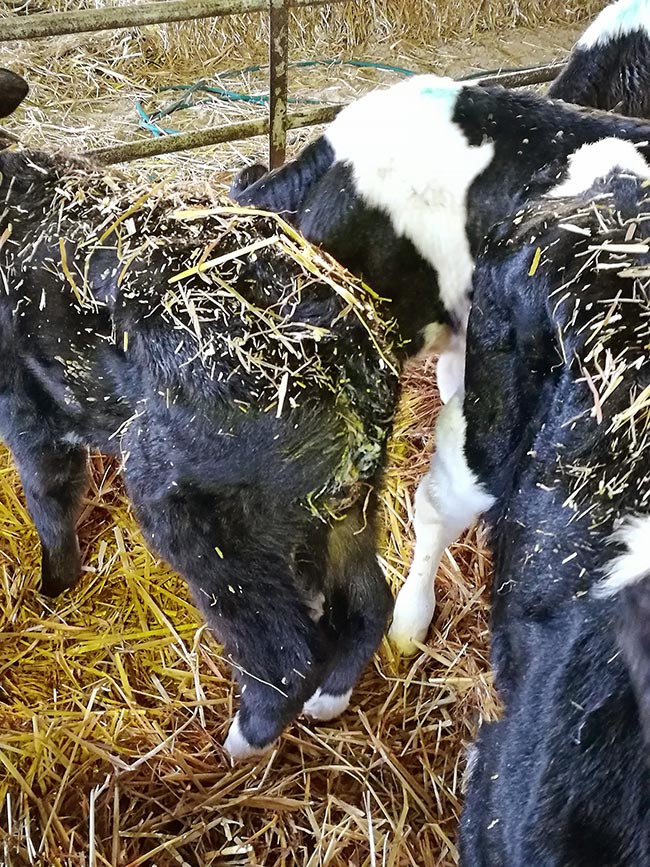
Some basic tips for spotting sick calves would be to ensure good lighting, so staff can easily see the rear end of calves to spot early signs of scour, as well as looking at bedding to identify pens with increased moisture. Additionally, encouraging staff to use their sense of smell can be helpful; pens containing calves with diarrhoea will have pungent odours, and this should not be ignored.
If an automatic calf feeder is used, information regarding reduced feed intakes and slower drinking speeds may be helpful. For staff who require a more regimented approach to identifying sick calves, using the Wisconsin calf scoring app can be helpful (McGuirk, 2008). This system has been in use for more than a decade and takes users through a step-by-step assessment of various calf health parameters to help identify animals requiring treatment. The system is available as a smartphone app, which may appeal to some users. Otherwise, a paper copy of the scoring system can be used.
Once sick animals have been identified, appropriate treatment standard operating procedures should be followed. Many practices have already developed these, with the general principles not differing between farms. Treatments centre around maintaining the hydration and electrolyte balance of a calf, while the calf’s own immune system deals with the causative pathogen.
Unless Salmonella is identified as the causative pathogen, antibiotics are not usually required and may, in fact, compound diarrhoea by causing further intestinal microflora dysbiosis. Isolation of affected calves is always beneficial – helping to reduce pathogen spread to the rest of the cohort, while allowing easy monitoring of feed intakes.
If calves are either standing or in sternal recumbency, and have a suckle reflex, they will be approximately 8% dehydrated and clinically suitable to receive oral rehydration therapy (ORT; Lorenz et al, 2011). The use of ORT must be additional to the calf’s normal milk feeds if it is still willing to suckle. However, if the calf refuses its normal milk, this too should be replaced by ORT. This means a calf receives at least four fluid intakes a day. If a calf is not willing to voluntarily suckle ORT, an oesophageal feeder or stomach tube can be used to feed it. Milk feeds, however, must not be administered by this route as milk will putrefy in the rumen and may lead to bloating.
The main aims of ORT are to promote plasma expansion, correct electrolyte imbalances, provide energy for sodium and water uptake from the small intestine, and act as an alkalising agent to address metabolic acidosis. Four types of ORT products are available, with each having slightly different components and overall usefulness (Table 1).
| Table 1. Summary of types of oral rehydration fluid for treating calf diarrhoea | ||
|---|---|---|
| Type of fluid | Components | Uses |
| First generation | Contain sodium, potassium and (insufficient) glucose. | Effective in treating dehydration associated with scour, but cannot correct acidosis. |
| Second generation | Contain bicarbonate or bicarbonate precursors, such as lactate, propionate or citrate, which consume hydrogen ions when metabolised. | Very effective at treating dehydration and acidosis caused by scour, but do not provide energy to the calf. |
| Third generation | Contain much higher concentrations of glucose. | As well as the electrolytes, it helps to counter energy deficits. |
| Fourth generation | Contain the amino acid glutamine. | Provides an energy source to facilitate sodium absorption from the small intestine, and to help sustain the intestinal villi form and function. |
In addition to ORT, NSAIDs can be useful to reduce the gastrointestinal cramping and discomfort associated with diarrhoea. This can improve voluntary feed intakes, which, in turn, has a positive impact in intestinal healing. However, care must be taken with NSAID use in severely dehydrated calves and with repeated treatments, as side effects – such as abomasal ulceration and renal impairment – may occur.
The final steps in an infectious disease outbreak investigation are to come up with prevention methods and monitor the outcomes when implemented on farm. The specific combination of recommendations will depend on the pathogen diagnosis and risk factors identified in a particular outbreak, and some of the most common points that often need to be addressed are discussed.
A dam vaccine is available for three of the most common diarrhoea pathogens (E coli, rotavirus and coronavirus). This vaccine must be given to the dam between 12 and 3 weeks pre-calving, and work by enhancing the colostral antibodies that are passed on to the calf. These vaccines can be very efficacious, but do hinge on farms having good colostrum management protocols. Colostrum management should always be evaluated when calf health issues are identified, with investigations into colostrum harvesting techniques, quality assessment and administration protocols being the main target areas.
Additional to the use of dam vaccines, transition cow management, as a whole, has a large impact on the cow’s ability to produce high-quality colostrum. Additionally, transition cow nutrition to control metabolic disease – especially milk fever – is important as this affects calving/dystocia and calf vigour. Calves that experience dystocia are at a higher risk of passive transfer failure and increased risk of developing diarrhoea.
Calving pen hygiene is also closely linked to outbreaks of diarrhoea in neonates – dirty, highly stocked calving pens have very high levels of coliforms present, as well as increasing the risk of Johne’s infection. It is important to clean calving pens out regularly – ideally every month – and to provide clean, dry bedding on a daily basis to minimise calf exposure to pathogens.
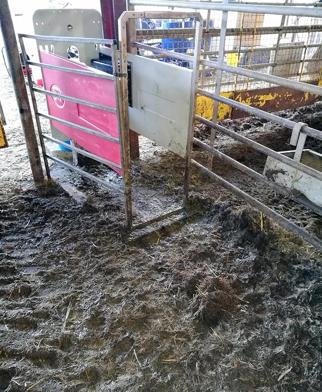
The trend of maintaining high levels of hygiene continues into the assessment of calf housing. Calves can be housed in numerous ways, but key aspects are to ensure pens have good ventilation and drainage to reduce pathogen loading in the environment. High-use areas, such as around automatic calf feeders (Figure 2), are hotspots for protozoa buildup, and should be cleaned and disinfected at regular intervals.
Also, avoiding the need for calves to feed off the floor, and keeping feed troughs free of faecal contamination, will help lower oocyst ingestion. Short-term prophylaxis can be used in outbreaks of cryptosporidium. Halofuginone lactate is licensed to prevent and treat this, but its use should not lead to complacency about the underlying management factors that actually cause the disease – hygiene.
Diarrhoea is a common presentation of the sick calf, but most outbreaks have common treatment strategies and risk factors that can be addressed. Treatment needs to focus on maintaining the hydration status of the animal, and prevention strategies should ensure environmental hygiene and colostrum management practices are optimised.