
15 Sept 2026
Kate Parkinson BVSc, MRCVS recounts the events, treatments and conclusions in two occurrences of this rare disease.

Kate Parkinson
Job Title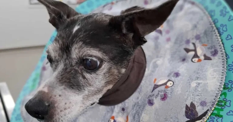
Figure 1. Patient A on initial presentation.
A 14-year-old female neutered fox terrier (patient A; Figure 1) belonging to a staff member presented two weeks following a routine dental and lump removal with sore footpads. Pre-anaesthetic bloods showed increased liver enzymes, with an otherwise normal profile.
At the postop check, a nurse noticed strange growths on all four feet which had not been present at the time of the dental. A veterinary check was advised and performed one week later. All four feet were affected.
Hyperkeratosis was present, and the dog’s feet smelled yeasty. The patient also had a history of allergic dermatitis.
At this stage, a flare up of allergy with potential hyperkeratosis was suspected. She was given her usual monthly injection of lokivetmab and a two-week course of antibiotic (cefalexin 15mg/kg twice daily for 14 days), as well as diluted chlorhexidine footbaths. This initially seemed to resolve the problem.
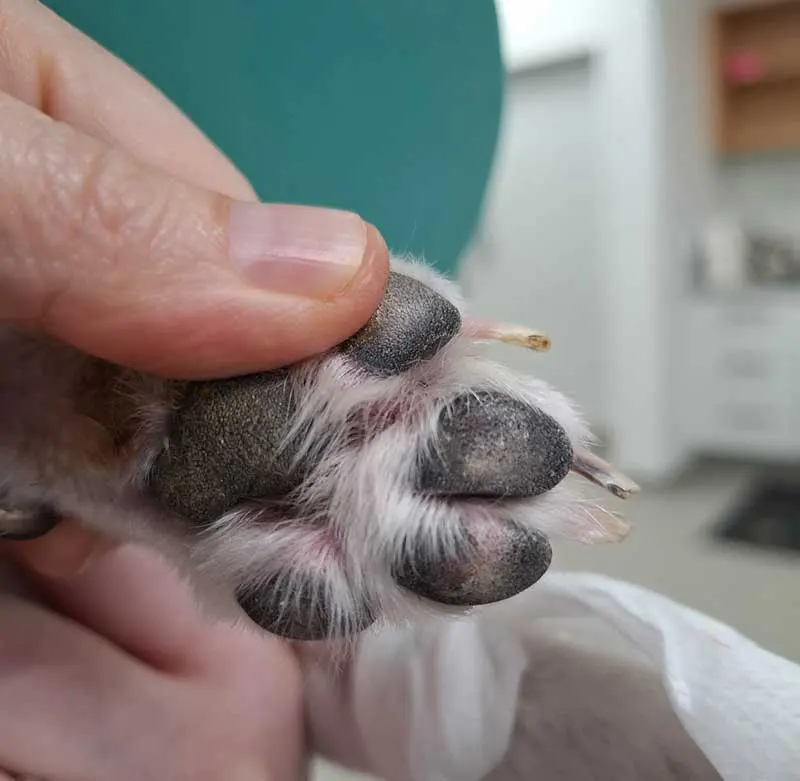
However, the dog re-presented three weeks later with reoccurring symptoms. In addition, she had developed more crusting lesions on the foot pads (Figure 2) and crusting at the lip commissures (Figure 3). Autoimmune disease was considered as a differential at this stage, as well as endocrine disease.
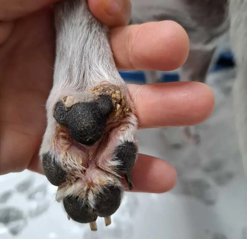
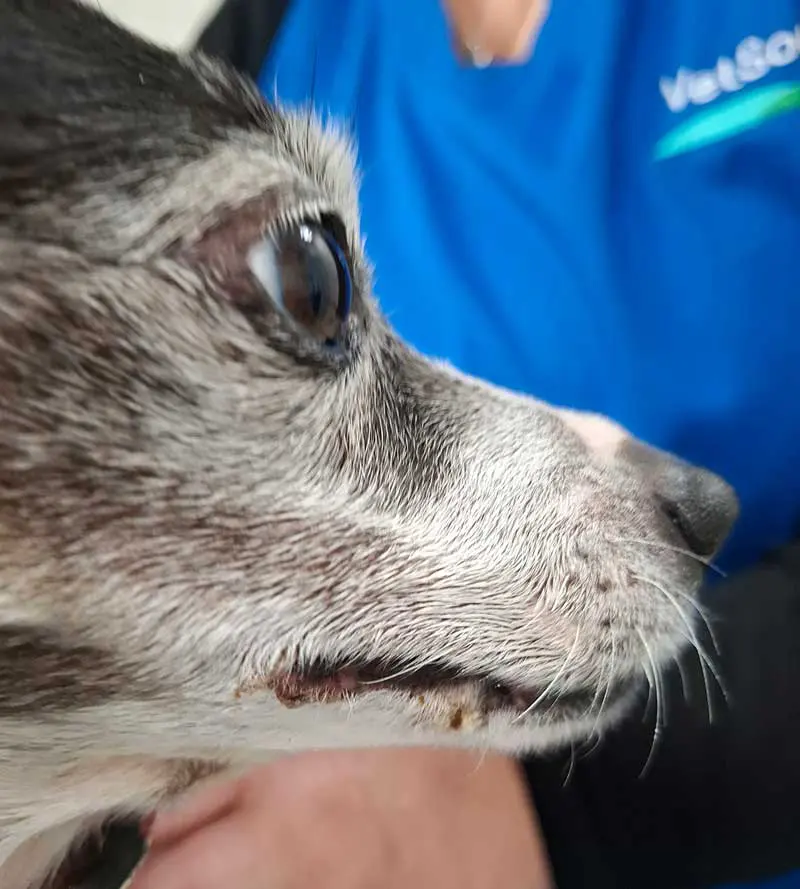
Further investigation was initially declined due to the dog’s age. Repeated footbaths (diluted chlorhexidine daily) and a second course of antibiotics resulted in limited improvement, with lesions remaining on the lip commissures and footpads.
Investigation was pursued seven weeks following initial presentation.
The patient was sedated with midazolam (0.3mg/kg) and methadone (0.3mg/kg) IV, and skin biopsies were taken from the footpads and muzzle (three in all). Each site was closed with a single cruciate suture. Blood samples were also taken and run in house for thyroxine and basal cortisol. These values were normal.
The skin biopsies displayed acanthosis with parakeratosis and coccoid bacteria. Lymphocytes and plasma cells were present in the dermis. This was consistent with a diagnosis of suspected hepatocutaneous syndrome with secondary surface bacterial and yeast infection.
Skin biopsies have the classic “red, white and blue” pattern, with red being parakeratotic hyperkeratosis, white vacuolated keratinocytes and blue basal cell hyperplasia (Almendros et al, 2019).
Hepatocutaneous syndrome (HCS) is a rare cutaneous manifestation of systemic disease (glucagonomas, enteropathies and hepatopathies in humans, and hepatopathies and pancreatic disease in dogs).
This is a metabolic syndrome characterised by hypoaminoacidaemia and aminoaciduria, which causes both superficial necrolytic dermatitis and hepatopathy. Though the precise mechanism remains unclear, it is thought that depletion of amino acids causes failure of the mechanisms essential for maintaining normal integument. Dogs with HCS are also predisposed to diabetes development.
HCS is most common in small-breed geriatric dogs, with a slight male predisposition. In conjunction with the characteristic skin lesions confirmed on biopsy, patients with HCS have hypoaminoacidaemia and distinctive hepatic changes on ultrasound scan.
An ultrasound scan was performed under mild sedation (midazolam 0.3mg/kg IM and methadone 0.3mg/kg IV, with alfaxolone IV).
The ultrasound scan displayed the typical “Swiss cheese” appearance of the entire liver, with hypoechoic nodules and a hyperechoic background, reflecting severe vacuolar hepatopathy (Loftus et al, 2021). The pancreas appeared normal, as well as all the other organs, except for the spleen, which had two hyperechoic nodules less than 1cm. The decision was made to monitor these given the severity of the HCS at this stage.
CT can also be used as method of diagnosis, though was not used in this case due to the convincing evidence presented by alternative diagnostic tests and limited availability (the closest vet CT was a seven-hour drive away).
Blood was submitted for an amino acid profile and a human laboratory. This was somewhat challenging to interpret given the format, but displayed widespread hypoaminoacidaemia.
At this stage, the case was discussed in rounds. A colleague had just diagnosed a similar case, which was highly unusual due to the rarity of this condition. This dog was a 13-year-old female neutered shih-tzu cross-breed (patient B) presented as a second opinion with progressive skin disease affecting the mucocutaneous junctions over the last four months, as well as occasional diarrhoea and vomiting.
The diagnosis was made following an ultrasound scan showing the characteristic hepatic signs, as well as a blood profile showing increased liver enzymes. Blood testing in this patient also showed widespread hypoaminoacidaemia.
Further advice was sought from a specialist regarding both cases.
The mainstays of treatment are amino acid infusions combined with a high-protein home-cooked diet and amino acid dietary supplementation.
A specialist nutritionist was consulted, and a high-protein home-cooked diet with amino acid supplementation was formulated, tailored to each patient’s nutritional needs.
A diet with more than 50% of metabolisable energy through protein has been shown to improve patient’s length and quality of life compared to commercial or plain home-cooked diets.
Both patients were treated with amino acid infusions through peripheral catheters. The infusion chosen due to high cost and low availability was Primene 10% (used in human medicine to provide nutrition to young children), which was sourced through a specialist wholesaler.
The infusion came in the form of 250ml bottles for single use only and only in packs of 10 bottles, the cost of which was initially split between both patients. Amino acid infusions such as Primene have a relatively high osmolarity, and though complications are rarely seen with infusion through a peripheral catheter, the infusion can be irritant if any leaks subcutaneously. A risk of allergic reaction also exists, and dogs should be closely monitored throughout the infusion process. The amino acid infusion was given at a rate of 25mg/kg over eight hours, with temperature, pulse and respiration recorded every 10 minutes for the first hour, then reducing to every 30 minutes throughout the day. No side effects were seen in either dog following the infusion.
Infusions are given every 7 to 10 days until clinical improvement (classified as resolution of skin lesions) is seen, then reduced to every three to six weeks depending on response.
Frequency of infusions varies and can be titrated according to clinical response. Some evidence exists that combining lipid with the amino acid infusion may improve response.
Hepatic supplementation may also be helpful in affected cases.
Patient A improved following the first infusion. Seven days after the first infusion, the lip commissure lesions had completely resolved and the paws were visibly less inflamed (Figure 4). She had gained 1.1kg in weight. This weight was maintained through two further infusions, although the paw pad lesions, though improved, persisted (Figure 5).
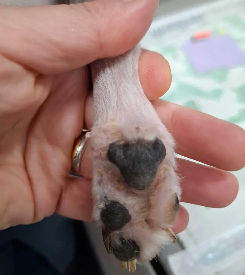
On week three, some thrombophlebitis resulted when her intravenous catheter slipped, though this resolved quickly and without treatment. On week four, (a public holiday), the decision was made to reduce infusions to every 14 days for reasons of cost and convenience. However, the following week, the patient’s pads were more irritated, and infusions were increased to weekly for a fortnight. After this, her skin improved, so twice-weekly infusions were continued. Clinical deterioration was noted if infusion frequency was decreased. At the time of writing, she has had 12 infusions.
Patient B tolerated the infusion well, but showed little improvement. Her appetite decreased, and her footpads and skin became progressively more painful. She was euthanised five days after the first infusion. Her owner kindly donated the remaining bottles of Primene to the surviving patient.
A diagnosis of hepatocutaneous syndrome carries a poor prognosis. Most dogs die or are euthanised within a few years of diagnosis. Studies are usually small due to the rarity of this condition. In a study of 41 dogs, two out of three patients died or were euthanised as a direct consequence of their diagnosis, while one out of three died of other causes. No difference in survival of dogs based on skin lesion status or weight was reported (Loftus et al, 2021). Dogs receiving two or more amino acid infusions survived significantly longer, with a mean survival time of 667 days compared to 168 days for dogs with less than two (Loftus et al, 2021).
Dogs fed a home-cooked, supplemented diet also survived longer. Optimally treated dogs received both two or more amino acid infusions, as well as a home-cooked, supplemented diet (mean survival 1,783 days) compared to variably treated dogs (any other treatment method, mean survival 214 days). The most infusions any dog received was 13 (Loftus et al, 2021).
Resolution of skin lesions can also reflect an improvement in hepatic appearance on ultrasound, with repeat abdominal ultrasound recommended six months following the start of treatment.
Subsequent development of diabetes mellitus may be an additional complicating factor.
These were interesting cases to diagnose and treat, with initial diagnosis complicated by the fact that patient A had a history of skin allergy and patient B presented as a second opinion. Although some improvement followed initial antibiotic courses, likely due to the treatment of secondary infection, the effects quickly waned.
Skin biopsy was diagnostic, with abdominal ultrasound, blood sampling and amino acid assays confirming the results. CT may also be used when available (Almendros et al, 2019).
Patient A responded to the amino acid infusion, though declined again when the treatment interval was decreased from 7 to 14 days. Patient B did not and was euthanised due to deteriorating quality of life. Both dogs were placed on a home-cooked, supplemented diet, though both dogs experienced issues with palatability.
Once diagnosed, cost was a major treatment limitation, with the amino acid infusion unavailable in single treatment bottles. The purchase of a box of 10 infusion bottles cost approximately NZ$3,000 (£1,295). The cost in this case could be shared between two clients, though to have two dogs present with this condition at the same time in the same practice was highly unusual.
Temperament of the dogs was another concern, as they were required to tolerate long periods of repeated hospitalisation. Patient A became increasingly more anxious when hospitalised and gabapentin (100mg by mouth half an hour before visits) was used to decrease her stress levels.
Home-cooked diet was a significant factor in length of survival time (Loftus et al, 2021). The diet could be batch cooked and refrigerated or frozen for up to one week. However, the diet was costly and time consuming to prepare, with nine different ingredients.
In conclusion, hepatocutaneous syndrome is a rare and interesting syndrome in dogs. Both cases presented initially for non-healing skin lesions which responded poorly to conventional approaches.
Diagnosis can be performed by a variety of methods. A combination of skin biopsies, abdominal ultrasound, blood testing and amino acid assays were used to diagnose both patients. Skin biopsies and abdominal ultrasonography are available in many practices. The author, a mediocre ultrasonographer, found the hepatic changes very easy to visualise and thoroughly rewarding. Amino acid assays, however, may not be readily available at veterinary laboratories.
The three mainstays of treatment are amino acid infusions, a nutritionist designed home-cooked diet with more than 50% of metabolisable energy through protein, and amino acid supplementation added to the diet.
Amino acid infusions are readily performed through a peripheral intravenous catheter, although require close monitoring for complications (thrombophlebitis or allergic reactions), and are easily achievable within a general practice.
Diet plans should be prepared by a specialist veterinary nutritionist, ideally one with experience in the treatment of hepatocutaneous syndrome. It may be possible, as in these cases, for the nutritionist to also provide the amino acid supplements.
Kate Parkinson graduated from the University of Bristol in 2006. Since graduating, she has travelled to more than 50 countries and worked as a veterinarian in Australia, New Zealand, and the UK. Kate currently works in small animal practice in Invercargill, New Zealand, where she combines her interests in writing and general small animal practice and writes occasional veterinary articles.