
1 Nov 2026
Deborah James RVN, discusses the intricacies of the stages of healing and dealing with treatment

Deborah James
Job Title
Image: kmls / Adobe Stock
Wound management is a daily occurrence in veterinary practices and is an area where RVNs play a crucial role – both in the management of the wound but also in the ongoing, day-to-day care of the wound and the monitoring for complications.
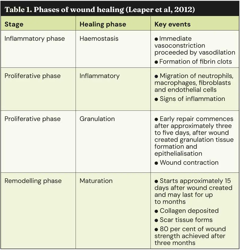
Four stages comprise the normal healing process, which is important for veterinary nurses to understand prior to being involved in wound management in practice. Table 1 summarises these stages (Leaper et al, 2012):
Frameworks such as patient safety or anaesthetic checklists are commonly found in veterinary practices and hospitals as a way to improve patient outcomes (Yon and Lilley, 2024). While no veterinary-specific frameworks for wound assessments are currently available, some are available from the world of human medicine that can be adapted for veterinary use.
The TIME framework was created 20 years ago to help improve the assessment and ultimately treatment of wounds in human medicine (Schultz et al, 2003). TIME stands for tissue, infection or inflammation, moisture and edge (of a wound) with some versions of it including an S for surrounding skin (Curtis, 2022).
This framework can then be integrated into a larger case decision system such as ABCDE (Moore et al, 2019); CASE (Wounds UK, 2018) and WAT (Dowset and Nyløkke, 2015). WAT stands for the wound assessment triangle that assesses the wound bed, wound edge and peri-wound skin and assesses areas of the wound that are overlooked in other frameworks such as TIME (Dowset and Nyløkke, 2015). CASE (cause, assess, select and evaluate) was adopted by Wounds UK (2018) in their best practice guidelines for assessing chronic wounds that integrates TIMES in the wound assessment phase.
ABCDE follows a similar pattern to those in nursing care plans – assess, plan, carry out and evaluate (Orpet, 2011). In this case, ABCDE stands for:
By using the TIME framework, veterinary nurses can assess different wound management techniques dependent on the patient presented to them.
In this initial section, nurses can evaluate the type of wound and the health of the tissue identified as viable versus non-viable tissue with the primary goals being removing dead tissue and promoting healthy issue growth (Lilley et al, 2025).
For any traumatic wounds, cleansing and lavage should take place that nurses can do under the supervision of the veterinary surgeon to remove contaminants which may be a barrier to the healing process (Lux, 2022).
Isotonic solutions are typically recommended for lavage (Davidson, 2015). However, alternative recommendations include chlorhexidine 0.05 per cent solution or povidone-iodine 0.1 per cent solution (Bell, 2021), as well as the addition of tris–ethylenediaminetetraacetic acid (EDTA) to improve antimicrobial properties (Davidson, 2015).
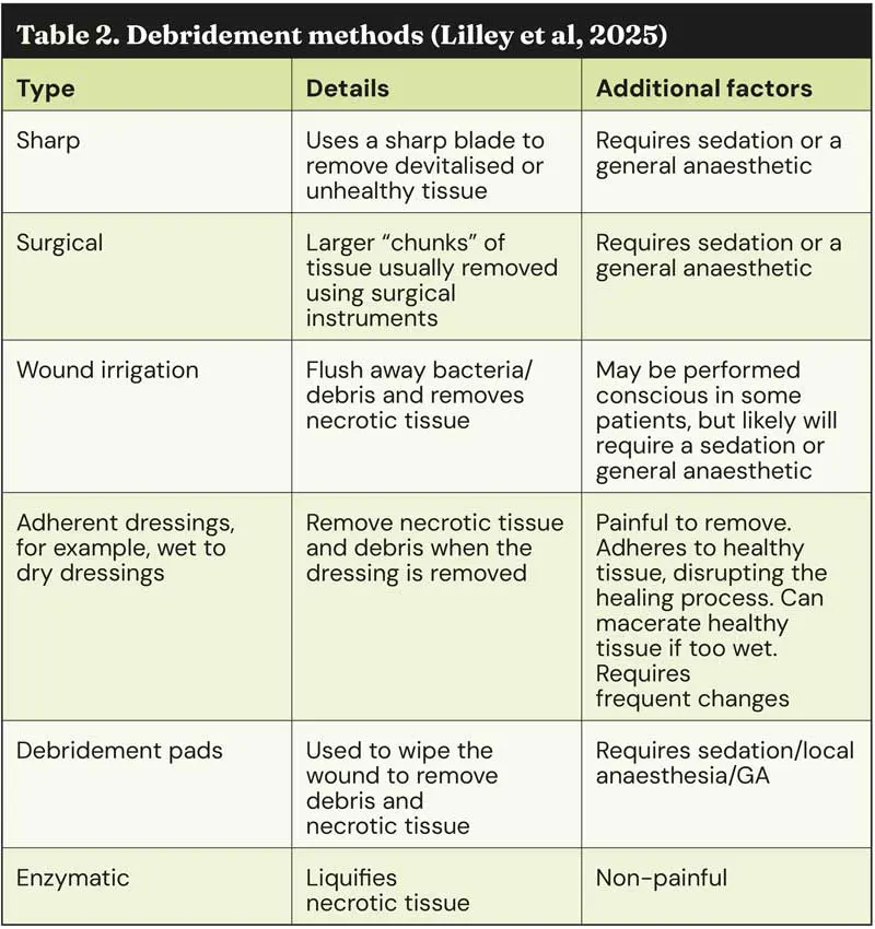
Once the wound has been lavaged, debridement is an essential part of the following process to remove any non-viable tissue and create the ideal environment for wound healing. Table 2 summarises the different debridement methods for nurses to consider and be aware of (Lilley et al, 2025):
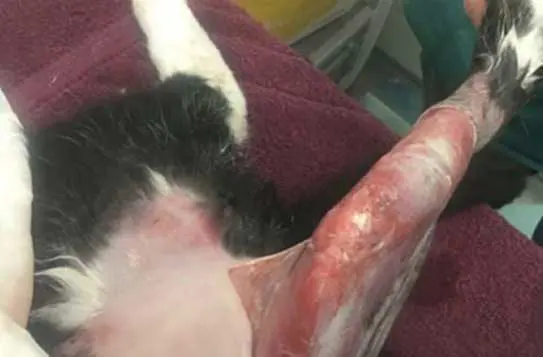
Negative pressure wound therapy (NPWT) is perfect for complex or chronic wounds and works by loosening necrotic tissue and facilitating infectious material, with studies demonstrating numerous benefits including increased blood flow, fewer complications during the healing process, faster wound closure, reduced necrosis and improved comfort levels for patients leading to shorter hospitalisation times and fewer visits to the practice (Buote, 2022). This is ideal for patients with open traumatic wounds such as degloving injuries (Figure 1), bites or large abrasions, surgical site infections and incisional dehiscence (Nolff, 2021).
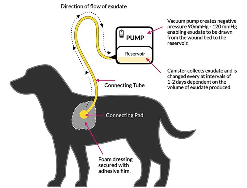
NPWT devices are powered using a vacuum pump at a negative pressure of 90mmHg to 120mmHg to remove exudate and move it to a reservoir that remains closed and retains the exudate until it is emptied every one to two days, dependent on the amount produced (Hollis, 2024). Figure 2 depicts this (Hollis, 2024):
As all nurses know, infection or inflammation on a chronic level can impair the healing process and biofilms present a challenge in this. By definition, a biofilm is a colony of microorganisms that group together and form a barrier within two to four hours continuing over two to four days (Heasler et al, 2019). The main challenge is that biofilms increase the tolerance to antibiotics and the body’s own healing mechanisms and requires regular debridement to penetrate (Bell, 2024). Biofilms are likely to be present if (Heasler et al, 2019):
If these indicators are present, RVNs can identify these and can use this information in discussions with the veterinary team with regards to the best course of action to take, as well as using it for an audit retrospectively – assessing when a biofilm could have been present, effective/non-effective protocols and use this information to inform future wound management approaches in the future. Manuka honey has long been used for its use in promoting a moist healing environment and reducing inflammation, but often has mixed findings in research. Honey aids the healing of infected wounds (Vogt et al, 2021), as well as improving tissue perfusion and reducing oedema, but is inconclusive when it comes to the healing speed (Repellin et al, 2021).
Wounds require a balanced moisture environment and selecting dressings is an important part of this. Maintaining the correct moisture levels within a wound is vital – it promotes healing and prevents further tissue damage. Insufficient moisture and the wound bed will dry out, delaying healing and increasing pain, while too much moisture will lead to the skin around the wound breaking down (Lilley et al, 2025).
NPWT, which has been discussed previously, is effective in managing moisture from large wounds with large amounts of exudate as it actively removes excess fluid while maintaining a balanced environment, while hydrogels or foam dressings are best for smaller wounds (Lux, 2022).
Veterinary nurses play a key role in monitoring these moisture levels by assessing for strike through or dryness within the wounds and liaising with the veterinary team about the appropriate dressings to be used.
Exactly what it says on the tin – examining if the wound edges are closing. If a wound isn’t closing then there may be underlying issues that need to be taken into consideration or further intervention may be required to encourage epithelialisation.
The easiest way to determine this other than by sight is to measure the wound to track it. Simple measuring techniques using a ruler are common place as it is simple and quick, but studies have shown that this method can actually overestimate a wound area by around 40 per cent – not as reliable as once thought (Rogers et al, 2010).
Planimetry is a more accurate option involving tracking wound edges and calculating surface area, but is often time consuming, costly and can require specialist equipment. Latest advances in technology and the capabilities of our smartphones have made planimetry more accessible as they are easy to use, no specialist equipment is required and ultimately they provide consistency with results (Bodea et al, 2021).
RVNs do this every day – monitor skin and prevent infection and secondary issues. Regular assessment enables maceration or erythema to be detected at an early stage and prevents complications or delays to recovery (Curtis, 2022).
Surrounding skin can be further protected through the application of barrier creams and sprays to create a protective layer and, by selecting non-adhesive dressings and applying adhesive removal sprays, skin trauma risks are reduced significantly.
By protecting this skin, RVNs play a key role in preventing secondary complications and support faster wound healing. More importantly, they can support the comfort levels of their patients.
Wound management within the role of RVNs is a vital, yet evolving, area. RVNs can expand their responsibility within practice by taking active charge of assessing wound healing phases, selecting appropriate dressings and applying bandages alongside vigilantly observing wounds for signs of bandage slippage, strike-through or sores – all while collaborating with the veterinary surgeons to ensure the best outcomes for their patients.
These patients, although time consuming, can be the most rewarding and fulfilling, enabling nurses to deepen their bond with their patients and their owners while fulfilling their professional needs to a high level.
Deborah James qualified from Hartpury College in 2017 with a BSc in veterinary nursing science and works as team leader at Vet’s Klinic. She has completed an ISFM certificate in feline nursing and is in her second year of her MSc in veterinary nursing at the University of Glasgow. She has two cats at home: Tipsy, a 10-year-old hand-reared cat, and six-year-old Frank. In her spare time, you can find Deborah snuggled with a book or teaching and participating in pole fitness and aerial arts.