9 Dec 2020
Diana Ferreira in the first of a two-part article, walks readers through the key elements in the work-up of pruritic canine patients.
Hypersensitivity conditions have in common the fact that all of them result in pruritus and inflammation. Most allergic hypersensitivity reactions present with a similar clinical pattern. Differentiation between the allergic reactions is, for this reason, challenging and should be based on a combination of thorough history, on the pattern of the clinical signs, and on ruling out all the potential causes of pruritus.
Because of the multiple diseases that can result in pruritus, the diagnostic approach to a pruritic dog must proceed in a stepwise fashion where parasitic causes are addressed first, along with the diagnosis and management of bacterial and yeast infections/overgrowth. This first approach is then commonly followed by exclusion/diagnosis of allergic components. The work-up has to be sequentially ordered and methodically performed, otherwise it will be unreliable and misleading conclusions will be drawn.
The term allergic dermatitis refers to an inflammatory skin disease caused by any type of hypersensitivity. Hypersensitivity conditions have in common the fact that all of them result in pruritus and inflammation.
The main allergic skin diseases that can affect dogs are:
Other allergic skin diseases – such as urticaria and angioedema, contact dermatitis, and bacterial and Malassezia hypersensitivity – although less common, should also be considered in a clinical setting. They will be outside the scope of this article.
Most allergic hypersensitivity reactions present with a similar clinical pattern. Initial pruritus, erythema, hair loss and papules may exist. With chronicity, patients can show cutaneous hyperpigmentation and lichenification.
Differentiation between the allergic reactions is, for this reason, challenging and should be based on a combination of thorough history on the pattern of the clinical signs, and on ruling out all the potential causes of pruritus.
Apart from allergic causes, many other diseases can result in pruritus and can mimic allergic skin disease. These include parasitic diseases such as sarcoptic mange, pediculosis, flea infestations, cheyletiellosis and trombiculiasis.
In addition to these, Malassezia or bacterial overgrowth and infections are usually superimposed on the allergic skin diseases, contributing significantly to the manifestation of pruritus.
Other less common pruritic diseases to consider are adverse drug reactions and epitheliotropic T-cell lymphoma. Rarely, pruritus may also be associated with neuropathies (for example, Chiari-like malformation and syringomyelia, and acral mutilation syndrome) or behavioural changes (for example, acral lick dermatitis). Very rare cases of pruritus have been reported in dogs associated with systemic causes (portosystemic shunt and pseudorabies).
Because of the multiple diseases that can result in pruritus, the diagnostic approach to a pruritic dog must proceed in a stepwise fashion where parasitic causes are addressed first, along with the diagnosis and management of bacterial and yeast infections/overgrowth. This first approach is then commonly followed by exclusion/diagnosis of allergic components.
The work-up has to be sequentially ordered and methodically performed, otherwise it will be unreliable and misleading conclusions will be drawn.
Obtaining a complete history, performing a detailed dermatological examination and performing the appropriate minimum diagnostic tests are the key elements in the work-up of a pruritic dog.
The history can provide very relevant clues about the primary cause of the clinical signs the animal is showing. Certain elements of the history may be particularly useful:
After scrutinising the history associated with the clinical signs, the animal must be observed.
A detailed dermatological examination gives us the distribution of the lesions and their characteristics. Both ear canals should also be examined to determine if associated ear disease exists.
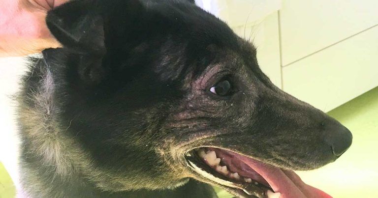
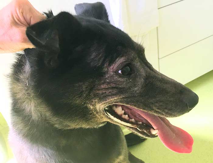
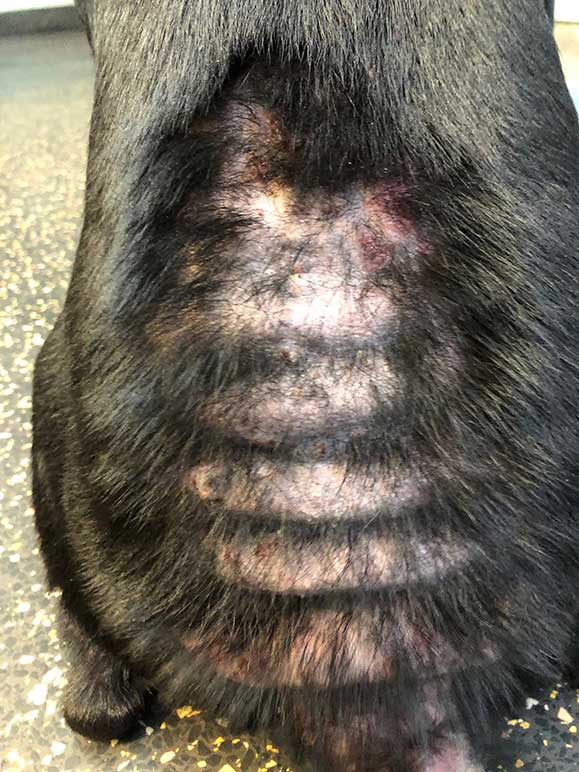
The distribution of the lesions may, in some cases, help differentiate the underlying disease. Some pruritic diseases have a typical clinical pattern. For example, ear, facial, feet, axillary and perianal pruritus is typical of atopic dermatitis and food allergy (Figure 1), while FAD affects mainly the dorsal/dorsolumbar areas (Figure 2).
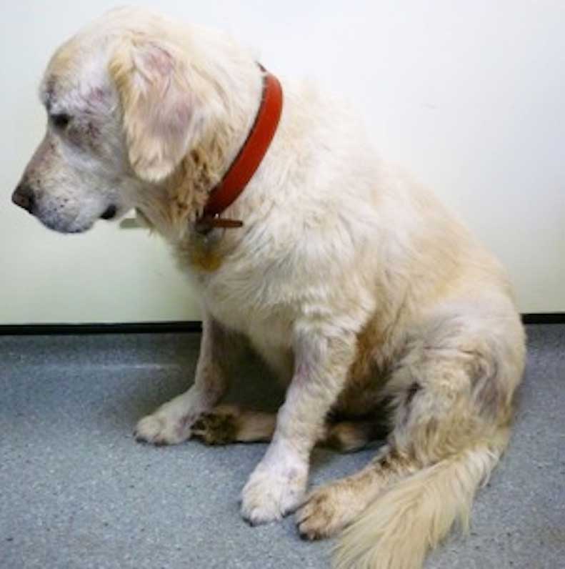
Sarcoptic mange, on the other hand, often affects the outer pinnae, elbows and hocks, and the chest region (Figure 3).
The diagnostic approach to a typical pruritic dog needs to be systematic. The first diseases to be eliminated and managed are bacterial and Malassezia infections/overgrowth, and parasitic. This implies performing cytology; skin scrapings; and microscopic examination of hairs, scales and cerumen.
Malassezia dermatitis and bacterial infection may mask the underlying cause and intensify the pruritus. It is, for that reason, important they are eliminated in the course of the diagnostic work.
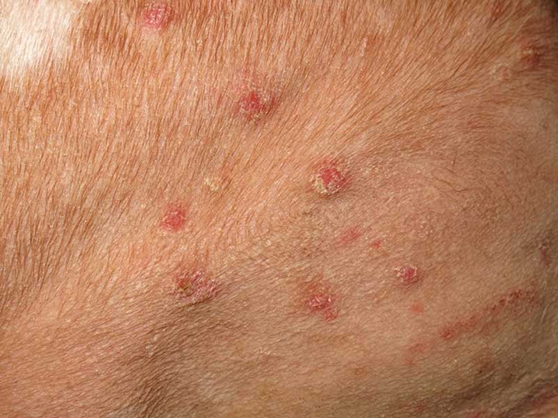
Cytology should be performed on all lesions that could suggest a secondary infection, such as papules, pustules, collarettes and scaling (Figure 4). If detected, these secondary infections should always be addressed. Topical antiseptic therapy is usually the first line of treatment.
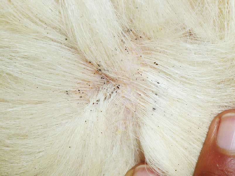
It is important to perform a good examination of the animal’s skin to look for fleas and flea faeces. Fleas can also be detected by combing the animal with a fine comb. If flea faeces are suspected (Figure 5), moistening of the suspicious black spots with water on white paper will reveal a red staining. This confirms the presence of flea faeces on the animal.
Intradermal skin testing with flea allergen may reveal wheal formation with immediate and delayed hypersensitivity, and can be used to support the diagnosis. However, a negative response does not rule out the possibility of flea bite hypersensitivity.
Serum in vitro testing for flea-specific IgE has variable accuracy and does not identify the animals that have delayed hypersensitivity reactions. Histopathology is non-specific and will reveal a pattern that can be seen in other hypersensitivity reactions (superficial perivascular inflammation, often containing eosinophils).
FAD is the most common allergic skin disease of dogs and cats. Clinical signs can be seasonal or year-round, depending on where the animal lives. In dogs, the classic presentation is a pruritic, papular, crusted eruption over the dorsal lumbosacral area and the caudal aspect of the hindlimbs (Figure 2). Alopecia, secondary seborrhoea, hyperpigmentation and lichenification can occur in chronic cases.
Skin scrapings can detect Sarcoptes, Trombicula, Cheyletiella and Demodex mites. Overpopulation with Demodex canis mites does not result in pruritus unless associated with bacterial infection. Lice can also be detected with skin scrapings. In the presence of scaling, scales should be analysed microscopically after being collected with tape.
The ear canals should also be examined and the cerumen analysed for the presence of Otodectes cynotis.
Because negative results in these tests do not exclude the possibility of ectoparasites, a therapeutic trial is recommended with a suitable insecticidal and acaricidal antiparasitic product.