30 Jun 2026
Chloe Fisher BVSc, PGDipVCP, DipECVN, MRCVS shares a practical approach for diagnosing feline neurological presentations.
Chloe Fisher
Job Title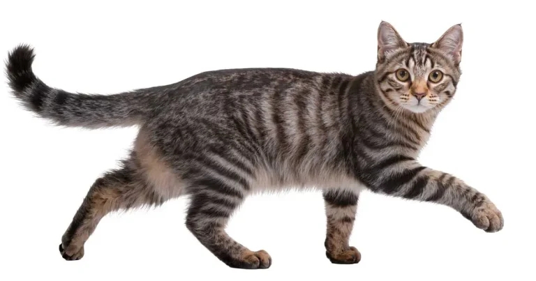
Image: Anneliese / Adobe Stock
The stumbling cat: a perplexing and somewhat rare clinical presentation. Our feline friends are indeed “not just a small dog”.
From subtle clinical signs to exam limitations, the approach to these cases can present a unique diagnostic challenge.
When presented with a cat with an abnormal gait, the clinician must first determine whether the problem is neurological. If so, the next steps are to neurolocalise the lesion and develop a prioritised list of differential diagnoses.
A key early distinction is between ataxia (incoordination) and paresis (reduced voluntary movement). Ataxia may be vestibular, cerebellar or proprioceptive in appearance, whereas paresis typically reflects spinal cord or neuromuscular disease.
This article provides a practical, structured approach to the “stumbling cat”, focusing on examination technique, neurolocalisation and the most relevant differential diagnoses encountered in UK general practice.
A concise and targeted history is invaluable. Particular attention should be paid to:
These features allow categorisation using the VITAMIN D framework (Table 1), which remains a useful clinical tool in everyday practice.
The neurological examination in cats requires patience and adaptation.
Stress and restraint can significantly alter findings; therefore, wherever possible, cats should be allowed to move freely within the consultation room prior to handling.
Observing the cat while spontaneously moving (including jumping, turning and navigating obstacles) can provide valuable information.
Owner-supplied video footage can also be extremely helpful in cases where amenability in the consultation room is limited.
In addition to providing a safe space and time to allow cats to navigate the room, further feline-specific considerations should be given to the following parts of the neurological examination.
The menace response is less reliable in cats than in dogs.
Accuracy improves when the contralateral eye remains uncovered and the stimulus is delivered from behind the patient (Abbasi et al, 2018).
Hopping and paw placement are typically the most informative proprioceptive tests in cats.
Results should be interpreted cautiously in any limb with a catheter or bandage.
Cutaneous trunci reflex has limited sensitivity in cats in the clinical setting. It is most consistently elicited by gently displacing the fur with a pen tip or haemostat rather than performing a skin pinch (Tsai and Chang, 2022).
Assessment for spontaneous and positional nystagmus can aid identification of vestibular lesions. In some cases, nystagmus may only become apparent with positional changes (such as head elevation or positioning the cat on its back).
Cervical ventroflexion occurs secondary to paraspinal cervical muscle paresis, leading to a reduced cervical muscle tone and, therefore, a flexed position of the neck. The lack of the nuchal ligament in cats predisposes them to developing this posture.
Common causes include hypokalaemia, hyperthyroidism, thiamine deficiency, immune-mediated polyneuropathy, cervical ischaemic myelopathy, acquired myasthenia gravis and feline infectious peritonitis (Karpozilou et al, 2025). Other features of neuromuscular disease include a stiff, stilted gait; dorsal scapular protrusion; exercise intolerance; generalised weakness; and difficulty jumping. A plantigrade stance is associated with diabetic neuropathy.
Gabapentin is frequently used to reduce stress during examination; however, it may alter gait and postural responses. Where feasible, gait should be assessed prior to administration (DuPont et al, 2024).
The ability to neurolocalise the presenting cat to one of the three major systems (intracranial, spinal cord or neuromuscular) allows refinement of the list of differentials and targeting of the diagnostic approach (Table 2).
A UK retrospective study reported intracranial disease in 44% of feline neurological cases, with spinal and neuromuscular disease accounting for 26.3% and 25.6%, respectively (Pisco and Gomes, 2026).
A wide range of conditions may result in ataxia or paresis in cats (Table 3). The following sections highlight commonly encountered differentials in general practice.
Immune-mediated polyneuropathy is an increasingly recognised condition which typically affects young cats and presents with symmetrical, progressive weakness, reported to involve all four limbs, or the pelvic limbs alone. Animals often show decreased or absent tendon reflexes in affected limbs.
Diagnosis is supported by electrodiagnostics (demonstrating a motor axonal polyneuropathy) and nerve biopsies. Prognosis is favourable, with many cats showing an improvement over several weeks (although, recovery may be prolonged). Relapses are common.
Feline infectious peritonitis remains the most common infectious disease affecting the feline central nervous system (CNS).
Neurological involvement occurs in up to 30% of cases and is most often associated with the non-effusive form. It most commonly affects young animals (younger than three years) from multi-cat households.
Clinical signs vary but commonly include ataxia, seizures and vestibular dysfunction. Concurrent abnormalities on general examination are common and should heighten suspicion.
The introduction of antiviral therapies such as GS-441524 has significantly improved outcomes (Dickinson et al, 2020). Relapse is possible.
Otitis media (with or without interna) is a common cause of vestibular disease in referral feline populations; males are overrepresented. A history of otitis externa or upper respiratory disease increases clinical suspicion.
Middle ear polyps are associated with the presence of Horner’s syndrome. Diagnosis is supported by advanced imaging and myringotomy.
Temporary worsening of neurological signs following myringotomy is not uncommon and owners should be warned accordingly.
It is worth noting that idiopathic vestibular syndrome is the second-most common cause of vestibular signs in cats, in a referral population. It is significantly associated with an improving clinical picture.
Meningiomas are the most common intracranial neoplasia in cats, accounting for around 55% to 60% of feline primary intracranial tumours. These tumours are typically solitary (although multiple meningiomas are documented in around 17% of cats) and slow growing.
Clinical signs are location dependent, but commonly include seizures, behavioural change, circling and ataxia (forebrain signs predominate).
Lymphoma is the second-most common intracranial tumour and the most common spinal neoplasm in cats. It often affects younger animals and may present with acute, progressive neurological signs, alongside systemic disease.
CNS lymphoma can be primary or secondary; therefore, screening is recommended in all suspected cases.
Intervertebral disc disease (IVDD) is less frequently encountered in cats compared to our canine patients; however, it still represents an important differential diagnostic category to consider. Males are overrepresented. The most common types of intervertebral disc herniations seen in cats include:
IVDE is the most common type of IVDD in cats, followed by IVDP and ANNPE. Clinical signs are dependent on the location affected, with the lumbar spine, followed by thoracolumbar, most commonly affected. Pain is commonly reported, with an otherwise normal general clinical examination expected. Onset is often peracute in cases of ANNPE, acute for IVDE and sub-acute to insidious in IVDP.
Advanced imaging techniques are required for diagnosis and, where applicable, surgical planning.
Not all cats presenting with gait abnormalities have neurological disease. Important differentials include:
A thorough general clinical examination, including assessment of peripheral pulses, blood pressure assessment and an orthopaedic examination, is essential in all cases.
Given the wide range of differential diagnoses considered, routine laboratory investigations for the stumbling cat should include:
CK is a sensitive but non-specific marker of muscle injury and may be mildly elevated in anorectic or stressed cats. AST has a longer half-life and may remain elevated for longer.
It is prudent at this stage in the article to also discuss the utility of testing for toxoplasmosis. In the author’s experience, toxoplasmosis is the most commonly mentioned (and tested for) differential in cats referred for further investigations with neurological presentations. Although Toxoplasma gondii infection is common in cats (around 30% to 55% of cats have antibody titres indicating exposure), clinical disease is very rare – particularly in the adult cat. Clinical disease is usually associated with reactivation of cystic stages, secondary to immunosuppression (for example, cats with FIV or FeLV), rather than after a newly acquired infection.
Cats with toxoplasmosis often show diffuse clinical signs involving the nervous (seizures, ataxia, myalgia), ophthalmic (uveitis), gastrointestinal (diarrhoea, anorexia, weight loss) and hepatobiliary (icterus) systems. Definitive diagnosis of toxoplasmosis requires detection of T gondii in body tissues or fluids. Tentative diagnosis can be based on rising IgM titres, together with exclusion of other differential diagnoses and positive response to treatment.
Feline neurological presentations can be challenging, but are often highly rewarding. A structured approach, combining careful observation, accurate neurolocalisation and targeted diagnostics, will allow the majority of cases to be appropriately managed within general practice or referred when necessary.
Use of some of the drugs in this article is under the veterinary medicine cascade.
Chloe Fisher works as a veterinary neurologist at Eastcott Veterinary Hospital. She is a European specialist in veterinary neurology. Chloe has a particular interest in feline neurological disease and education.