31 Oct 2022
Diana Ferreira outlines some of the therapies available for managing this common and incurable skin condition in dogs.
Diana Ferreira
Job Title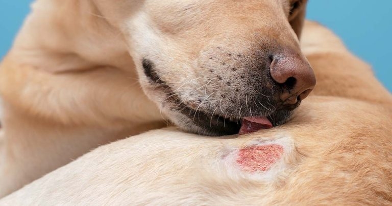
Image © Юлия Усикова / Adobe Stock
Atopic dermatitis (AD) is one of the most common inflammatory and pruritic conditions in dogs. The pathogenesis of canine AD is complex and multifactorial.
The susceptibility to develop clinical disease is, however, associated with multiple genetic and environmental factors. Sensitisation to environmental and/or food allergens – microbial or from insects – leads to skin inflammation, with activation of resident cells and local production of inflammatory mediators1.
Several factors are known to contribute to the development of sensitivities. These include epidermal barrier dysfunction, bacterial and yeast skin infections, psychogenic factors and concomitant skin diseases1. These factors are inter-related, and it is not clear if they represent the cause of sensitisation by themselves or if they are a consequence of the allergic disease1.
The diagnosis of AD in dogs is based on the presence of characteristic clinical signs and exclusion of all diseases that result in a similar clinical presentation.
The diagnosis, therefore, is clinical and no need exists to carry out complementary tests – such as intradermal or serological ones – although these may contribute to making clinical and therapeutic decisions1.
AD is a chronic, lifelong disease. Clinical management is dependent on many variables that characterise the clinical signs in one specific patient, ranging from severity of the clinical signs to the extent in which they are affected.
For this reason, treatment has to be tailored to each individual and is invariably multimodal1.
Traditionally, management of canine AD focused on reactive treatments, where therapeutic approach was aimed at the active inflammatory process after it had already become well established in the skin. It discontinued once clinical signs abated2. However, in the past decade, this approach was complemented by a proactive therapy approach1-4.
This approach will promote the remission of the skin lesions to reduce the residual subclinical cutaneous inflammation that is maintained after reactive therapy to prevent or delay their relapse4.
In the proactive therapy approach, the amplitude of anti-inflammatory action of a specific drug is favoured instead of the effectiveness time4.
Their long-term goal is to avoid the development of chronic inflammatory changes in the skin that can become much more difficult to reverse5.
The therapeutic approach of the allergic dog is divided in to two different treatment phases2.
Phase I is where a reactive approach is adopted with the goal of inducing rapid remission of the clinical signs. This is the acute or flare phase, where the dog exhibits an increase in pruritus, whether as a first manifestation of the condition or in the context of a relapse.
In this phase, acute and/or chronic skin lesions will be present, and often with a significant degree of inflammation2.
During the reactive approach, fast-acting and broad-range anti-inflammatory drugs would be preferred. Including, therefore, almost always a glucocorticoid (Panel 1).
In this case, oral prednisone or prednisolone at a dose between 0.5mg/kg/day and 1mg/kg/day, or oral methylprednisolone between 0.4mg/kg/day and 0.8mg/kg/day, are most frequently used1.
Glucocorticoids have the advantage of a fairly rapid action and not being an expensive treatment; however, the side effects that can result – such as polyuria, polydipsia, polyphagia, obesity, muscle and skin atrophy, behavioural changes, increased risk of urinary infections and iatrogenic hyperadrenocorticism – represent an important disadvantage1.
In this phase, the use of a short-acting oral glucocorticoid is more logical than that of a topical one. Topical glucocorticoids are, however, very useful in conjunction with systemic formulations to treat highly inflamed or markedly lichenified local skin lesions2.
However, if the initial inflammatory condition of the patient is mild, oclacitinib could be used instead, in monotherapy or with a topical glucocorticoid to amplify its anti-inflammatory effect (Panel 2)2.
Oclacitinib has been shown to be an effective and fast-acting treatment, representing an acceptable alternative to glucocorticoids. The recommended regimen is 0.4mg/kg to 0.6mg/kg twice daily for the first two weeks of treatment, followed by a once daily regimen5.
Occurrence of adverse effects associated with its administration is very infrequent, which represents a great advantage over glucocorticoids. Adverse effects are seen in two per cent of patients, and include anorexia, vomiting and diarrhoea5.
Many patients experience an aggravation of symptoms when the regimen is altered from twice a day to once a day, gradually returning to an acceptable level of itching over time5.
In the case of glucocorticoid use, authors recommend to use it in such a dose and for a length of time that will ensure complete resolution of the clinical signs to guarantee only minimal inflammation remains in the skin. Reducing treatment when inflammation is still present can result in a rapid aggravation of the clinical signs2.
Once the patient has been free of clinical signs for several weeks, then the proactive phase treatment approach can be instituted2.
The drugs will also be chosen depending on the severity of its clinical disease. If mild, then topical glucocorticoids and injectable biologics, such as lokivetmab, can be considered (Panel 3)2.
Lokivetmab is a caninised monoclonal anti-IL-31 antibody. IL-31 is a recently identified inflammatory mediator that appears to play a key role in the development of pruritus in the atopic dog9.
This drug is administered subcutaneously and is intended to block the action of circulating IL-31. This therapy is effective in approximately 70% of patients10. It is also a safe treatment, as it is associated with a very low rate of adverse effects.
The most frequent adverse effects reported are lethargy and vomiting11.
Proactive therapy with topical glucocorticoids is defined as the treatment of areas more susceptible to inflammation for two consecutive days per week, with or without visible inflammation3. A recent trial confirmed the benefit of proactive use of a topical spray of hydrocortisone aceponate in atopic dogs, with a nearly fourfold increase in median time to flare compared to placebo with no adverse events6.
However, if the patient’s degree of inflammation is historically moderate to severe and rather generalised, then it is recommended to intensify the anti-inflammatory treatment, with long-term drugs that have a broader anti-inflammatory action, such as oclacitinib or ciclosporin (Panel 4)7.
Ciclosporin is given at a dose of 5mg/kg/day. Compared with glucocorticoids, ciclosporin is equally effective and has a lower frequency of adverse effects; however, its maximum action takes between two to three weeks to occur.
Gastrointestinal adverse effects, including vomiting and diarrhoea, can be identified in approximately 20% of patients, and loss of appetite12. These adverse effects are self-limiting and lead to definitive discontinuation of the drug in only a small percentage of patients12.
After an initial treatment period of four to six weeks, if efficacy is maximal, the administration regimen may be decreased. Either the dose is decreased by 25% or the frequency of administration to alternate days. This decrease can be repeated gradually every four to six weeks until the minimum effective dose is found.
In the proactive approach phase, other interventions can help prevent acute flares8. These include:
This therapy is considered the only therapeutic intervention that can alter the course of the disease and should always be an option to consider in animals with indication for it.
It is an individualised treatment, based on the results of intradermal skin testing and/or IgE serology.
The efficacy of immunotherapy is observed through the lower use of symptomatic treatments; however, a delay occurs before improvement of around three to nine months1.
The management of atopic dermatitis must always be multimodal, adapted to each specific patient and re-evaluated frequently.
Although no cure exists for atopic dermatitis, many advances have been made in recent years regarding not only therapeutic options, but also in the approach to the use of the available drugs.
A proactive treatment approach will aim to control patients with AD long-term, preventing chronic inflammatory skin changes, less need of flare management interventions and, overall, providing better outcomes.
Diana Ferreira
Job Title