
7 Jul 2026
Dee Mullowney MVB, CertSAM, PGDip(VCP), MVetMed, DipECVIM-CA, MRCVS and Sam Taylor BVetMed, CertSAM, DipECVIM-CA, MANZCVS(medicine of cats), FRCVS cover diagnosis and treatment of these two conditions, which, despite being very different, tend to affect older pets.
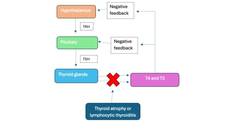
Figure 1. Regulation of thyroid hormone concentrations is controlled under a simple negative feedback loop called the hypothalamic-pituitary-thyroid axis. In primary hypothyroidism, thyroid atrophy or lymphocytic thyroiditis results in reduced thyroxine (T4) and triiodothyronine (T3), leading to loss of negative feedback at the hypothalamus and pituitary, causing increased thyroid-stimulating hormone (TSH) production.
Disease of the thyroid is an area where cats and dogs differ significantly. In this article, the authors will discuss canine hypothyroidism and feline hyperthyroidism to bring readers up to date on diagnosis and management of these very different conditions.
Canine hypothyroidism is a common endocrine disorder, with reported annual prevalence of 0.23% in UK primary care practice (O’Neill et al, 2022), and it can be challenging to diagnose and manage.
Thyroid hormones thyroxine (T4) and the more potent triiodothyronine (T3) are produced in the thyroid gland under the control of the hypothalamus and the pituitary gland.
Thyrotropin-releasing hormone (TRH) is released from the hypothalamus and acts on the pituitary gland to stimulate release of thyroid-stimulating hormone (TSH). This hormone acts under a simple negative feedback loop, where circulating T4 and T3 decrease further release of TRH and TSH (Figure 1).
Thyroid hormones have positive inotropic and chronotropic effects on the heart, are necessary for lipid synthesis and metabolism, and can stimulate erythropoiesis.
Primary hypothyroidism, caused by either lymphocytic thyroiditis or thyroid atrophy, is the most common form of the disease.
Secondary hypothyroidism, which results from pituitary disorders such as pituitary neoplasia or hypophysectomy (surgical removal of the pituitary), accounts for less than 5% of cases of hypothyroidism.
Tertiary hypothyroidism, which occurs secondary to a problem in the hypothalamus, is extremely rare. Lymphocytic thyroiditis and thyroid atrophy occur in roughly equal frequency, and thyroid biopsy would be required for definitive diagnosis, but lymphocytic thyroiditis can be presumptively diagnosed based on the presence of thyroglobulin autoantibodies (TgAA). Only one-third of TgAA-positive dogs developed clinical hypothyroidism in a one-year follow-up study (Egbert et al, 2024), so hypothyroidism should never be diagnosed based on a positive TgAA titre alone.
Hypothyroidism is usually diagnosed in middle-aged to older dogs, although predisposed breeds (the English setter, Gordon setter, golden retriever, Dobermann, old English sheepdog, Rhodesian ridgeback, cocker spaniel, and boxer) tend to develop the disease earlier in life. The predisposed breeds have a high prevalence of TgAA positivity, and a familial link is present in other breeds including the great Dane, beagle, Swedish hovawart and giant schnauzer.
Hypothyroidism is more common in neutered dogs – especially females (Sundburg et al, 2016).
Thyroid hormones have extensive effects throughout the body, so hypothyroidism can result in a variety of clinical signs (Panel 1).
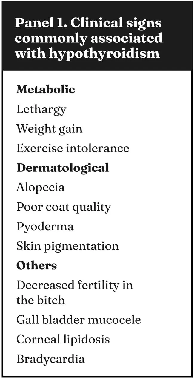
Because signs can be vague, insidious and non-specific, hypothyroidism is frequently misdiagnosed (Travail et al, 2024). Metabolic features such as lethargy, heat-seeking behaviour, general weakness, shivering and exercise intolerance occur in about 80% of cases. Obesity or weight gain occurs in 44% of cases (Dixon et al, 1999), although hypothyroidism is a relatively uncommon cause of obesity and weight gain in the general dog population.
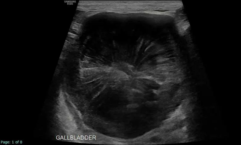
An association exists between hypothyroidism and the development of gallbladder mucocele, and 26% of dogs with gallbladder mucoceles are diagnosed with hypothyroidism (Figure 2; Aicher et al, 2019).
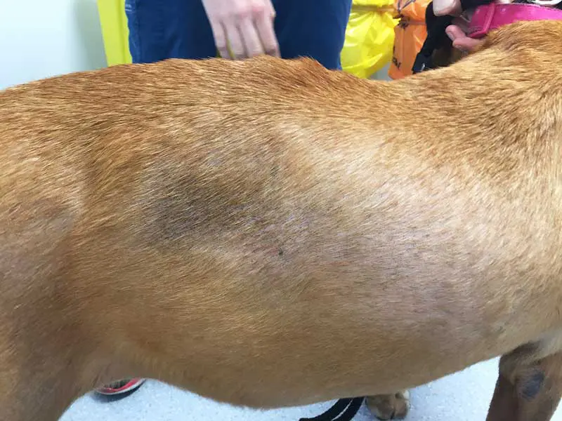
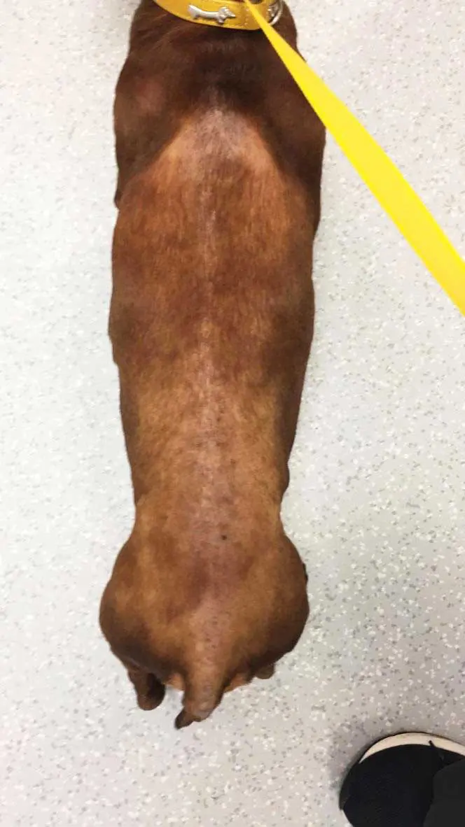
Dermatological features occur in 70% of affected dogs and include alopecia, poor coat quality, hyperpigmentation and failure to regrow hair after clipping. Alopecia is typically non-pruritic and can occur over the flanks or in areas of friction such as under the collar, or in extremities such as the tail (Figure 3).
Thyroid hormones have positive ionotropic effects and stimulate myocardial function. However, cardiac abnormalities are rarely attributed to hypothyroidism. About 15% of hypothyroid dogs develop subclinical bradycardia. No clear causative link exists between hypothyroidism and dilated cardiomyopathy (DCM) in the Dobermann (Beier et al, 2015). Routine screening for hypothyroidism in dogs with DCM with no other clinical signs of hypothyroidism is not recommended.
Less common features include reproductive features including reduced fertility in the bitch and ocular features such as corneal lipidosis and reduced tear production.
Hypothyroidism has been associated with various neurological manifestations including facial nerve paralysis and laryngeal paralysis, although the link is not proven. Hypothyroidism is sometimes reported as a potential cause of megaoesophagus.
Although 8.8% of dogs with megaoesophagus are reported to have hypothyroidism, no proven causal link exists (Gaynor et al, 1997; Haines 2019). Hypothyroidism has also been reported as a cause for behavioural problems in dogs, but again, no evidence exists to support this link (Hrovat et al, 2019). Hypothyroidism does not cause polyuria or polydipsia.
Several changes occur commonly in hypothyroid dogs. These include:
Clinicians have several diagnostic pitfalls to be aware of when attempting to achieve a diagnosis of hypothyroidism (Panel 2).
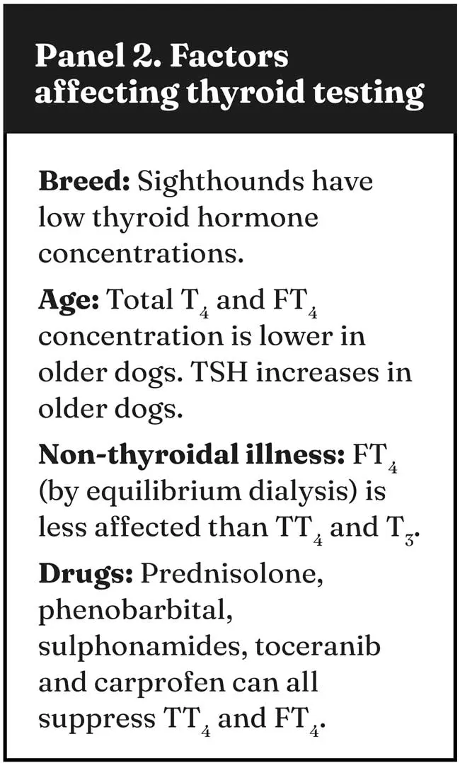
Sighthounds are known to have lower thyroid hormone concentrations, with 90% of greyhounds and 25% of whippets with a total T4 (TT4) below the reference range. Total T3 concentrations are not reduced in the greyhounds (Shiel et al, 2007). TSH concentration is less affected by breed. TT4 and free T4 (FT4) tend to be lower in older dogs while TSH tends to be higher. Non-thyroidal illness can affect thyroid testing. TT4 and T3 are commonly reduced in dogs with non-thyroidal illness, but FT4, measured by equilibrium dialysis, is less affected by non-thyroidal illness (Mooney, 2008). Certain drugs can reduce serum concentration of TT4 and FT4; however, sulphonamides and toceranib can closely mimic hypothyroidism and result in reduced TT4, FT4 and increased TSH concentrations. Care should be taken in evaluating dogs receiving these medications for hypothyroidism.
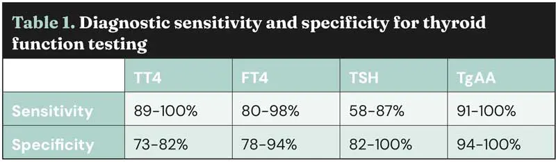
The gold standard test for diagnosis of primary hypothyroidism is TSH stimulation test, but TSH preparations are not readily available. Diagnosis usually involves a combination of history, signalment, physical examination, haematology, biochemistry and thyroid testing including TT4, FT4, TSH and TgAA. Sensitivities and specificities of these tests can be found in Table 1. It is also worth noting that when measuring TT4 concentrations, in-house analysers using ELISA method are not as reliable as validated radioimmunoassay at an external lab (Lurye et al, 2002).
Hypothyroid dogs require lifelong thyroid hormone supplementation. Tablet and oral liquid formulations of levothyroxine are available. The starting dose is usually 0.02mg/kg by mouth every 24 hours, although twice-daily dosing might be required in some cases. Metabolic signs tend to improve within days to weeks of starting treatment, but dermatological signs can take months to normalise.
Key points include:
Feline hyperthyroidism is the most common endocrinopathy in cats, frequently affecting older cats. It can be straightforward to manage, but more complex when cats also have chronic kidney disease (CKD) or other comorbidities.
With cats living longer, they may be hyperthyroid for many years, so choosing the right treatment for the individual cat and caregiver is important.
In a process opposite to that of canine hypothyroidism, in feline hyperthyroidism excess thyroid hormone is produced autonomously, independent of TSH (Figure 4).
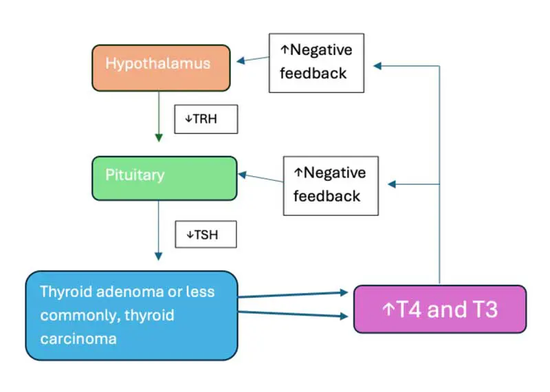
The increased negative feedback lowers TSH, which can be useful diagnostically in difficult cases.
Hyperthyroidism in most cats is caused by a thyroid adenoma, and in 1% to 3% of cases, a thyroid adenocarcinoma. Importantly, this is a progressive process, with a period when the cat’s thyroid gland(s) contain some hyperplastic tissue but T4 is normal, and ongoing exposure to factors associated with hyperthyroidism (such as low iodine, environmental thyroid-disrupting chemicals) can then result in nodules becoming an adenoma-secreting thyroid hormone. Evidence also suggests this process can continue and even result in a carcinoma in a minority of cases (Peterson et al, 2016).
Several studies have examined causes of hyperthyroidism, and no clear single factors have been identified. In general, nutritional deficiencies and excesses, and toxins or thyroid-disrupting chemicals, have been implicated, and TSH receptor mutations and mutations in inhibitory G protein expression identified in some cats.
Factors such as feeding canned food, fish, liver and giblet-flavoured canned food, soy isoflavones in dry food, iodine excess or deficiency, low selenium, various chemicals, herbicides and pesticides have been implicated. It is likely a combination of factors acting synergistically over many years that affects thyroid hormone metabolism, eventually leading to hyperthyroidism.
Most cats with hyperthyroidism are older, with an average age of 13 years, but the disease is diagnosed in cats younger than 10 years, and with increased vigilance of the condition, cats are diagnosed at an earlier stage and, therefore, younger age.
A decreased risk of hyperthyroidism has been identified in the Tonkinese, Abyssinian, British shorthair, Burmese, Siamese and Persian breeds, and an increased risk for longhaired and non-purebreds (Crossley et al, 2017).
Clinical signs of hyperthyroidism are familiar to clinicians, with the “classic” hyperthyroid cat presenting with polyphagia, polyuria/polydipsia, vomiting, diarrhoea and weight loss.
On clinical examination, hyperthyroid cats may be in reduced body (and muscle) condition, with a poor haircoat, tachycardia and a palpable goitre. Severe cases can present with signs of congestive heart failure, but early detection means we diagnose cats that remain in good (or even excessive) body condition and have few clinical signs. The term “apathetic” hyperthyroidism has been applied to cats with hyperthyroidism but without typical signs and actually lethargic and anorexic.
Importantly, these cats likely have other comorbidities rather than a different form of hyperthyroidism, and management should focus on looking for, and managing, these conditions. Common comorbidities include:
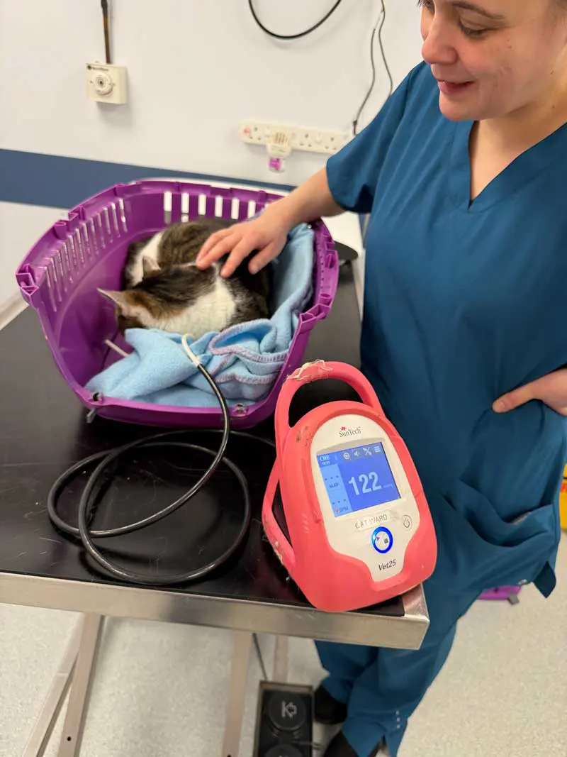
Importantly, cats with hyperthyroidism should have blood pressure assessment (Figure 5), as up to 25% of affected cats may be hypertensive and this can be diagnosed after restoration of euthyroidism.
Some cats – particularly with early hyperthyroidism – may have minimal changes in blood results. Some typical findings include:
Many cats with hyperthyroidism (more than 90%) are diagnosed simply by documenting a high T4, and this is an accurate test without false positives.
Generally, bench-side tests agree with external laboratory testing, but be aware of limitations with your machine and, ideally, confirm results at an external laboratory – particularly if the result is unexpected. FT4 is the active form of thyroid hormone and can add additional information; however, results must be interpreted with the T4 and the clinical signs of the patient. It is possible to have false-positive FT4 results in euthyroid cats with other diseases, so importantly, a diagnosis of hyperthyroidism should not be made based on solely an elevated FT4 (Peterson et al, 2001).
As cats have a period of “subclinical” hyperthyroidism before they develop clinical signs of disease, tests can be conflicting with a low TSH but normal T4 and FT4. Figure 6 shows the potential results for thyroid testing in different situations.
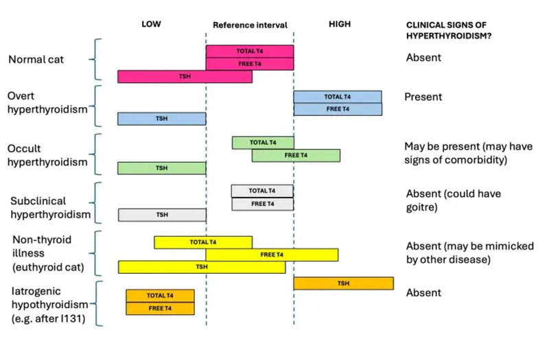
Cats with “occult” hyperthyroidism may have signs of the condition but a normal T4. Such cats often have another non-thyroid illness lowering the T4 into the reference interval, such as CKD. They may require treatment of hyperthyroidism, but should be investigated for other diseases, and comorbidities also managed.
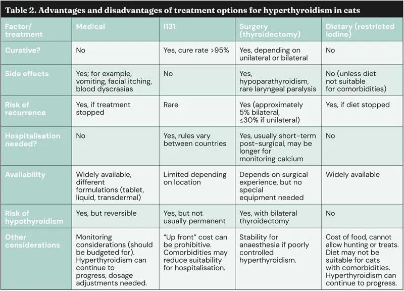
Treatment options for hyperthyroidism include medical management, radioactive iodine (I131), surgical thyroidectomy and dietary therapy. All have advantages and disadvantages, and should be discussed with caregivers.
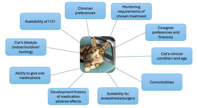
No “perfect” treatment exists and it should be individualised. Figure 7 covers some considerations that may affect choice of therapy for a hyperthyroid cat. Table 2 discusses some advantages and disadvantages of each treatment choice. It is important to investigate comorbidities that could affect response to treatment and quality of life (for example, managing dental disease and providing analgesia for degenerative joint disease).
Hypothyroidism in dogs and hyperthyroidism in cats obviously require very different management, but both tend to affect older dogs and cats, and for both groups treatment should be tailored to the individual animal and caregiver, and comorbidities considered.
All affected animals can have an excellent quality of life when their endocrinopathies are well managed.
Dee Mullowney graduated from University College Dublin in 2010 and spent eight years in mixed and small animal practice across Ireland, New Zealand, and Australia. She then moved to the UK to complete a rotating internship and residency in small animal internal medicine at the RVC. In 2022, she became a diplomate of the American and European colleges of veterinary internal medicine and an RCVS-recognised specialist. She currently works at Lumbry Park Veterinary Specialists.
Sam Taylor completed a Feline Advisory Bureau residency at the University of Bristol and was awarded the European Diploma in Veterinary Internal Medicine in 2009. In 2011, she became an RCVS-recognised specialist in feline medicine and in 2019 was made a fellow of the RCVS for outstanding contributions to the profession. Sam is a consultant for the International Cat Care Veterinary Society and works as an Internal Medicine Specialist at Lumbry Park Veterinary Specialists. She is currently completing a PhD in feline health and welfare.