
4 Aug 2026
Dan White MSc, BSc RVN, VTS(Dermatology), NCertSAN, C-SQP covers diagnosis and management of this issue in dogs, including options for treatment.

Dan White
Job Title
Image: kobkik / Adobe Stock
Pruritus, also known as itching, is a sensation that causes the desire to scratch (Sauvé, 2023) and is the most common symptom recognised in dermatology patients (Espadale and Santoro, 2021).
Scratching can be somewhat beneficial in some patients, in removing irritants such as insects off the skin, but in most cases scratching causes skin damage that can cause defects in the barrier that protects the body, allowing for further complications such as secondary skin infections to arise.
Cytokines are signalling proteins that are released by skin and immune cells such as lymphocytes in the areas of affected skin (Garcovich et al, 2021). They bind to receptors on sensory neurons in the skin, which causes pruritus. Interleukin 31 (IL-31) is the main cytokine that is associated with pruritus in dogs; other cytokines linked to pruritus and inflammation are IL-2, IL-4, IL-6, IL-13 (Tater, 2021).
Obtaining an accurate and comprehensive medical history is essential, and will help when patients present to you with skin disease.
Pruritus is often caused by parasites, skin infection and allergy (atopic dermatitis and cutaneous adverse food reaction; CAFR), and the location and degree of itching is important when assessing your patient. See Panel 1 for important questions to ask owners.

Owners’ perception of itching may differentiate from a veterinary professional’s, so clarifying the frequency, duration and location(s) of itching can help when it comes to your clinical examination, diagnosis and prescribing decisions. Numerical rating scales (Figure 1) and other scales such as the pruritus visual analog scale (PVAS; Figure 2) can help the owner during the consultation to make their quick assessment on the severity of the itch on their pet. This scale can then be used every time the owner visits the practice.

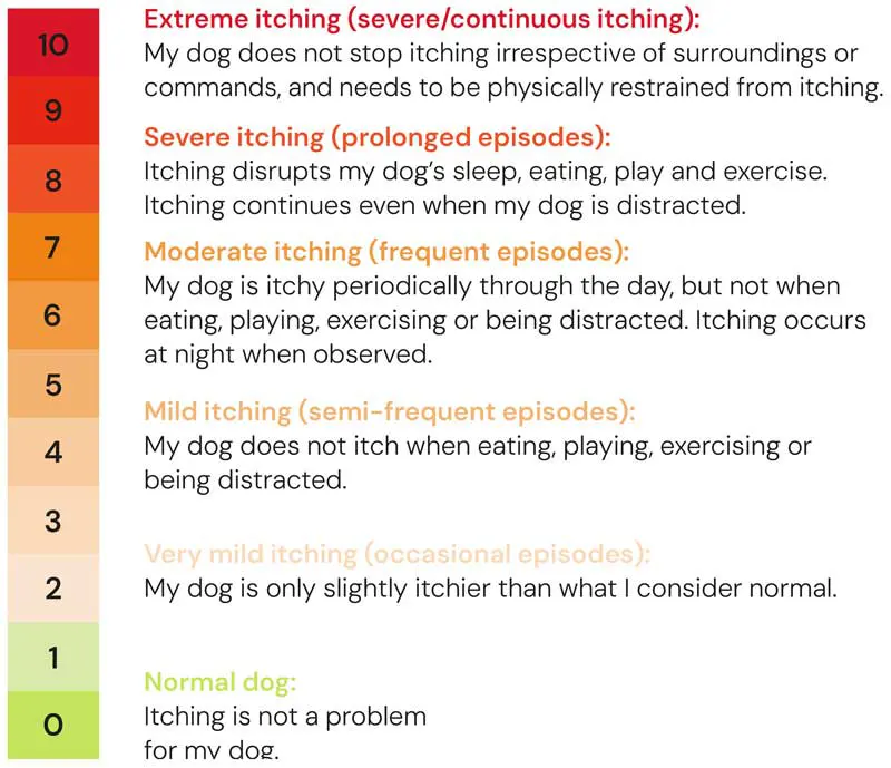
A study by Young in 2019 showed that using the PVAS alongside showing the owner the previous score improves the owner’s perception of pruritus in their dog. Comparing previous scores given to us by the owner can help guide veterinary professionals on whether the current treatment is beneficial.
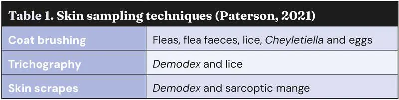
It is also important to clarify whether your owner is using effective parasite control, what products they are using, how frequently they are administering them and when was the last application. These questions, alongside some appropriate skin samples such as coat brushings, trichograms (hair plucks) and skin scrapes, will help you rule out ectoparasites (Table 1).
Pruritus management can vary depending on a lot of factors:
Ensuring regular, effective parasite control is essential to rule out ectoparasites playing a role in pruritus. Atopic patients that are repeatedly exposed to flea bites are more likely to develop a flea saliva hypersensitivity (Hensel, 2021). Isoxazolines such as afoxolaner, fluralaner, sarolaner and lotilaner are very effective anti-parasiticides (Defalque, 2022). They have a broad insecticidal spectrum with an onset of action in two to four hours for fleas (Datz, 2018), and although recent concerns exist for the overuse of parasiticides, their use is recommended in pruritic or atopic patients.
Once parasites had been excluded, ruling out secondary infection/microbial overgrowth would be your next stage of investigation. Often, this may be done concurrently while administering ectoparasite control.
Skin inflammation can cause dysbiosis of the natural skin flora and microorganisms, which includes yeast and bacteria. This dysbiosis can cause pruritus, which in turn leads to primary and secondary skin lesions. Improving the skin barrier is also essential in patients that have pruritic and allergic skin disease. Other causes of secondary infection could include endocrinopathies (hypothyroidism and hyperadrenocorticism) and follicular dysplasia; for example, colour dilution alopecia.
All surface and superficial secondary skin infections should receive some topical antimicrobial therapy to minimise the need for systemic therapy. Topical therapy can replace systemic antibiotics to support good antimicrobial stewardship with the increasing emergence of multidrug-resistant bacteria (Frosini and Loeffler, 2020). Typically, chlorhexidine is the most common and effective topical antimicrobial we use. Hypochlorous acid is also used frequently by the author – especially in patients with methicillin-resistant Staphylococcus pseudintermedius infections, and good response is seen.
A 2005 study using a skin spray showed that ingredients such as occlusive agents, humectants and lipids help improve, restore and hydrate the skin barrier after antiseptic bathing (Pseftogka et al, 2025).
Prednisolone is a very effective and quick treatment for controlling pruritus. An anti-inflammatory and anti-pruritic dose for dogs would range from 0.5mg/kg to 1mg/kg once a day until clinical signs are controlled, and then it should be tapered to the safest and lowest dose and frequency (Hensel, 2021).
The more common side effects seen with prednisolone are polyuria, polydipsia and polyphagia – especially when given daily – so the preferred frequency of administration would be an every-other-day dosing if long-term treatment is required (Marsella, 2013a).
Methylprednisolone is very similar to prednisolone, although the dose is 0.4mg/kg to 0.8mg/kg in dogs. Methylprednisolone is reported to have fewer side effects such as polyuria and polydipsia due to its lower mineralocorticoid activity, so is useful in dogs that do not tolerate prednisolone (Brooks, 2023).
Repeated treatment with injectable glucocorticoids is not recommended due to the risks of side effects and the inability to withdraw the medication once it has been administered (Hensel, 2021).
We do, however, have special liquid formulations of prednisolone that can be ordered from specialist veterinary pharmaceutical manufacturers if oral tablets are challenging for owners to administer.
A couple of topical glucocorticoids are available, such as a hydrocortisone aceponate spray or a triamcinolone acetonide spray. Both sprays can be useful for localised areas of pruritus and acute flares (Hensel, 2021). They can be used short term for one to two weeks, but if used long term then side effects such as scaling, cutaneous atrophy, comedones and cysts can be seen (Hensel, 2021).
However, in the author’s experience, using these sprays a couple of times week for long-term management of localised pruritus is generally well tolerated, and these sprays can be helpful to reduce the systemic treatment.
Oclacitinib is a first-generation JAK inhibitor. It is a fast-acting antipruritic and is highly selective for JAK1 and has some effect on JAK3 (Patterson, 2025).
The dose is 0.4mg/kg to 0.6mg/kg and it is initially given twice a day for 14 days before reducing to once-daily dosing. Patients must be at least 12 months old and weigh 3kg or more. Rebound itch can be seen in some patients when once-daily dosing is initiated; if this happens, alternative treatment options should be considered if it does not settle. Continued twice-daily dosing is off-licence and not recommended due to side effects such as leukopenia, hypercholesterolaemia and proteinuria (Hensel, 2021). Regular blood monitoring and urinalysis is advised every three to six months when patients are on JAK inhibitors long term.
Oclacitinib has been available now for 12 years, along with long-term safety data that makes some clients feel more reassured when discussing treatment options for their dog.
Ilunocitinib is a newer second-generation JAK inhibitor recently licensed in the UK. It is also a fast-acting antipruritic, but clinically, through the author’s experience at a specialist referral hospital, it has shown to have more anti-inflammatory properties compared to the other JAK inhibitors. It is a non-selective JAK inhibitor targeting JAK1, JAK2 and TYK2. The dose is 0.6mg/kg to 0.8mg/kg once a day, and patients must be at least 12 months old and weigh 3kg or more. A 112-day comparative study with oclacitinib and ilunocitinib showed 77% of dogs on ilunocitinib achieved clinical remission compared to 53% on oclacitinib, showing that 24% more dogs improved with ilunocitinib treatment (Forster et al, 2025). This seems to be reflected in the author’s experience.
Atinvicitinib is a second-generation JAK inhibitor and is the newest JAK to the market, becoming available in April 2026. It is a fast-acting antipruritic and is the most selective drug for JAK1, with it being reportedly 10 times more selective to JAK1 than JAK2, JAK3 and TYK2 (MSD Animal Health, 2026). Current research suggests blocking JAK1 promotes a greater antipruritic effect, as IL-31 is more frequently associated with JAK1 (Sato et al, 2025); however, IL-31 can still activate through JAK2.
The dose of atinvicitinib is 0.8mg/kg to 1.2mg/kg once a day and it can be used in patients from six months of age that weigh more than 3kg, which is a great benefit when compared to other JAK inhibitors when you have patients younger than 12 months old presenting with pruritus.
Ciclosporin is a long-term medication for patients with atopic dermatitis and pruritus (Archer et al, 2014). It can take four to six weeks to reach its peak therapeutic effect, so it is not recommended for acute flares. Combination therapy is usually suggested initially with ciclosporin for the first two weeks; pairing with glucocorticoids or a JAK inhibitor such as oclacitinib can help the acute flare while the ciclosporin starts to take effect (Hensel, 2021).
The dose is 5mg/kg orally every 24 hours until the patient’s clinical signs are under control (Hensel, 2021). The dose of ciclosporin can be reduced if patients are well controlled to achieve the lowest dose possible for maintenance. Gastrointestinal side effects can be seen with ciclosporin such as vomiting and diarrhoea; to reduce the risk of side effects, it can be given with food (Hensel, 2021). The gastrointestinal side effects are usually transient and normally seen in the first two weeks of starting ciclopsorin (Marsella, 2013b).
Monitoring blood screening such as biochemistry and haematology is usually advised every six months with patients receiving this medication long term (Marsella, 2013b). The administration of inactivated or live vaccines should also be avoided during ciclosporin treatment (Nuttall et al, 2014).
A temporary two-week withdrawal interval before or after the vaccinations is recommended (Nuttall et al, 2014).
Lokivetmab is a canine monoclonal antibody that is given monthly by subcutaneous injection. It binds to IL-31 before it can attach to skin neuron receptors, which helps prevent the transmission of the pruritus sensation (Hensel, 2021).
The subcutaneous UK dose is 1mg/kg once a month and can be used in dogs of all ages if they are at least 3kg in weight (Zoetis, 2017). Itch relief can occur within 48 hours, and the duration can last for up to four to eight weeks in some patients (Hensel, 2021). This treatment unfortunately has no anti-inflammatory action and, therefore, may be less beneficial in patients with skin inflammation.
A benefit of this medication is that it can be used as an additional therapy alongside other more potent anti-inflammatory medication, only if an additional antipruritic medication is required.
Essential fatty acids (EFAs), such as omega-3 and omega-6, can be useful as adjunctive treatment for patients with allergic skin disease to aid skin barrier health (Hensel, 2021).
Atopic patients have a skin barrier dysfunction that leads to increased water loss, allergen penetration and infection susceptibility (Espadale, 2016). Another thought is that atopic patients have decreased lipids in the skin barrier, so supplementing EFAs could help with the lipid deficiency (Espadale, 2016).
It is usually suggested that EFAs are given for at least three months before they can be assessed, as their onset of action is slow and it can take time to see the benefits (Hensel, 2021). One study by Saevik in 2004 found that EFAs can have a steroid sparing effect to allow the dose of glucocorticoids to be reduced to the lowest effective dose. More research is needed with EFAs and pruritic/allergic skin disease, but supporting the skin barrier and function is an important feature when managing patients with atopic dermatitis.
Antihistamines can be used as a treatment for dogs with mild pruritus (Hensel, 2021); however their evidence is lacking, with some studies reporting they are not effective at all (Hsiao et al, 2016). If they are used, then they should be given continuously and proactively before a flare begins, to prevent the effects of histamine once it is released (Olivry et al, 2015). No licensed antihistamine is available for dogs.
Antihistamines alongside glucocorticoids can also be beneficial with a steroid sparing effect (Hensel, 2021). The antihistamine with some evidence in dogs is cetirizine or hydroxyzine, which is metabolised into cetirizine (Banovic et al, 2020). The suggested dose of cetirizine is 1mg/kg orally every 12 to 24 hours, and the suggested dose of hydroxyzine is 2mg/kg orally every 12 hours (Hensel, 2021).
Pruritus is the most common clinical sign with patients that have underlying allergies such as CAFR (MSD Veterinary Manual, 2025).
CAFR can develop at any age, and other clinical signs can include urticaria, pyoderma, soft faeces, flatulence and diarrhoea (Jackson, 2023). The prevalence of CAFR in dogs is estimated around 9% to 40% of patients presenting pruritus (Mueller et al, 2016). A 2019 review by Olivry and Mueller showed that almost 40% of dogs developed signs of CAFR by one year of age. It is also estimated that one-third of patients which have been diagnosed with atopic dermatitis also have CAFR (Olivry and Mueller, 2019).
Performing an 8 to 10-week diet trial, once we have ruled out parasites and secondary infection, is recommended to exclude dietary involvement with the pruritus (Mueller, 2021). CAFR cannot be ruled out with an allergy blood test (Tater, 2021), as they are highly unreliable when testing against food (Marsella, 2013b).
It is normally suggested to continue systemic treatment during the diet trial and then, when coming to the end of the diet trial, try to withdraw the medication. Client compliance is the biggest issue when trying to perform a diet trial (Mueller, 2021), so it is important to make sure the patient is comfortable during this process.
If the pruritus reoccurs once the medication is withdrawn alongside the special diet, then you can conclude that food does not play a role in the pruritus and we can exclude this as a differential, and conclude the patient has atopic dermatitis.
If patients remain comfortable once the medication has been withdrawn, then it is recommended to reintroduce all aspects of the diet for up to 10 days to see if the pruritus returns, which in some patients can be as quick as one to two days (Marsella, 2013b). If the pruritus does return, then you can conclude that your patient has a cutaneous food reaction and stabilise them back on to the diet used for the diet trial. Most common food allergens in dogs are beef, diary and chicken (Mueller et al, 2016).
Suitable diets for performing a diet trial can include diets with a hydrolysed protein or a novel monoprotein, such as horse or rabbit, but discussing with the owner, and finding a protein their dog has never been exposed to, is important to perform the diet trial as efficiently as possible.
Allergen-specific immunotherapy (ASIT) is the only treatment available that treats the cause of atopic dermatitis (Nextmune, 2025). It can be given as a subcutaneous injection or sublingual drop. It helps to desensitise the hypersensitivity that the patient has to environmental allergens; for example, house dust/storage mites or tree, grass and weed pollens. Variable data from multiple studies exists regarding success rates, but the data suggests that 50% to 80% of patients will respond to treatment (Reinbacher, 2025; Fennis, 2022).
In one study by Fennis in 2022, out of 664 dogs, 31.5% were well controlled on ASIT alone, 28.5% were controlled but needed a lower dose of medication alongside the immunotherapy, and 40% of dogs did not achieve enough benefit to continue the treatment.
This highlights that around 60% of patients benefited from ASIT when it was given for at least nine months; however, other studies suggest that success rates can increase to as high as 80% when patients are treated for at least 12 months with ASIT (Reinbacher, 2025).
Dan White works as a nurse specialist in dermatology at a referral hospital in Bristol. He is the first and only nurse in Europe to achieve the Veterinary Technician Specialist in Dermatology qualification. Dan enjoys performing dermatology procedures, intradermal skin testing, skin biopsies and video otoendoscopy. He likes managing patients with multi-resistant ear infections and seeing his patients improve.