14 Sept 2020
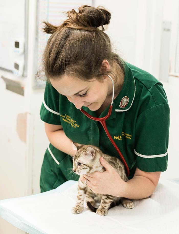
Lou Northway walks readers through the stages of, and considerations around, anaesthetising cats and dogs with cardiac conditions.
Lou Northway
Job Title
Heart disease affects approximately 15% of cats (Fuentes et al, 2020) and 11% of dogs (Parker et al, 2006).
In some patients, the disease may have already been investigated before anaesthesia, therefore giving more information on pathophysiology and enabling optimal perianaesthetic management.
A recent study showed that of 40 client-owned cats, 26 had audible murmurs and 4 had a gallop rhythm (Clark et al, 2020). Twenty-six of the 40 cats had their anaesthetic plan changed based on the results of a preanaesthetic echocardiogram. The study also emphasised that an echocardiogram is required to accurately assess the level of anaesthetic risk, as some cats with murmurs had minimal haemodynamic disturbances while others were of greater significance.
However, some patients may have a previously undetected heart murmur or arrhythmia on a preanaesthetic clinical examination, and the possible cause must be considered. A knowledge of the different types of heart disease more commonly found in certain breeds is helpful.
More recent studies of feline heart disease have found that hypertrophic cardiomyopathy is commonly seen in middle-aged, non-pedigree male cats, as well as pedigree cats such as Maine coons (Fuentes et al, 2020).
In dogs, chronic valvular disease is more commonly seen in smaller breeds, while dilated cardiomyopathy is more often seen in large breeds of dog, such as great Danes, boxers and Dobermanns (Dukes McEwan, 2000).
Reducing stress and anxiety in cardiac patients is vital. Stress results in the release of catecholamines, which can induce cardiac arrhythmias, therefore reducing cardiac output and increasing myocardial oxygen demand.
In addition, tachycardia should be avoided in any patients where structural cardiac defects exist, such as hypertrophic cardiomyopathy (thickened myocardium with reduced chamber size), as this will reduce stroke volume because the reduced diastolic time means less time exists for the heart to refill with blood.
Also, patients with valvular dysfunction are more likely to develop regurgitation if heart rate is greatly increased or decreased, which will affect stroke volume (Robinson and Borgeat, 2016).
Speaking to owners before their pet’s procedure may help ensure stress is minimised. If the pet is of a nervous disposition, it may be appropriate to admit the animal close to the procedure time, therefore minimising the time waiting in the kennel environment.
Many patients with heart disease will be on long-term medication, so it is important to check with the veterinary surgeon if the patient should receive its medication on the day of its anaesthetic. For example, some commonly used drugs – such as angiotensin‑converting enzyme inhibitors – may exacerbate hypotension during anaesthesia.
IV catheters should be placed in all patients undergoing anaesthesia to enhance patient safety and aid emergency drug administration. A discussion with your vet about the patient, procedure and possible adverse events (hypotension or arrhythmias, for example) should be discussed and planned for in advance.
Preoxygenation is strongly recommended to reduce the chances of hypoxaemia and cardiac ischaemia during induction. A study by McNally et al (2009) showed that when patients were preoxygenated, it slowed the onset of desaturation. Using a face mask rather than simple flow-by with the end of the breathing system is far more effective (Ambros et al, 2018).
It is worth noting, however, that preoxygenation should only be performed if tolerated by the patient, to keep stress to a minimum. If the patient is not tolerating a mask over its muzzle then flow-by may be a better option.
A thorough patient assessment should be performed, including:
Most anaesthetic and analgesic drugs will cause some degree of cardiovascular depression; they will affect autonomic function and reduce vasomotor tone, which will affect both cardiac output and blood pressure (Robinson and Borgeat, 2016).
Our aim is to maintain cardiac function and cause minimal haemodynamic disturbances.
The vet will prescribe a balanced anaesthetic protocol, which should include a multimodal analgesic approach to reduce anaesthetic requirements for both induction and maintenance of anaesthesia.
Balanced anaesthesia involves the use of a smaller dose of a combination of drugs to achieve the three key components of anaesthesia – unconsciousness, analgesia and muscle relaxation. When different drugs are used together they can have a synergistic effect, meaning much lower doses can be used, resulting in less dose-dependent adverse effects.
We must not forget that volatile anaesthetic agents – such as isoflurane and sevoflurane – are drugs, too, and will reduce both cardiac contractility and systemic vascular resistance, which may lead to hypotension.
As VNs, we should aim to administer the minimum concentration possible. Knowledge of the minimum alveolar concentration (MAC) – a measure of each agent’s potency (Table 1) – can be used as a guide. When multimodal analgesia is used (including local anaesthetic techniques), adequate anaesthesia can often be maintained with concentrations below MAC.
| Table 1. Canine and feline minimum alveolar concentration (MAC) for volatile anaesthetic agents | ||
|---|---|---|
| Volatile agent | Feline MAC | Canine MAC |
| Sevoflurane | 3% | 2.3% |
| Isoflurane | 1.7% | 1.3% |
| Adapted from Pang DSJ (2016). Inhalant anaesthetic agents. In Duke-Novakovski T, de Vries M and Seymour C (eds), BSAVA Manual of Canine and Feline Anaesthesia and Analgesia, BSAVA, Gloucester: 209 with permission. | ||
Many discussions always occur on what drugs are “safe” to use, but as Robert Smith MD once said: “There are no safe anaesthetic agents, no safe anaesthetic procedures; only safe anaesthetists.” Therefore, we need to think about what drugs are available and how each of them affects cardiovascular function.
Table 2 shows different drugs and how each affects the cardiovascular system. It is worth remembering that all drugs have dose-dependent effects – the higher the dose, the more likely you are to see changes in physiological variables.
Multimodal analgesia involves giving two or more analgesic agents, each of which targets different parts of the pain pathway. Regional anaesthesia is useful because it blocks transmission of all painful stimuli to the spinal cord. However, local anaesthesia is only effective for a few hours after administration and it is, therefore, important to give additional analgesics to target other parts of the pain pathway to reduce nociception and central sensitisation when it wears off.
| Table 2. Different drugs and how each affects the cardiovascular system | |||||
|---|---|---|---|---|---|
| Drug | Cardiac output | Heart rate | Mean arterial pressure | Systemic vascular resistance | Arrhythmic effects |
| Acepromazine | – | O | –– | –– | Antiarrhythmic |
| Medetomidine | – | ––– | (Biphasic) + then – | (Biphasic) ++ then – | Bradycardia/AV block |
| Methadone | O | –– | + | + | Bradycardia/AV block |
| Buprenorphine | – | – | O | + | None likely |
| Butorphanol | O | – | – | O | None likely |
| Propofol | – | – | –– | –– | None likely |
| Alfaxalone | O | + | O | O | None likely |
| Isoflurane | + | ++ | –– | –– | None likely |
| Sevoflurane | – | O | –– | – | None likely |
| Ketamine | + | + | + | + | Ectopic arrhythmias |
| Diazepam or midazolam | O | O | O | O | None likely |
| + = increase / – = decrease / O = negligible or minimal effects | |||||
| Robinson R and Borgeat K (2016). Cardiovascular disease. In Duke-Novajovski T, de Vries M and Seymour C (eds), BSAVA Manual of Canine and Feline Anaesthesia and Analgesia (3rd edn), BSAVA, Gloucester: 283-313 with permission. | |||||
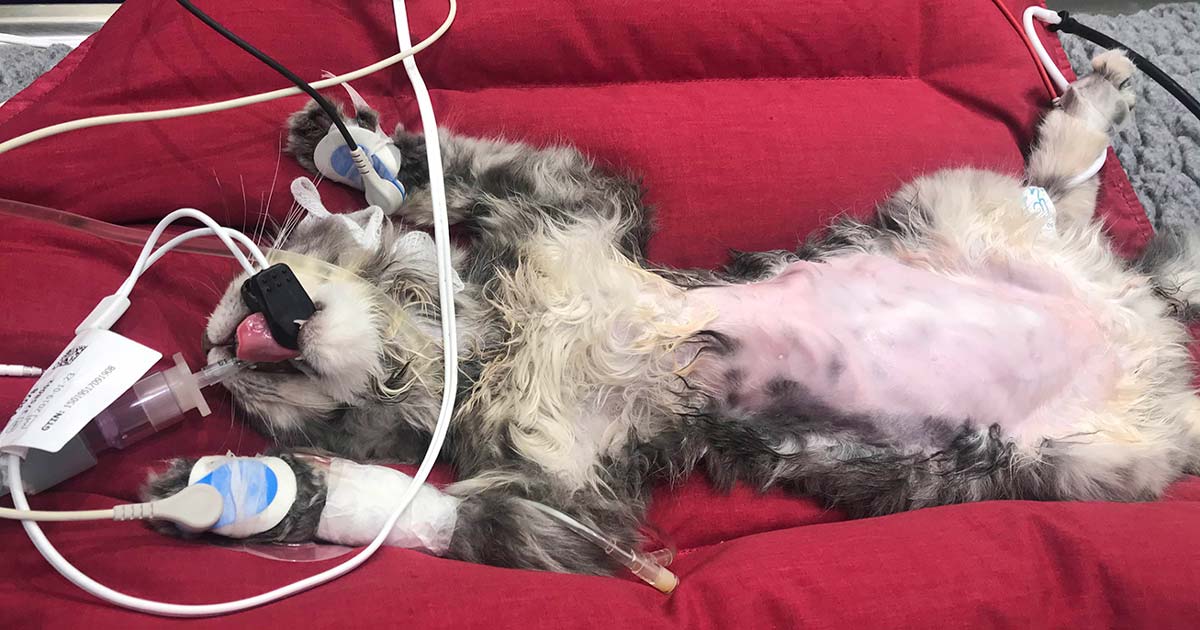
Before induction of anaesthesia, it is helpful to attach as much monitoring equipment as possible, unless this causes stress to the patient.
A baseline pulse rate, respiration rate, blood pressure and ECG is very useful to help us assess later if the patient is compensating or decompensating during anaesthesia. How far from the patient’s “normal” (even if it is was abnormal) is it now?
Capnography is a vital additional piece of monitoring equipment as it provides information indirectly on cardiac output and circulation.
If cardiac output suddenly reduces (due to reduced contractility, heart rate or stroke volume) then so, too, will pulmonary perfusion and you will see a decrease in end-tidal CO2 values, as long as ventilation does not also change. This may be a sudden decrease and an early indicator if the patient is in cardiovascular compromise.
Hypercapnia (increased end-tidal CO2) can result in tachycardia, arrhythmias and increased myocardial oxygen demand, so should be avoided.
Pulse oximetry provides information on both the percentage of available haemoglobin saturated with oxygen, and perfusion of peripheral tissues. If the heart is unable to pump the blood to the peripheral tissues, we may see a reduced oxygen saturation.
The amplitude of the plethysmograph can also indicate vascular tone and pulse quality, and should be observed alongside the numerical values. Some more modern pulse oximeters will also give a perfusion index – a non-invasive measure of peripheral perfusion that can be measured continuously.
We monitor blood pressure based on the theory that if blood pressure is normal, so is tissue perfusion. However, hypotension is always abnormal and must be addressed.
It is essential to try to maintain normotension during anaesthesia. Table 2 shows how administered drugs may affect cardiac output, contractility and systemic vascular resistance; a knowledge of this will help both the VN and vet navigate the troubleshooting process on how to address the hypotension.
If the patient is bradycardic then treating the bradycardia should be the priority. If the heart rate is acceptable then we should consider the other components of the blood pressure equation (Panel 1). A decrease in systemic vascular resistance is common because isoflurane and sevoflurane are potent vasodilators. Therefore, reducing the volatile agent can often improve blood pressure.
Blood pressure = heart rate × systemic vascular resistance (SVR)
(Heart rate: normal, bradycardic or tachycardic?) (SVR = vasodilation or vasoconstriction?)
Cardiac output = heart rate × stroke volume
(Stroke volume is influenced by preload [venous return], afterload [mainly SVR] and contractility)
Temperature should be monitored closely and hypothermia should be avoided. Severe hypothermia can cause arrhythmias, reduce cardiac contractility and, if the patient shivers on recovery, can increase oxygen demand.
Active warming techniques – such as the use of warm air systems – is advantageous, as well as ensuring your patient does not become hypothermic preoperatively.
ECG monitors the electrical activity of the heart, but not an indicator of cardiac output. Cardiac patients are more prone to arrhythmias primarily through cardiac dysfunction, although they may also be drug induced.
As VNs, we should always palpate a pulse when observing an ECG, as a complex on the screen does not always mean it is generating an actual cardiac contraction and pulse. If you see an ECG complex yet do not palpate a corresponding pulse, this is known as a pulse deficit. If the arrhythmia is severe, and affecting heart rate and blood pressure, the vet will often prescribe antiarrhythmic drugs such as lidocaine.
Alongside our advanced monitoring machines, RVNs should continue to closely monitor our patients using their hands-on skills, observing pulse rate and quality, capillary refill time, mucous membrane colour and respiratory rate.
Regular thoracic auscultation throughout the procedure is also recommended, observing for changes in heart murmurs and to ensure no development of pulmonary crackles, which may be indicative of pulmonary oedema. Any adverse events or observations should be recorded on the anaesthetic record for a timeline of events.
No anaesthetic should ever be viewed as “routine”, but when anaesthetising patients with diagnosed or suspected cardiac disease, it is imperative we discuss and plan for every eventuality ahead of the procedure. Consider the patient in front of you – is it one of the “known breeds” over-represented for cardiac disease?
A thorough understanding of the disease pathophysiology, drugs and their effects on the cardiovascular system is essential to treat any adverse effects accordingly.