Allele: one of two or more versions of a genetic sequence at a particular region on a chromosome. An individual inherits two alleles for each gene: one from the dam and one from the sire.
29 Jun 2023
Matthew Keane and Emily Dutton review latest information on genetic mutations associated with this affliction.

Image: © Dmytro S / Adobe Stock
A genetic component has been identified or suspected in several different cardiac diseases, such as dilated cardiomyopathy (DCM), myxomatous mitral valve disease (MMVD), arrhythmogenic right ventricular cardiomyopathy (ARVC), subaortic stenosis (SAS) and tricuspid valve dysplasia.
This review article outlines and focuses on the genetic basis associated with common congenital and acquired cardiac disease.
In human medicine, many genetic mutations have been identified that are associated with the development of cardiac disease.
Dilated cardiomyopathy (DCM) has been associated with mutations in more than 50 genes1.
The genes identified are involved in a variety of functions, such as calcium/sodium-handling, transcription factors, desmosomal proteins, cytoskeletal proteins, sarcomeric proteins and nuclear envelope proteins1.
Any mutation affecting any of these functional groups can lead to the development of DCM.
Autosomal: having to do with any of the 38 pairs (dogs) or 18 pairs (cats) of chromosomes found in most cells. Autosomal chromosomes are numbered 1 to 38 (dogs) or 1 to 18 (cats). The sex chromosomes (X and Y chromosomes) determine whether an individual is male or female, and are not considered autosomal chromosomes.
Autosomal dominant inheritance: one of the ways a genetic trait or a genetic condition can be inherited. In autosomal dominant inheritance, a genetic condition occurs when a variant is present in only one allele (copy) of a given gene.
Autosomal recessive inheritance: one of the ways a genetic trait or a genetic condition can be inherited. In autosomal recessive inheritance, a genetic condition occurs when one variant is present on both alleles (copies) of a given gene.
Chromosome: a structure found inside the nucleus of a cell. A chromosome is made up of proteins and DNA organised into genes. Each cell normally contains 39 pairs (dogs) or 19 pairs (cats) of chromosomes.
Familial: a phenotype or trait that occurs with greater frequency in a given family than in the general population.
Gene: the basic unit of heredity passed from parent to offspring. Genes are made up of sequences of DNA and are arranged, one after another, at specific locations on chromosomes in the nucleus of cells. They contain information for making specific proteins that lead to the expression of a particular physical characteristic or trait.
Genome-wide association study: this method surveys the entire genome for genetic polymorphisms, typically single nucleotide polymorphisms (SNPs; pronounced “snips”), that occur more frequently in cases (animals with the disease or trait being assessed) than in controls (people without the disease or trait).
Incomplete penetrance: penetrance refers to the likelihood that a clinical condition will occur when a particular genotype is present. A condition is said to show incomplete penetrance when some individuals who carry the pathogenic variant express the associated trait while others do not. Also called reduced penetrance.
Locus: the physical site or location of a specific gene on a chromosome.
MicroRNA (miRNA): a type of RNA found in cells and blood. MicroRNAs are smaller than many other types of RNA and can bind to messenger RNAs (mRNAs) to block them from making proteins.
Missense mutation/variant: a genetic alteration in which a single base pair substitution alters the genetic code in a way that produces an amino acid that is different from the usual amino acid at that position. Some missense variants (or mutations) will alter the function of the protein.
Mutation: a change in the usual DNA sequence at a particular gene locus. Mutations (including polymorphisms) can be harmful, beneficial, or neutral in their effect on cell function. The term variant is sometimes used as a synonym for the term mutation.
Segregation analysis: the process of fitting formal genetic models to data on expressed disease characteristics (phenotype) in biological family members to determine the most likely mode of inheritance for the trait or disease under study.
Single nucleotide polymorphism (SNP): a DNA sequence variation that occurs when a single nucleotide (adenine, thymine, cytosine or guanine) in the genome sequence is altered, and the particular alteration is present in at least 1% of the population.
Variable expression: variation in the manner in which a trait is manifested. When variable expressivity is present, the trait may vary in clinical expression from mild to severe.
X-linked dominant inheritance: refers to genetic conditions associated with mutations in genes on the X chromosome. A single copy of the mutation is enough to cause the disease in both males (who have one X chromosome) and females (who have two X chromosomes).
X-linked recessive inheritance: refers to genetic conditions associated with mutations in genes on the X chromosome. A male carrying such a mutation will be affected, because he carries only one X chromosome. A female carrying a mutation in one gene, with a normal gene on the other X chromosome, is generally unaffected.
Due to identification of these genes, genetic testing of individuals with a familial history of DCM can be performed as a monitoring tool2. Genetic testing consists of assessing around 50 genetic loci, and affected individuals may have more than one affected locus3.
In contrast to our canine patients, gene penetrance has been described in human patients whereby the gene affected, or the type of mutation, may lead to altered disease expression or severity4-5.
Studies into hypertrophic cardiomyopathy (HCM) in human patients first demonstrated that a mutation in the gene coding for the sarcomere protein MYH7 was identified in a high proportion of familial HCM cases6. Subsequent mutations were identified in further sarcomeric proteins – including myosin binding protein C3 (MYBPC3), the most frequently mutated gene identified in human HCM7-10.
Further mutations in genes encoding calcium signalling proteins and Z-disc proteins have also been identified in human patients11-12. HCM in cats was first identified in 1977, and subsequent research identified similar echocardiographic and anatomical findings to that of HCM in humans13-14.
Familial inheritance, as found in humans, has also been documented in a population of Maine coon cats15. Mutations in the gene responsible for sarcomeric protein MYBPC3 have been identified in Maine coons and ragdolls16-17. A case report has also documented a mutation in MYH7 in a domestic shorthair cat18. These findings suggest a similar genetic basis of HCM in both human and feline patients.
However, a high prevalence of the MYBPC3 mutations in non-affected cats has been reported, suggestive that other genetic or environmental factors may be indicated19.
Mutations in the ALMS1 gene have also more recently been identified in sphynx cats, resulting in the development of hypertrophic cardiomyopathy, although this may require validation in the European sphynx population20.
One of the key steps to improve health in breeding programmes has been the introduction of genetic testing, which allows rapid identification of predisposed individuals prior to breeding from them. Collating data for investigation of a possible genetic basis in a given breed is important, and a need exists for more prospective studies; for example, progressive retinal atrophy is a disease in veterinary patients where single gene mutations in different breeds have been identified as responsible for the development of this condition21.
This has allowed for the development of commercial DNA tests that can identify affected cases, unaffected cases or subclinical cases prior to disease expression22.
Further to this, this has enabled breeders to select appropriate breeding partners to decrease expression of this disease.
Identification of similar targets in cardiac disease could help identify affected breeding animals for common acquired or congenital disease.

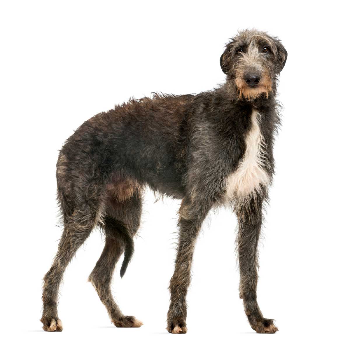
DCM is a common acquired cardiac disease identified mostly in large breed dogs, such as the Dobermann, great Dane, Irish wolfhound, deerhound and Newfoundland23-28.
DCM is described as an idiopathic condition leading to a primary myocardial disorder, and resulting in poor systolic function and secondary ventricular dilation. Electrocardiographic abnormalities may also be noted with this disease process29.
DCM is a diagnosis of exclusion and, therefore, other conditions that may lead to poor systolic function and a dilated heart must be excluded, such as myocarditis, hypothyroidism, volume overload, taurine deficiency and nutritional cardiomyopathy30-33.
Another condition that can mimic the DCM phenotype is that of tachycardia-induced cardiomyopathy, where persistently elevated heart rates may result in left ventricular dysfunction34.
Primary DCM is believed to be genetic in origin and hereditary, passing from one generation to the next. The exact mechanism leading to the expression of a DCM phenotype remains unknown. It has been suggested that individual breeds may have their own genetic mutations leading to the development of DCM.
Different dog breeds may also have similar or the same causative genetic disorders. It has been suggested that DCM may have “incomplete penetrance” and “variable expression”. This means that not all the dogs with the same genetic mutation will go on to express a DCM phenotype, and that the same mutation may not result in the same disease expression, which may explain the different clinical signs and disease severity we see in patients with DCM35.
The Dobermann is a breed overrepresented for developing DCM, with a reported cumulative prevalence of 58%36.
Dobermanns with DCM may have left ventricular dilation or cardiac arrhythmias, and these can either occur together or separately. Screening protocols for this breed, therefore, include both echocardiography and a 24-hour ambulatory ECG37.
Initially, it was suggested that male Dobermanns were more likely to develop DCM than female Dobermanns38. A further study has found this is not the case, and no difference exists between gender and developing DCM.
The same study also found that a difference does exist between disease expression in male and female Dobermanns, with female Dobermanns more likely to develop ventricular arrhythmias and male Dobermanns more likely to develop changes during echocardiography sooner in the disease process36.
DCM in the Dobermann is considered to be a hereditary disease, inherited as an autosomal dominant trait39. Cardiac genes that have been associated with the development of hereditary DCM in humans have been studied in Dobermanns, but none of these genes have been consistently linked with the development of DCM in this breed40.
It has been documented in an American population of Dobermanns that a splice site mutation in a gene encoding for pyruvate dehydrogenase kinase 4 (PDK4) has been associated with DCM in the Dobermann35. This was not consistent with Dobermanns from Europe, as it was shown that no association was found between the development of DCM and this mutation in this population41.
The difference between the two studies may be reflective of different genetic populations between the US and Europe.
Further studies have also identified a missense variant in the “titin gene” in Dobermanns with dilated cardiomyopathy and sudden cardiac death42. Titin is the largest protein in the body, and it contributes to both passive stiffness and active contraction of the myocardium through unfolding and refolding of its numerous domains in response to tension.
The results of microRNA (miRNA) analysis in Dobermanns affected with DCM has shown a difference in expression compared to healthy animals (142-3p, 144, 21, let-7c and 92a); however, the results published have not been statistically significant43. It is suggested this may be due to a small sample size.
Further research into miRNAs may highlight cardiac biomarkers that can be used in the clinical setting for diagnosis of DCM.

Reported prevalence of DCM in the Irish wolfhound varies from 24.2% to 29%, with male dogs overrepresented25, 44-45.
It is suspected that inheritance of DCM is not due to a simple monogenic model, but rather a mixed monogenic-polygenic with the involvement of a sex-dependent allele44.
As well as DCM, Irish wolfhounds have been shown to have a high prevalence of atrial fibrillation (AF)25, 46. An association between AF and the development of DCM has been demonstrated25, 47. AF can also occur in isolation without resulting in the development of DCM25, 47-48.
A study performed in North American Irish wolfhounds with AF identified a genetic basis to AF with a high heritability estimate for developing AF and pedigree analysis suggesting a dominant mode of inheritance49. However, no causative genetic mutations for the development of AF have yet been identified.
The mode of inheritance of AF demonstrated by studies has some differences to that of DCM, as an X-linked inheritance of AF has been ruled out, whereas it remains a consideration for the inheritance of DCM44, 49. A polygenic mode of inheritance of AF has not yet been excluded49.
At present, no genetic loci have been found that are responsible for the development of AF. Future genome-wide association studies (GWAS) to identify whether AF is due to a single gene or a number of genes are needed.
Further to this, GWAS could also identify whether AF and DCM share the same or similar genetic loci.
Tafazzin is a protein that is highly expressed in the cardiac myocardium and is produced by the TAZ gene, which is an X chromosome-related gene. As male Irish wolfhounds are overrepresented in the population, one study evaluated the role of this gene in the development of DCM in this breed. The study found that TAZ was unlikely to result in the development of DCM50.
Two GWAS in the Irish wolfhound have demonstrated multiple loci that are associated with the development of DCM48, 51. In these two studies, single nucleotide polymorphisms (SNPs) were identified that were associated with the development of DCM. However, it was found that only three of the five SNPs (on chromosomes 1, 21 and 37) identified in European Irish wolfhounds were found in Irish wolfhounds from the UK, and only one had the same allele associated with the development of the disease.
It may be that the differences between the genetic backgrounds of these two populations may explain the difference between these two studies. Both studies identified that it is likely multiple genes are involved in the inheritance of this condition, and causal genetic factors exist that remain to be identified.
This is supported by the evidence that some loci were associated with DCM in some animals, but not throughout the whole population48.
The reported prevalence of DCM in the Newfoundland is around 10%27.
Analysis of a family of Newfoundlands from the UK demonstrated an autosomal dominant mode of inheritance with incomplete penetrance52. In this study, a genome-wide linkage analysis of more than 200 markers failed to identify any linkage.
Further studies using the same family of Newfoundlands, plus further families of Newfoundlands, investigated 15 candidate genes for a cause of DCM and found no evidence any of these genes were associated with the development of DCM in the breed53.
A GWAS performed in Newfoundlands identified some genetic loci of interest, but nothing of genome-wide significance54. It is thought that future genetic sequencing may identify genetic loci of significance54.
Pedigree analysis of American great Danes with DCM found that the disease may be inherited as a recessive trait linked to the sex-determining chromosome X55. As a result of this study, recommendations were advised to avoid breeding from affected dogs.
Pedigree analysis performed in the UK was indicative of an autosomal dominant inheritance pattern, although polygenic inheritance as suggested in other breeds could not be excluded. This study identified that an X-linked recessive inheritance was not present in the population of great Danes studied in the UK24.
A study has also identified that great Dane dogs affected with DCM have an abnormal expression of triadin and calstabin 2, both of which are components of the cardiac ryanodine receptors (RyR2)56. This has resulted in the identification of candidate genes for for future studies.
A GWAS performed in great Danes identified some genetic loci of interest, but nothing of genome-wide significance54. It is thought that future genetic sequencing may identify genetic loci of significance54.
A juvenile form of DCM has been identified in Portuguese water dogs57-58. This form of DCM affected puppies born to healthy parents.
An autosomal recessive pattern of inheritance was initially suggested based on pedigree analysis57.
Further studies performed segregation analysis, which were consistent with an autosomal recessive pattern of inheritance58.
A mutation in the phospholamban gene (R9H) has been identified in Welsh springer spaniels, resulting in a dilated cardiomyopathy phenotype with left ventricular dilation, arrhythmias and poor systolic function59.
This population of Welsh springer spaniels studied also had a high incidence of sudden cardiac death and the mutation was a result of a single base pair change59. Inheritance of this mutation was dominant, with a high degree of penetrance suspected59.
Early onset DCM in the standard schnauzer and giant schnauzer has been associated with an autosomal recessive mode of inheritance as a result of a two-base pair deletion60-61. The result of this deletion is a mutation of the RNA-binding motif protein 20 (RBM20), which is responsible for the splicing of cardiac genes, including the gene for titin61.
This study showed that homozygosity for the RBM20 variant in the standard schnauzer was highly associated with DCM and an 80% shortened lifespan.
The RBM20 variant was also identified in giant schnauzer dogs in the same study, and was associated with DCM and premature death.
The authors, along with professors Jo Dukes-McEwan, Lucy Davison and Ottmar Distl, are carrying out research assessing whether a genetic basis to DCM exists in the deerhound. The study is called “GENetic associations with dilated cardiomyopathy in DEERhounds”; the GENDEER Study.
Discovering a genetic basis to primary DCM in the deerhound may help in the discovery of a DNA test for DCM in this breed. A genetic test or panel of tests could aid diagnosis of DCM in individual dogs.
The team is looking for help collating samples from DCM-affected cases and would request any surplus blood from deerhounds to be sent to the authors in ethylenediaminetetraacetic acid for future DNA analysis (more information and a consent form can be found online).
The team is very grateful to PetSavers and the Veterinary Cardiovascular Society for their generous funding of this study. For any questions regarding the GENDEER Study, or if you have a case that may be suitable, email [email protected]
Previously termed “boxer cardiomyopathy”, arrhythmogenic right ventricular cardiomyopathy (ARVC) is a myocardial condition of boxer dogs manifested by fibro-fatty infiltration of cardiomyocytes, ventricular premature complexes and ventricular tachycardia62.
It has been demonstrated that ARVC has an autosomal dominant inheritance pattern with age-related penetrance63. Reduced expression of the cardiac ryanodine receptor protein (RyR2) and its messenger RNA was found in dogs with ARVC, but no genetic linkage with the RyR2 gene was identified64.
Reduced expression of calstabin 2 messenger RNA has also been documented in boxers with ARVC in comparison to healthy controls or Dobermanns with DCM. However, no causative genetic mutations to explain this reduced expression have been identified65.
Immunofluorescence studies have been conducted to evaluate the structure and composition of the intercalated disc in boxers with ARVC. These studies documented abnormalities of the intercalated discs in affected dogs (including loss of gap junction plaques, remodelling of intercalated disc structures, and a general loss of molecular integrity of the intercalated disc), but no mutations were identified to affect the proteins that were evaluated66.
It is suggested the abnormalities identified are more a reflection of the disease process rather than being the causal elements.
A GWAS conducted in American boxers with ARVC identified a mutation responsible for a reduced production of messenger RNA for the protein striatin67. This mutation was associated with an eight-base pair deletion on chromosome 17. The mutation was identified in 57 out of 61 boxers affected by ARVC (either homozygous or heterozygous), but was also found in unaffected control boxers (all heterozygous).
A mutation in striatin was also identified in boxers from the UK; however, no significant difference was noted between affected dogs or healthy dogs, suggesting that this mutation did not explain the occurrence of ARVC in the UK population68-69.
Research conducted by Cattanach et al (2015) identified that all cases of ARVC could be traced back to an initial population of boxers based in America, which were the dogs studied by Harpster (1983) to initially describe this disease62, 69. This is suggestive that the disease is the same between the US and the UK, and the mutation in the striatin gene is not responsible, as this mutation has been documented in both the affected and non-affected population.
Further to this, some dogs in the affected group did not have the striatin mutation.
Studies have also been performed to assess an autoimmune aetiology of ARVC. In human patients, autoantibodies to desmoglein-2 were present in almost all patients with ARVC, while they were absent in nearly all patients without disease or with other cardiac conditions, such as DCM or HCM70. Similar findings were not documented in boxers with ARVC as autoantibodies to desmoglein-2 were detected in dogs with ARVC, dogs with DCM, dogs with myxomatous mitral valve disease (MMVD) and even in healthy patients71.
In Rhodesian ridgebacks, an inherited ventricular arrhythmia with no evidence of structural heart disease has been identified72. This disease has been associated with a mutation in the QIL1 gene that is the result of a single base pair mutation73. An autosomal recessive mode of inheritance is suspected, although an autosomal dominant mode of inheritance with incomplete penetrance has not been excluded73.
A similar disease process has been identified in German shepherd dogs with no evidence of underlying structural cardiac disease74. Pedigree analysis is suggestive of single gene defect and/or polygenic inheritance with incomplete penetrance74.
Further genetic analysis supports a genetic component to this disease process; however, no causative genetic mutation has been identified as of yet75.
MMVD is the most common cause of congestive heart failure in dogs76. The disease is characterised by the progressive degeneration of both atrioventricular valves, but particularly affects the mitral valve77.
A familial basis to MMVD was suspected due to the evidence of a predisposition to the early development of the disease in some breeds, and similarities in the age of onset between parents and offspring78-80. Predisposed breeds include the cavalier King Charles spaniel, whippet, poodle, dachshund, cocker spaniel and Chihuahua77, 81.
Between healthy dogs, and mildly or severely affected dogs, a number of genes and microRNAs have been identified that are differentially expressed between these groups82-89.
In 2010-11, products of a cavalier King Charles spaniel breeding scheme had a 69% reduced risk of having a murmur compared to dogs that were not products of the breeding scheme. Also recorded was a 36% reduced risk of having mitral valve prolapse compared to non-products of the breeding scheme suggestive of a genetic involvement90.
GWAS have been performed in patients with MMVD; however, our understanding of responsible individual variants or genetic loci remains limited91-94. SNPs in the 5-HT transporter gene have been identified in Maltese dogs with MMVD; however, these SNPs were not identified in cavalier King Charles spaniels95-96.
Candidate genes near the nebulette (NEBL) gene have been suggested in dachshunds97. NEBL is responsible for the coding of cardiac proteins97. However, the results of this study are yet to be validated in further populations.
Research into exosomal miRNAs has documented changes in certain miRNA expression in dogs that develop MMVD (miR-9 and miR-599), and dogs that develop congestive heart failure (miR-181c and miR-495)98.
It has been previously suggested that miRNAs may be involved in the development of MMVD. It was found that miR-17, miR-20a, miR-30d and let-7c are significantly down-regulated in disease mitral valves compared to normal valves98-99. Evidence also exists that expression of miRNA may change as dogs progress through American College of Veterinary Internal Medicine (ACVIM) guidelines for stages of the disease.
Differences in down-regulation or up-regulation of certain miRNA has been documented throughout different ACVIM stages, suggestive that miRNAs may be useful as a future biomarker diagnostic tool or for monitoring treatment response100-102.

Subaortic stenosis (SAS) is a common condition in large breed dogs, with certain breeds characterised by a ridge of fibrous tissue below the aortic valve that leads to a left ventricular outflow tract obstruction103.
Large breed dogs such as the boxer, golden retriever, Newfoundland, German shepherd dog and Rottweiler are over-represented104.
Due to the reported increased incidence in certain breeds, a genetic component to the disease was suspected. Heritability of the condition has been demonstrated in both Newfoundlands and golden retrievers105-107.
An autosomal recessive mode of inheritance has been documented in Rottweilers, golden retrievers, dogue de Bordeaux and bullmastiffs108-109, whereas an autosomal dominant mode of inheritance has been shown in Newfoundlands110.
These studies also demonstrated that affected offspring could be produced by unaffected parents, and vice versa.
In Newfoundlands, a genetic variant has been identified in dogs affected with SAS, affecting the phosphatidylinositol-binding clathrin assembly protein (PICALM). A GWAS did not identify a locus associated with the development of subaortic stenosis in Newfoundlands, but identified the region where the PICALM variant with a single codon insertion was located110.
It is reported that this variant was present in 80.6% of the Newfoundland population and, therefore, it is likely that a polygenic inheritance pattern for SAS exists, rather than the PICALM variant being solely responsible.
The Labrador retriever is documented as being predisposed to congenital malformation of the tricuspid valve, as well as the dogue de Bordeaux109, 111-112.
Famula et al (2002) investigated the heritability of tricuspid valve dysplasia in a population of Labrador retrievers111. Findings of the study suggested the disease was indeed inheritable, which led to breeding recommendations to avoid the breeding of affected dogs or dogs closely related to affected dogs.
It was not possible to identify a major locus responsible for the expression of tricuspid valve dysplasia in this study. Further studies identified that the disease could be mapped to chromosome 9 in the Labrador retriever112.
The French bulldog and bulldog have been reported as having the highest prevalence of pulmonic stenosis108. Pedigree analysis is suggestive of an autosomal recessive pattern on inheritance in bulldogs108.
A GWAS has been performed in French bulldogs, suggesting the possible involvement of a region of chromosome 34113. The region of chromosome 34 that was affected involves two genes involved in embryonic cardiac development113.
Many different genetic mutations have been linked with both canine and feline heart disease, which has furthered our understanding of these disease processes and has enabled the development of genetic tests that may be used in breed screening.
However, many of these diseases may be polygenic in nature or exhibit incomplete penetrance; therefore, care must be taken in interpreting these test results.
Furthermore, some of the genetic mutations demonstrated are present in some populations (for example, North America) but not in other populations (for example, Europe).
To summarise, the authors would express caution in interpreting genetic test results. Genetic diversity may be limited when selecting against a particular disease or trait – particularly when the prevalence of the disease being tested for is high.