14 Jul 2020
Francesco Cian discusses the case of a 1.5-year-old German shepherd dog seen by the referring vet for a well-circumscribed, non-ulcerated, 2.5cm in diameter cutaneous lesion on the hindlimb footpad.
Francesco Cian
Job Title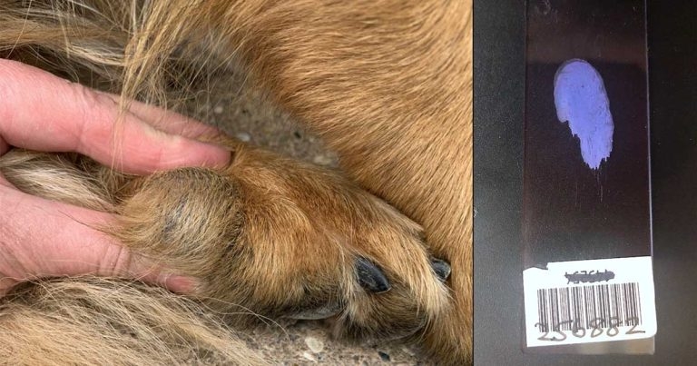
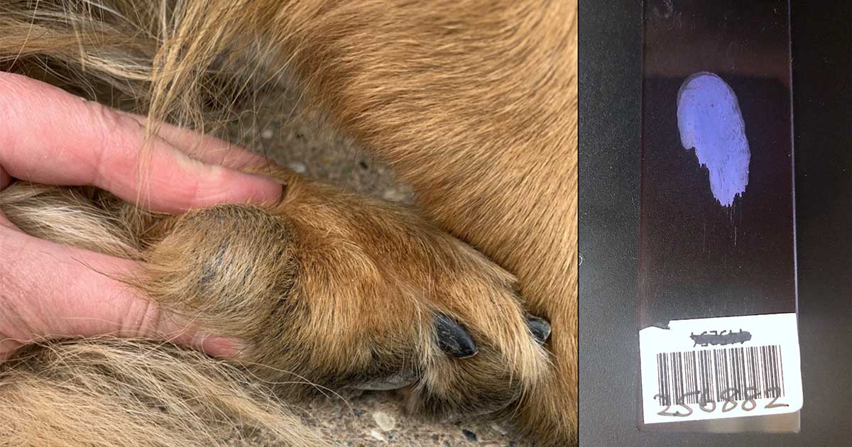
Cutaneous nodule on the dog’s hindlimb footpad.
A 1.5-year-old German Shepherd dog was seen by the referring veterinarian for a well-circumscribed, non-ulcerated, 2.5cm in diameter cutaneous lesion that had been present on the dog’s hindlimb footpad for a few weeks.
The dog was otherwise clinically well. A fine needle aspiration of the mass was performed as part of the diagnostic process.
The aspirate yielded variable amounts of chalky white material and, after staining the smear, it acquired a characteristic lavender colour. The sample appeared hypocellular on microscopic examination.
A lightly basophilic background was present, with large numbers of fine, small, clear, crystal-like structures with angular borders. Occasional large mononuclear cells with vacuolated cytoplasm – likely macrophages – were also noted.
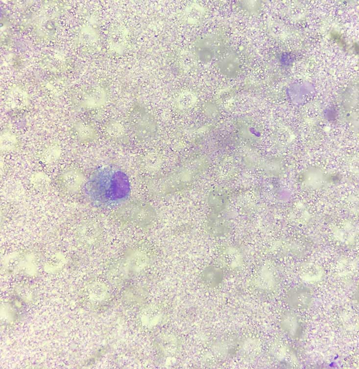
A diagnosis of calcinosis circumscripta was given.
These cytological features, together with the signalment and the clinical presentation, were indicative of calcinosis – in particular, calcinosis circumscripta.
The mass was excised and submitted for histopathological examination, which confirmed the initial diagnosis.
Calcinosis is a condition characterised by the deposition of mineral (mostly containing calcium) in the dermis, subcutis and, rarely, the epidermis. This may elicit a pyogranulomatous inflammation as calcium acts as a true foreign body.
Clinical presentation may vary from pale yellow to white papules, plaques or distinct nodules.
Mineralisation of soft tissues has been classified into four types according to underlying factors:
Two major clinical forms of calcinosis have been described in domestic animals.
Calcinosis cutis is a term mainly used to describe dystrophic mineralisation that develops in association with iatrogenic hyperglucocorticism or endogenous hyperadrenocorticism. The exact pathogenesis is unclear. It usually presents with lesions in multiple regions of the body. Large-breed dogs appear over-represented.
Calcinosis circumscripta (from Latin – limited, confined) is a term used to describe a more localised process and is mainly observed at the level of pressure points or areas of trauma. It often affects young growing dogs with enhanced calcium and phosphate metabolism, including German shepherd dogs.
Cases of calcinosis circumscripta may also result from severe chronic inflammation and neoplasia. A few reports in the veterinary literature also describe localised forms of calcinosis affecting the foot pads of dogs with renal failure. This can also result in a widespread visceral and vascular mineralisation.
In the case of calcinosis due to trauma or unknown causes, single lesions can be cured surgically.
Treatment and prognosis for the other forms mainly depend on the underlying cause and its resolution/control.
Cytological features of calcinosis are quite characteristic. Knowing the different mechanisms of mineralisation and the clinical presentation may help identify the causative process to select the most appropriate treatment.