
1 Sept 2026
Bartosz Ropelewski PGCert(SAS), MRCVS covers clinical presentation, diagnosis and treatment of this infection in dogs.
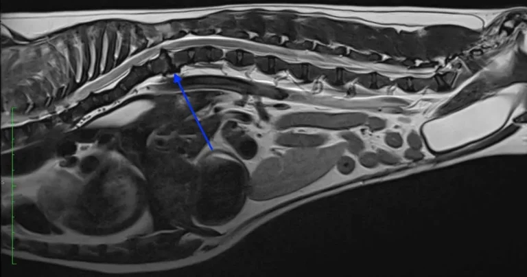
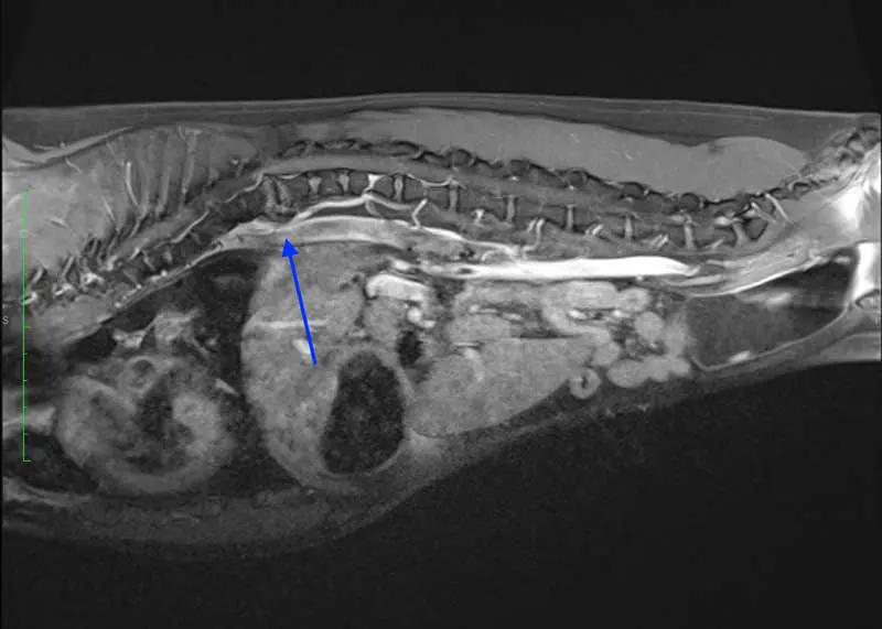
The preferred imaging technique for diagnosis of early cases of discospondylitis is MRI scan, although CT can be useful for assessment of the early changes affecting spinal columns, endplates and soft tissues.
Discospondylitis is an infection of the intervertebral disc and cartilaginous endplates. The infectious process initially starts in the endplates and then extends towards the intervertebral disc (Burkert et al, 2005).
Multiple pathophysiological pathways of contracting this infection can be identified, and both iatrogenic and autogenic sources of this infection can be distinguished. (Remedios et al, 1996).
One of the most common ways the infection spreads is through the bloodstream from the primary distant infection site (Kerwin, 2015).
Initial sites of infection may include: an infected wound, abscess, respiratory infection, endocarditis, urinary tract infection, migrating plant material, skin infection, and epidural injections. Spinal surgery can also predispose an animal to develop discospondylitis (Siems et al, 1999; Thomas, 2000; Canal et al, 2016). Pathogenic organisms commonly localise within the venous channels of the vertebral endplates due to the slow venous blood flow, before infiltrating the adjacent intervertebral disc (Thomas, 2000).
Discospondylitis can be caused by a bacterial or fungal infection. The most commonly isolated bacteria responsible for this type of infection is Staphylococcus aureus and Staph intermedius (Ruoff et al, 2018).
Other possibilities include:
Immunosuppressed animals have a much higher predisposition for developing this condition compared to healthy individuals. (Thomas, 2000; Barta et al, 1985).
Other predisposing factors include:
The clinical presentation of discospondylitis may be different depending on the extent of the disease and location of the affected spinal segments. The predominant symptom present in the majority of dogs (up to 80%) is spinal pain (Thomas, 2000).
Clinically, up to 30% of patients will develop pyrexia, anorexia and weight loss, or other signs of potential systemic disease (Thomas, 2000).
Discospondylitis can cause various degrees of neurological deficits, ranging from mild ataxia to non-ambulatory paraparesis (Gilmore, 1987).
Typically, clinical presentation has a progressive nature. Initially, owners may report the animal is reluctant to exercise; however, acute presentation can also be seen (Thomas, 2000).
Due to the non-specific presentation of this condition, differentials should include intervertebral disc disease, neoplasia, vertebral physitis, vertebral osteomyelitis and spondylosis deformans (Côté, 2015).
Full clinical examination, including neurological and orthopaedic assessment, should be performed. Patients affected by unspecific presentation, including pyrexia, neurological deficits and spinal pain, will require a blood test including haematology, biochemistry, blood culture, immunoassay for C- reactive proteins, urinalysis, and urine culture.
It is very important to include serology for Brucella canis in the blood panels. It has been estimated that up to 10% of dogs affected by discospondylitis may be affected by B canis (Kerwin et al, 1992). One of the tests used to confirm this diagnosis is an agar gel immunodiffusion. Cases displaying neurological deficits will also require a CSF analysis.
Survey radiographs or more advanced imaging, including CT or MRI, will be required to confirm or rule out discospondylitis. Radiographs can be used as a sole modality to confirm diagnosis. Radiographic evidence of infection can include irregularities on the surface of the vertebral endplates; lumbosacral junction (L7-S1) is the most typically affected (Thomas, 2000).
It is possible that early symptomatic cases may not display typical radiographic features for the first four to six weeks of the disease (Moore, 1992). Typical radiographic features depend on the stage of the disease and may include mild changes affecting the vertebral endplates in early stages and progress to more severe remodelling including endplates lysis. This eventually will lead to extensive bone sclerosis, osteophyte formation and fusion of the vertebrae (Hurov et al, 1978).
In cases of chronic progression, patients will develop spinal pathological fractures, spinal column collapse and spinal deformities (Thomas, 2000).
In early cases of discospondylitis, where changes can be very subtle, radiography may not be sufficient to pick up early signs of the disease. In these cases, more advanced imaging modalities including CT and MRI are recommended. CT can be useful for assessment of the early changes affecting spinal columns, endplates and soft tissues.
The preferred imaging technique for diagnosis of early cases of discospondylitis is MRI scan. It has been estimated that in human medicine, MRI has a 94% specificity and 96% sensitivity for the diagnosis of discospondylitis (Jevtic, 2004).
MRI scans using gadolinium contrast enable the very early detection of the spinal infection (Quesnele et al, 2012).
Cases unresponsive to conservative management, including broad-spectrum antibiotics, patients with neurological deficits, cases displaying radiographic features suggesting significant vertebral structural destruction/spinal instability, and dogs with negative blood and urine culture, will require disc biopsies (Ozuna and Delamarter, 1996).
Surgical biopsies can be collected via CT-guided needle aspiration or fluoroscopy guidance (Fischer et al, 1997). Patients affected by vertebral instabilities caused by bone destruction may require decompressive surgery, including hemilaminectomy, bone grafting and surgical stabilisation.
For thoracic and lumbosacral spine stabilisation, we can use screws and polymethylmethacrylate (Cabassu and Moissonnier, 2007; Font et al, 2013).
Stabilisation of the bacterial discospondylitis located in the lumbar spine will require use of screws with antibiotic-impregnated cement (Cabassu and Moissonnier, 2007).
Conservative management requires the use of a specific type of antibiotics, depending on the results of the bacteriology. In the case of negative culture results, it is recommended to start the patient on first-generation cephalosporins as the first line of treatment (Kerwin, 2015).
Depending on the clinical response, additional antibiotics may need to be added. Second-line antibiotics may include metronidazole, marbofloxacin or clindamycin. Improvement should be seen within a week from the initiation of the second-line treatment (Thomas, 2000). All patients should receive sufficient analgesia. It is very important to implement strict exercise restrictions – especially in patients affected by significant bone destruction and spinal instabilities.
Duration of the treatment is very individual and depends on the degree of bone and soft tissue involvement. It has been estimated that average treatment time to complete symptom resolution is 53 weeks (Kerwin, 2015).
It is highly recommended to perform imaging every four to six weeks (Motta, 2009).
The prognosis for bacterial discospondylitis is generally good.
A 2025 systemic review reported treatment success rates of approximately 69% for medical and 75% for surgical management (Vallios et al, 2026).
Bartosz Ropelewski qualified in 2006 from the University of Warmia and Mazury in Olsztyn, Poland. Following graduation, he completed a nine-month rotational internship at a veterinary hospital in Gdańsk. Bartosz then spent 12 years working in a first-opinion practice in south Manchester, where he developed a strong interest in orthopaedic and soft tissue surgery. This led him to undertake further postgraduate study, achieving a Certificate in Small Animal Surgery in 2016, followed by advanced practitioner status in 2017. In 2018, Bartosz joined a busy hospital in Nottingham, where he further expanded and refined his surgical expertise. He has a particular interest in neurosurgery, brachycephalic obstructive airway syndrome and reconstructive surgery. A significant proportion of his caseload involves neurosurgical patients, and he has extensive experience in the investigation and surgical management of spinal disorders, including intervertebral disc disease and other neurological conditions requiring advanced surgical intervention. Bartosz joined Rutland House Referrals in 2021.