1 Nov 2022
David Walker provides a summary of this disease – more commonly known as Alabama rot – including clinical appearance, diagnosis, treatment and prognosis.
David Walker
Job Title
About 75% of cutaneous and renal glomerular vasculopathy skin lesions are found on distal limbs.
Cutaneous and renal glomerular vasculopathy (CRGV) is a disease of unknown aetiology, characterised by ulceration of the distal extremities in dogs. It is variably associated with clinically significant renal azotaemia secondary to acute kidney injury (AKI).
CRGV was first reported in greyhounds in the US in the 1980s. Since 2012, more than 285 dogs across the UK have been identified with clinicopathological findings similar to those reported in greyhounds in the US. Cases have been identified in 47 of 48 counties in the UK.
A range of breeds have been identified with CRGV in the UK, suggesting the disease does not solely affect greyhounds. Hounds, gundogs and pastoral dogs appear to be at highest risk.
Cases have been identified across the whole of UK and a seasonal distribution appears to exist, with 91% of cases occurring between November and May.
Affected dogs in the UK have generally been walked in woodland areas; pasture is the habitat least associated with CRGV occurrence, which suggests a livestock-related pathogen (for example, Escherichia coli) is unlikely to be involved. Increasing relative probability of CRGV presence has been associated with increasing mean maximum temperatures in winter, spring, and autumn; increasing mean rainfall in winter and spring; and increasing mean temperature in spring.
The median age of affected dogs is 5 years (range 0.5 to 14.8 years) and females are more likely to be diagnosed with CRGV, as are all neutered dogs.
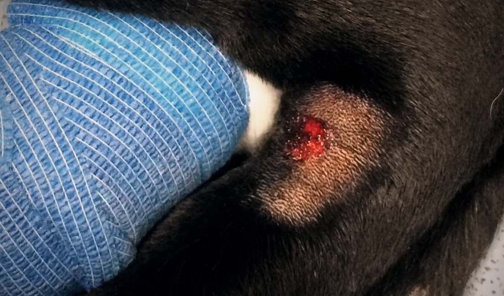
Skin lesion appearance is variable. Lesions range from small, superficial abrasions (0.5cm), to large areas of full-thickness ulceration and necrosis (larger than 30cm) with surrounding erythema and oedema. Skin lesions are often well demarcated and circular, and lesions affecting digits may appear similar to pododermatitis or paronychia.
In one study, most dogs (80.9%) were presented with a skin lesion on a limb or shoulder – the digits being the most common location.
A proportion of dogs remain clinically well and recover from skin lesions uneventfully without developing detectable AKI. In one study, 21.7% of dogs in contact with a dog with CRGV were reported to develop skin lesions without developing AKI.
Clinical signs in dogs that develop AKI can include vomiting, diarrhoea, polyuria/polydipsia, oliguria, anuria, lethargy, anorexia, hypothermia, icterus and petechiae. Neurological signs were identified in 18.6% of dogs during illness and in 1.1% at presentation.
The median time from development of skin lesions to AKI is 3 days (range -4 to 45 days; some dogs develop AKI prior to the development of skin lesions).
The most common haematological abnormality when a patient is presented with AKI is thrombocytopenia (83.9% of patients; median 49 × 103/µL; range in dogs with AKI 0 × 103/µL to 500 × 103/µL); anaemia was present in 28.7% of patients (median 0.32L/L; range in dogs with AKI 0.22L/L to 0.7L/L).
Blood smear examination may identify evidence of microangiopathic haemolysis – signs of fragmentation injury were present in 34.5% of dogs in one study, with schistocytes the most commonly reported morphologic abnormality.
The most common biochemical abnormalities, aside from azotaemia, are hyperbilirubinemia (51.9% of patients with AKI; median 16µmol/L, range 0µmol/L to 603µmol/L) and elevated alanine transaminase activity (71.8%; median 149.5U/L; range 18U/L to 1,722U/L).
Findings on urinalysis include:
Reduced or absent urine production is common in dogs with CRGV that develop AKI (42.6% of dogs at presentation). The aetiology of CRGV remains unknown at this time and common causes of AKI should be considered – and excluded – when presented with a dog that may be suffering from CRGV.
Antemortem diagnosis of CRGV can be challenging because no single, non-invasive diagnostic test is available. A combination of skin lesions and AKI with or thrombocytopenia and/or hyperbilirubinaemia are most likely to help reach a presumptive diagnosis of CRGV.
A definitive diagnosis of CRGV is made by histopathological assessment of the kidneys and/or skin.
The major renal histopathological lesion reported in CRGV is thrombotic microangiopathy (TMA). TMAs are characterised by inflammation and damage to vascular endothelium, leading to widespread formation of microthrombi and resultant consumptive thrombocytopenia.
Dermal histopathology commonly identifies non-specific changes, including ulceration of the epidermis, with coagulative necrosis of the subjacent dermis. Fibrinoid necrosis and thrombosis of the small dermal arterioles (diagnostic for a TMA process) are sometimes observed and, when present, support a diagnosis of CRGV.
As the aetiology of CRGV is unknown, treatment of azotaemic CRGV should be focused on management of AKI and the skin lesions.
Treatment of patients with AKI should be aimed at limiting further renal damage and enhancing cellular recovery, and the most important aspects of therapy are:
The prognosis for dogs with CRGV and clinically significant AKI appears to be poor, although with intensive management some dogs have survived and regained normal renal function.
The question remains as to whether CRGV is an emerging disease or one that was previously unrecognised.