29 Apr 2025
Lotfi El Bahri DVM, MSc, PhD explains the toxic effects of this bacteria found in and around bodies of water, and how veterinarians can administer treatment to pets that have been poisoned.
Lotfi El Bahri
Job Title
Image: Andre Engelhardt / Adobe Stock
Cyanobacteria are also known as blue-green algae due to their algal-like morphology, ability to photosynthesise, and massive growths, with colour ranges from the familiar blue-green, water green or brownish-green scum depending on the type of algae.
They are a group of Gram-negative bacteria commonly growing in fresh, brackish, estuarine and marine ecosystems. Growth is influenced by environmental factors including hot weather (between 20°C and 30°C), high light intensities, turbidity, and high levels of nitrogen and phosphorus due to contamination with fertiliser run-off or direct manure.
Elevation of temperature and increased atmospheric carbon dioxide levels (climate change) would also promote growth and development of cyanobacterial harmful blooms, and may enable their further expansion to new geographical regions.
Toxic blooms are increasing during the past decades in number, frequency and severity throughout the world. In general, 50% to 75% of bloom isolates can produce toxins, often with more than one toxin being present. Not all types of algae are deadly, but it is difficult to tell which are poisonous without specific analysis. It is safest to consider all algae blooms to be dangerous and avoid them altogether.
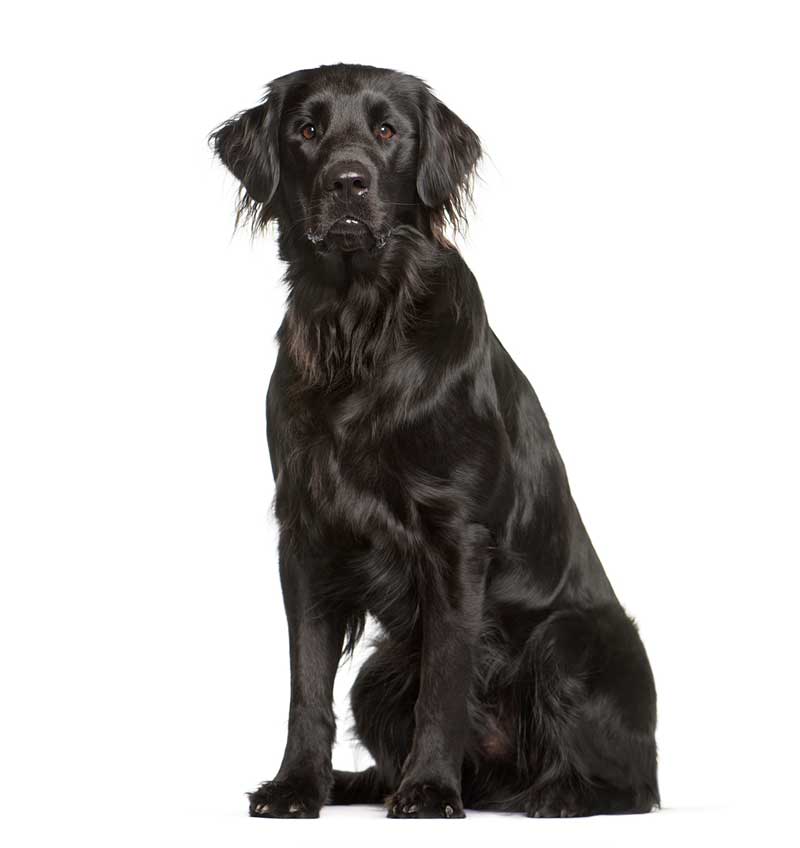
Toxic harmful algal blooms are capable of producing cyanotoxins, highly potent biotoxins implicated in lethal poisonings in dogs. Numerous cases of lakes and ponds contaminated by toxic blue-green algae around the UK have been reported. Imrie reported in August 2022 of poisoning by anatoxin-a in a two year old flat-coated retriever thought to have licked a dead fish, or material close to it, at the water’s edge of Wimbleball Lake on Exmoor in Somerset.
A planktonic cyanobacterium produces several types of cyanotoxins including anatoxins (ATX), microcystins (MCs), nodularins (NODs) and cylindrospermopsins (CYNs). For example, the intraperitoneal median lethal dose (IP; LD50) of anatoxin-a(s) is 20µg/kg in the mouse.
NODs and MCs are extremely toxic compounds with IP LD50 in the mouse ranging from 25µg/kg to 150µg/kg. CYNs have an IP LD50 in the mouse of 2.1mg/kg. Dogs in particular are attracted to blue-green algae. Ingestion of a dose of 60g of bloom algae containing anatoxin-a is sufficient to kill a 25kg dog.
Cyanobacteria toxicosis occurs when dogs drink contaminated water or by accidental ingestion during swimming in contaminated ponds, lakes, rivers, pools and streams by an algal bloom, or by eating toxin-producing algae at the beach. Dogs also lick algae caught in their fur after being in the water. Small exposures, such as a few mouthfuls of algae-contaminated water, may result in fatal poisoning.
Toxicosis can also occur in dogs following ingestion of blue-green algae (Aphanizomenon flos-aquae) health dietary supplements contaminated by MCs.
According to their mechanism of toxicity, three groups of cyanobacterial toxins can be classified: neurotoxins, hepatotoxins and nephro/hepatotoxins.
Anatoxins can be divided into anatoxin-a ([ATX]-a) and anatoxin-a(s) ([ATX]-a[s]): two potent neurotoxins. They are produced by several genera cynobacteria including Anabaena (Anabaena flos-aquae), Plankothrix, Oscillatoria and Aphanizomenon species. The letter “s” in (ATX)-a(s) corresponds to the characteristic symptom of salivation observed in toxicosis.
(ATX)-a, also known as the “very fast death factor” (death within two to seven minutes in mice by IP administration), is a bicyclic amine alkaloid with a small molecular weight (165,23 daltons), fully soluble in water. Its methylated derivative homoanatoxin-a presents very similar properties. (ATX)-a is a potent inhibitor of the enzyme acetylcholinesterase (AChE) by binding to neuronal receptors of acetylcholine. It is a nicotinic (cholinergic) agonist, which irreversibly binds to acetylcholine receptors of neuromuscular junctions and neurons of the CNS.
(ATX)-a is between 7 and 136 fold more potent than nicotine. The toxins mimic the neurotransmitter acetylcholine by activating the flow of sodium ions which induces muscle contraction.
Muscle cells are normally allowed to return their resting state following the degradation of acetylcholine by AChE. However, (ATX)-a cannot be degraded by this mechanism and, therefore, continues to act on muscle cells, producing continuously over-stimulated muscles causing muscular twitching leading to paralysis, respiratory arrest and death. The LD50 orally in mice is 5mg/kg.
At low pH levels, and in the absence of light, (ATX)-a may persist in the aquatic environment for a few weeks. (ATX)-a is rapidly degraded by sunlight and at elevated pH (more than 10).
ATX-a(s) is a guanidine methylphosphate ester (organophosphate), a small molecule (252,21 daltons), soluble in water, which causes prolonged inhibition of peripheral neuronal AChE, but not brain AChE because it is unable to cross the blood-brain barrier. Acetylcholine carries the signal across the gap between nerves and nerves to muscle.
ATX-a(s) inhibits AChE, preventing the removal of acetylcholine, and nerves and muscle become hyperexcited. ATX-a(s) is approximately 10 times more potent than ATX-a. ATX-a(s) is believed to have muscarinic effects on the cardiovascular and respiratory systems. It decomposes rapidly in basic solutions, but is relatively stable in neutral or acidic solutions (pH 3 to 5).
MCs and NODs are both hepatotoxic monocyclic peptides, water soluble, unable to directly penetrate the lipid membranes, with the same mechanism of toxicity. They are the most widespread class of toxins, often implicated in cyanobacterial toxicosis.
MCs mainly produced by Microcystis aeruginosa show several variants of its basic structure, with more than 300 variants ranging in molecular weight from 900 daltons to 1,100 daltons. Furthermore, MCs contain two variable L-amino acids. Among the different variants, the most common and potently toxic are MC-LR, containing the amino acids leucine (L) and arginine (R), and MC-LA, which contains the amino acids leucine (L) and alanine (A).
Particular strains of the cyanobacteria Nodularia spumigena are responsible for the production of NODs. At least 10 variants of NODs are known, ranging in molecular weight from 750 daltons to 850 daltons; the most important of these is NOD-R.
Structurally, the MCs are monocyclic heptapeptides (seven amino acids), while NODs are monocyclic pentapeptides (five amino acids). Both MCs and NODs contain in their structure a common side chain, closely related to their toxicity, a hydrophobic unsatured amino acid known as ADDA (3-amino-9-methoxy-2,6,8-trimethyl-10-phenyldeca-4-6-dienoic acid). ADDA can be used to detect and quantify MCs and NODs independent of the variant type. Total ADDA of MCs and NODs congeners concentrations in the liver, bile, urine, and blood can be determined by using ELISA.
As a result of their cyclic structures, MCs and NODs are extremely stable and resist common chemical breakdown such as hydrolysis or oxidation under conditions found in most natural water bodies. The toxins can break down slowly at 40°C, at either very low pH (less than 1) or high pH (more than 9). MCs and NODs break down slowly in full sunlight – especially when water-soluble pigments are present. These toxins are also resistant to proteases, such as pepsin, trypsin and collagenase. MCs and NODs can even persist after boiling, indicating that cooking is not sufficient to destroy the toxins.
To protect consumers from adverse health effects caused by these hepatotoxins, the World Health Organization (WHO) has proposed a provisional upper limit for MC-LR of 1ppb (µg/L) in drinking water. The WHO has established a tolerable daily intake (TDI) for humans of MC-LR of 0.04µg/kg/day. A TDI is an estimate of the amount of a substance in food that can be ingested every day over a lifetime without appreciable health risk.
Following ingestion, MCs and NODs are absorbed in the ileum and actively enter into the liver. They require active transport mediated by organic anion transporter polypeptides (OATP1B1, OATP1B3, OATP2B1), also known as the bile acid transporters. These transporters are significantly expressed in the liver.
MCs and NODs induce hepatotoxicity through potent specific inhibition by very low concentrations (IC50, or half-maximal inhibitory concentration in nanomolar values) of active sites of serine/threonine protein phosphatases PP1 and PP2A. The primary acute effect of protein phosphatase inhibition is hyperphosphorylation of many cellular proteins, including the hepatocellular cytoskeleton. Excessive phosphorylation induces profound damage to the major components of the cytoskeleton (actin microfilaments, intermediate filaments) on hepatocytes, disruption of liver cell structure leading to intrahepatic haemorrhage or hepatic failure, clotting deficits, inability to maintain adequate blood glucose via conversion from stored glycogen in the liver, hyperkalaemia due to potassium release from damaged hepatocytes, and hypovolaemic shock. The colorimetric protein phosphatase inhibition assay can be also used for detection and quantification of MCs and NODs.
Several planktonic cyanobacteria species can produce CYNs, the most representative being Cylindrospermopsis raciborskii, Umezakia natans, Aphanizomenon ovalisporum, Rhaphidiopsis curvata, Anabaena lapponica and Anabaena bergii. CYNs have a markedly different structure and mode of toxicity than MCs and NODs.
CYN is an alkaloid formed from tricyclic guanidine attached with hydroxymethyl uracil with a polar sulphate group, and highly water soluble, with a molecular weight of 415 daltons. The uracil moiety is required for toxicity.
Two structural variants exist: the epimer, 7-epi CYN, which is relatively more toxic, and 7-deoxy CYN, lacking the hydroxyl group, which is relatively less toxic.
CYNs have significant resistance to high temperatures, sunlight, pH extremes and biological degradation. The kidney is the organ most sensitive to CYNs toxicity following acute exposure. Renal lesions include acute degeneration and necrosis of proximal and distal convoluted tubules and collecting ducts. Toxic effects occur also to the liver, including lipid accumulation within cells followed by hepatocellular necrosis. CYNs also affect a number of vital organs including the heart, lungs and spleen.
The toxicity mechanisms appear to be diverse: irreversible protein synthesis inhibition, binding covalently to DNA (strand breakage), reduced glutathione depletion and reactive oxygen species generation. The LD50 of CYNs indicate a delayed toxicity: 2mg/kg IP in the mouse after 24 hours, but 0.2mg/kg after 5 days.
The clinical signs for cyanobacteria toxicosis will vary depending on the specific toxins in the water. However, a wide range of clinical signs can be present – particularly if different toxins from one or more cyanobacterial species are simultaneously involved.
Clinical signs following oral exposure to neurotoxins include an abrupt onset (within 30 minutes of exposure) of acute neurological signs including: weakness, vomiting, tremors, muscle rigidity, paralysis of the muscles of the hindlegs, seizures, difficulty in breathing, and sudden death due to paralysis of respiratory muscles, within a few minutes of the start of clinical signs.
Following oral exposure to a (ATX)-a(s), clinical signs are similar to those of (ATX)-a, but also include acute onset of excessive secretions (salivation, lachrymation, urination or defecation), convulsions, irregular breathing, and death within one hour. Laboratory values also indicate the decrease of whole blood cholinesterase activity.
Clinical signs following oral exposure to hepatotoxins usually begin within one to four hours, including: lethargy, weakness, pallor of mucous membranes (hepatic haemorrhage), acute vomiting and bloody diarrhoea. Death is due to hypovolaemic shock.
Laboratory values indicate marked elevations of hepatic enzyme activity, including aspartate aminotransferase (AST; normal values between 10U/L and 34U/L), alanine aminotransferase (ALT; normal values between 10U/L and 130U/L), and hyperbilirubinaemia (total bilirubin; reference range in normal dogs 1.71µmol/L to 5.13µmol/L).
In relation with hepatic failure, severe coagulopathy is noted with thrombocytopenia (PLT: reference range of platelet counts in normal dogs between 175,000/µL to 500,000/µL blood) and abnormally prolonged coagulation times such as prothrombin time (PT; reference range in dogs is 11 to 14 seconds) and activated partial thromboplastin time (APTT; reference range 60 to 93 seconds). Hypoglycaemia (reference range of blood glucose levels in normal dogs less than 5.6mmol/L to 13.9mmol/L) and hypoalbuminaemia (reference range of albuminaemia in normal dogs between 2.6g/dL and 3.5g/dL) are also noted.
Clinical signs of nephro/hepatotoxins are similar to hepatotoxins and also include: excessive thirst, increased urination and ataxia.
Laboratory values indicate kidney damage with elevated blood urea nitrogen (reference range in dogs 2.5mmol/L to 9.6mmol/L), serum creatinine (reference range 44µmol/L to 159µmol/L) and low urine specific gravity (normal ranges from 1.001 to 1.080). Laboratory values indicate also liver damage, increase in AST and ALT, severe coagulopathy with thrombocytopenia and prolonged coagulation times such as PT and APTT.
No antidote exists for the toxins produced by blue-green algae. Treatment involves intensive, supportive care focused on affected organ systems.
Most affected animals can develop life-threatening clinical signs or die acutely. Recovery of cyanobacteria poisoning in dogs can take weeks or months. Dogs that do survive from cyanobacteria will usually have chronic liver and kidney disease, as well as other possible complications. Treatment will depend upon the type of cyanobacterial toxin involved.
Controlling seizures: An IV catheter should be placed. To control seizures, diazepam (0.5mg/kg to 2mg/kg IV bolus) should be administered and repeated, if necessary, within 20 minutes (serum half-life in dogs is 2.5 to 3.2 hours) up to three times in a 24-hour period, or 1mg/kg to 2mg/kg rectally. Do not give diazepam intramuscullary. This is contraindicated in patients with severe liver disease.
Alternatively, administer lorazepam (long-acting benzodiazepine 0.2mg/kg IV bolus, because of its high affinity for benzodiazepine receptors in the CNS) or midazolam 0.2mg/kg to 0.4mg/kg IV, which may be repeated once. When IV access is not available, midazolam can be administered by IM because it is rapidly absorbed by this route. Barbiturates and propofol are contraindicated (respiratory depression). Ketamine used alone causes muscle rigidity and could potentially exacerbate seizures, while valproic acid is not recommended (serum half-life in dogs is between 1.5 and 2 hours). Alternatively, administer levetiracetam 20mg/kg IV slow bolus.
If seizures persist or recur, 2% to 2.5% concentrations of isoflurane alone with oxygen can be used. For maintenance, use 1.5% to 1.8% concentrations of isoflurane in oxygen.
Attention to the airway and breathing is paramount. Affected animals should be intubated and provided artificial respiration with oxygen during convulsions. ECG and continuous cardiac monitoring should be performed.
Treating acute respiratory distress: The first treatment is immediate oxygen supplementation by high-flow oxygen therapy (HFOT). HFOT is the administration of warm, humidified oxygen via nasal prongs.
HFOT is an effective method of delivering high volumes and pressures of oxygen. HFOT bridges the gap to mechanical ventilation, is less invasive than ventilation, and is generally more cost effective. It allows the delivery of higher flow rates of oxygen (4L/min to 60L/min with some devices). The flow rate is set to meet or exceed the inspiratory flow demand of the patient.
In normal mesocephalic dogs (for example, the golden retriever, Labrador retriever and German shepherd dog), the average flow demand is approximately 500ml/second to 1,000ml/second. Brachycephalic dogs (for example, the boxer and the bulldog) may have lower flow demands due to naturally occurring airway obstructions.
Controlling muscarinic signs: When (ATX)-a(s) is suspected, administer atropine (sulfate), an anticholinergic agent, to control muscarinic signs such as hypersalivation, increased bronchial secretions and bradycardia. The dosage is 0.2mg/kg to 0.5mg/kg. Atropine can be given repeatedly until cessation of hypersalivation.
Treating vomiting: Severe vomiting should be treated by antiemetics such as maropitant (neurokinin-1 antagonist) 1mg/kg IV every 24 hours. Alternatively, administer a serotonergic antagonist, such as ondansetron 0.5mg/kg to 1mg/kg IV slow bolus (two to three minutes), every 8 to 12 hours; or dolasetron 0.6mg/kg IV slow bolus every 24 hours. Metoclopramide (dopaminergic antagonist) is contraindicated (excitatory effects can occur).
Wash the dog off in fresh water immediately, so the pet doesn’t lick cyanobacteria from the coat.
Owing to the rapid onset of clinical signs, induction of emesis is contraindicated. Activated charcoal is also contraindicated in convulsing or comatose animals.
Treating hepatic failure:
Treating hypovolaemic shock: Hydroxyethyl starch (hetastarch) is a synthetic colloid derived from a waxy starch composed mostly of amylopectin. It expands the circulating plasma volume. The expanded plasma volume may last for 24 hours or longer. Hydroxyethyl starch is available as a 6% solution in saline. It should be infused slowly at a rate of 10ml/kg/day to 20 ml/kg/day.
Treating hypotension: Administer dopamine, an endogenous catecholamine, the immediate precursor to norepinephrine, at 1µg/kg/min to 3µg/kg/min CRI.
Treating hyperkalaemia: Severe hyperkalaemia with ECG changes or symptoms requires rapid correction to prevent significant cardiovascular complications. The treatment of hyperkalaemia requires a combined treatment approach. It is essential to monitor potassium.
Treating GI injury: Administer proton pump inhibitors omeprazole (0.5mg/kg to 1g/kg IV every 12 hours) or pantoprazole (1mg/kg IV).
Wash the dog off in fresh water immediately, so the pet does not lick cyanobacteria from its coat.
Emesis should be induced only if the patient is asymptomatic. Administer apomorphine 0.03mg/kg IV, 0.04mg/kg IM or 0.025mg tablet crushed and dissolved in physiological saline. Instil in the conjunctival sac and rinse with water after emesis has occurred. Alternatively, use xylazine (1.1mg/kg SC or IM).
To prevent further absorption of MCs and NODS from the intestinal tract, use activated charcoal (1g/kg to 4g/kg) mixed with water to make a 20% slurry (1g/5ml water) via a nasogastric tube as soon as possible post-ingestion, and after the airway is secured.
Remove the amount of the hepatotoxins being reabsorbed into the intestines with a high degree of enterohepatic circulation by administration of colestyramine. It is an anion exchange resin which binds with high affinity for bile acids in the intestinal tract and forms an insoluble complex that is excreted in the faeces. Colestyramine comes in a powder form. It is recommended in dogs at an oral dose of 0.3g/kg to 1.0g/kg twice a day for four days. The powder should be given before feeding if possible or mixed with a tasty wet substance such as canned food. Colestyramine has been effective up to seven days post-exposure. It has a wide margin of safety (no systemic effects).
Some human formulations contain xylitol and should not be used in dogs.
The antibiotic rifamycin SV – particularly effective against a variety of Mycobacterium species and Staphylococcus aureus – is a potent inhibitor of all human liver OATPs at therapeutic serum concentrations. Rifamycin SV can be administered to reduce hepatotoxin uptake and enterohepatic toxin circulation in patients suffering from intoxication with an OATP-mediated substrate transport, such as MCs.
Treating dehydration: Intravenous infusion is the preferred means of delivering fluids to severely dehydrated animals. Fluid therapy with IV sterile sodium chloride 0.9% should always be the first-line management.
The volume (ml) of fluid needed to correct dehydration is calculated from the following formula:
Percentage of dehydration × bodyweight (kg) × 1,000
Percentage dehydration can be estimated by examining mucous membrane moisture, skin turgor, eye position and corneal moisture, as described in Table 1.

It is recommended that one-fourth to one-half of the estimated dehydration deficit be replaced over the first two to four hours, with the remaining dehydration deficit and maintenance isotonic volumes administered over the subsequent 20 to 22-hour period. Adequate hydration and urine output (1ml/kg/hour to 2ml/kg/hour) should be ensured.
Treating kidney failure: Administer mannitol, osmotic diuretic, to improve urine output: 0.25g/kg to 1g/kg IV (slow bolus over 10 to 20 minutes) of 20% to 25% solution.
It is necessary to ensure that the patient is rehydrated prior to starting therapy (should be avoided if anuric). Mannitol very easily crystallises at room temperature, so it should be kept in an incubator, and given with an injection filter.
Treating hepatic failure: Similar to hepatotoxin poisoning.
Treating hyperkalaemia: Similar to hepatotoxin poisoning.
Similar to hepatotoxin poisoning.
A nutraceutical is a nondrug substance produced in a purified or extracted form, and administered orally to provide agents required for normal body structure and function. Administration of nutraceuticals may be needed to support waning liver function.
S-adenosyl-methionine, an endogenous molecule produced from the amino acid methionine, has hepatoprotective properties at the recommended dose of 20mg/kg/day orally. N-acetylcysteine (NAC) is rapidly metabolised to cysteine, which is an essential amino acid precursor required for the synthesis of glutathione. NAC can reduce liver damage; use 70mg/kg/day/orally.
Preventing the risk of dog poisoning by cyanobacterial toxins is important: