19 Aug 2019
In the second of a two-part series, Peter Kukadia looks at bacterial infection of the skin and pitfalls when discussing this complex condition with clients.
Peter Kukadia
Job Title
Image: © Willee Cole / Adobe Stock
Part one of this series discussed some potential pitfalls in client communication that may be encountered in general practice. This article will focus on pyoderma and some pitfalls that can be avoided when dealing with worried owners.
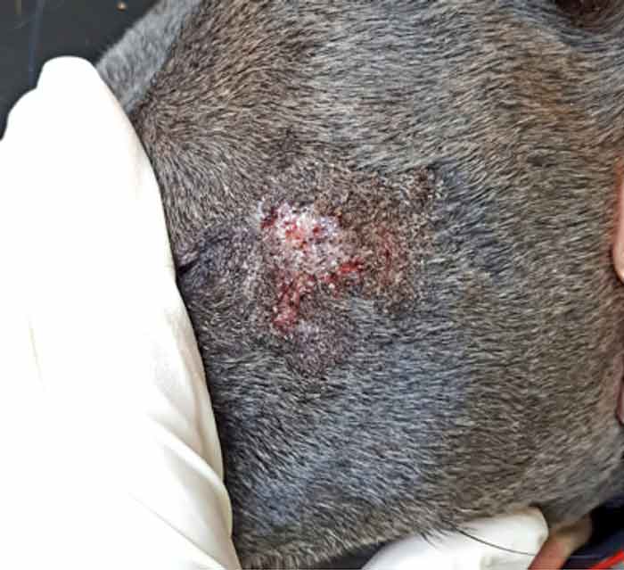
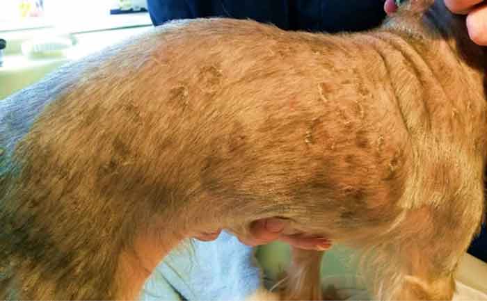
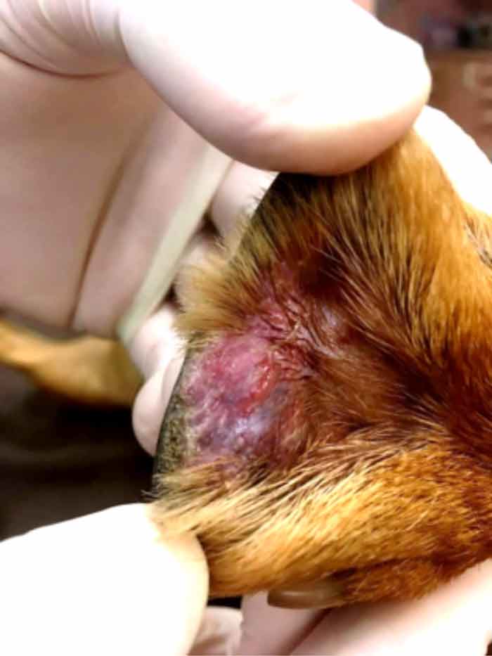
Pyoderma is a bacterial infection of the skin characterised by the formation of pus. It was the second most frequent dermatological presentation to UK practices in one survey (Hill, 2006) and, as alluded to in the aforementioned scenario, owners may raise suggestions in relation to how it should be clinically treated.
A successful approach towards appropriately managing bacterial infection would be dependent on the clinician identifying the characteristic lesions, identifying the anatomical location of the skin that is affected (surface, superficial and deep) and by demonstrating infection by cytology (Loeffler and Lloyd, 2018). On diagnosis, many pets will commence therapy at their first visit to the practice. Some instances will arise, however, where patients will have a considerable delay in their diagnosis and this can be due to concurrent corticosteroid therapy masking pruritus and inflammation (in the author’s experience) and/or due to the infection presenting in an atypical manner (Gortel, 2013).
A “recurrent” infection may be perceived as such, yet it is possible the original pyoderma might not have been completely resolved due to a treatment failure. The former may manifest as an infection that would have completely resolved following antimicrobial therapy, but has recurred weeks or months later. A pyoderma returning a few days following cessation of therapy is more than likely to be due to a treatment failure.
Treatment failures with pyoderma can raise questions of professional competence among some owners, despite the clinician adopting a diligent and empathetic approach. Such feelings may arise from the owner due to a sense of urgency to treat the infection as understandably, there may be concerns about the zoonotic potential. This frustration may be further augmented by the patient continuing to suffer and by more visits to the practice incurring further financial costs.
Treatment failures have been attributed to antibacterial resistance, poor owner compliance (Van Vlaenderen et al, 2011), a failure to address the underlying cause(s) of the pyoderma and inappropriate antibiosis (Ihrke, 2005). With the latter, inappropriate selection, dose and duration of antibiotic therapy has been implicated.
An opinion poll hosted by the author (Kukadia, 2018) revealed about 15% of first opinion practitioners surveyed would prescribe systemic antibiotics for the minimum recommended time duration of three weeks and beyond for superficial pyoderma. This is lower than the 57% of vets in the same survey who stated they would prescribe for a total of 14 days. Some clinicians firmly held the view that only 7 days of antibiotics would be necessary for treating superficial pyoderma, with concerns about resistance if prescribed beyond this time period.
It is absolutely vital clients understand that in companion animals, antibiotic/topical therapy for pyoderma will invariably be of a longer duration than a course of antibiotics given for skin and soft tissue infections in humans. A course of flucloxacillin will typically be prescribed for seven days in humans (National Institute for Health and Care Excellence, 2018; Francis et al, 2016) and based on this, it is reasonable to assume owners may wish to stop medicating their pets seven days or so after commencing treatment in an attempt to prevent overdosing.
Superficial and deep infections should be treated for a minimum of three weeks (with an extra week beyond clinical cure) and six weeks (with an additional two weeks past clinical cure), respectively (Meuller, 2014; Miller et al, 2012). Topical treatment is recommended for surface and superficial infections with deep infection necessitating use of systemic antibiotic therapy.
One pitfall to avoid when managing pyoderma is ignoring the underlying cause for the infection. The implications are that infection will most likely recur with a negative impact on the welfare of the pet.
In accordance with the meteor model, it is wise to set realistic expectations for the owner at the outset, and state that successful resolution of a bacterial infection can potentially take many weeks and investigating the underlying cause for the infection may take longer. In the current economic climate, some clients may feel uneasy about returning for rechecks and consenting to further investigations, while knowing it would be in the pet’s best interests to do so. Aforementioned, this can strain the owner/pet relationship, and may result in the owner being upset and anxious. It would be useful to pre-empt any complaints in this regard by informing them of the costs of the initial and follow-up treatment plans (Panel 1).
For a more comprehensive list, the author directs readers to standard dermatology textbooks. Alternatively, referral to a veterinary dermatologist is advised.
The following is a list of various tests and treatments that may be included on a cost estimate that would be prepared for the client at the first consultation:
* The author considers this to be mandatory trial treatment when working up cases of pyoderma.
** Indications for culture and sensitivity are discussed in an article by Beco et al (2013).
Client and patient compliance should be regarded as an essential consideration when managing pyoderma.
It is important to ascertain the owners’ willingness to medicate their pet as this will invariably affect the quality of life of the animal. Options for treating pyoderma include topical and/or systemic therapy. Of the former, many antiseptic preparations are available in the UK in the form of creams, gels, foams, shampoos, sprays and wipes. These aid to improve patient and owner compliance. In contrast, the range of formulations available for systemic antibacterial therapy are limited to oral antibiotic and injectable therapy. Long-acting injectable cefovecin (a third generation cephalosporin) has been shown to be more effective and cheaper than the use of oral amoxicillin/clavulanic acid in one study (Van Vlaenderen et al, 2011), and its use may help to decrease both owner and patient distress.
The author’s two articles have discussed some of the clinical and non-clinical pitfalls that may occur when managing canine atopic dermatitis and pyoderma in practice. Some owners will fully acknowledge the complexity of dermatological disease, whereas some may need greater communication with regards to managing their expectations.
The human/animal bond is a positive relationship between the owner and their pet, and disruption of this may provide the impetus for any complaints raised with the practice. The meteor model article aims to amalgamate this bond with the clinical factors of the case and aids to improve client communication, which, in turn, leads to positive impacts on case management and patient welfare.
The details given in this article are based on a combination of evidence-based medicine and personal observations made by the author. In the event of a concern raised by a client, readers are advised to follow their practice protocol, which may involve contacting their professional indemnity insurer.
Peter Kukadia
Job Title