18 Nov 2025
Abbe Crawford BVM&S, BSc, PhD, MRCVS outlines the steps vets must take to identify, stabilise, investigate and manage seizure episodes in dogs and cats – a subject she covered in a session at London Vet Show 2025.
Abbe Crawford
Job Title
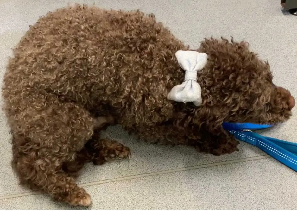
A generalised tonic-clonic seizure in a five year old cross-breed dog, characterised by lateral recumbency, clonic movements of all limbs, jaw chomping, salivation, urination and loss of awareness.
Seizures are a common emergency presentation in both dogs and cats. If left untreated, they can progress to status epilepticus with concurrent hyperlactataemia, autonomic derangements, hypoxia, hypotension, hyperthermia, rhabdomyolysis and irreversible brain injury.
A vital first step is distinguishing seizure activity from other possible causes of an acute neurological presentation, such as a paroxysmal dyskinesia, vestibular dysfunction, systemic weakness (for example, secondary to cardiovascular disease), pain and narcolepsy or cataplexy. A generalised seizure is typically characterised by lateral recumbency, rigid extension (tonic phase) and/or paddling movements (clonic phase) of the limbs, with hypersalivation, urination or chewing movements.
The dog/cat is unaware of its surroundings and cannot be distracted from the seizure. This ictal event is then followed by a postictal phase in which the animal is disoriented and potentially non-visual, ataxic and aggressive. The postictal phase can last anywhere from a few hours to multiple days.
Paroxysmal dyskinesias are increasingly recognised in both dogs and cats, with episodes characterised by abnormal postures, increased muscle tone and often sustained flexion of one or multiple limbs. The dog or cat is conscious and aware of the episode, but often appears anxious. The episodes can be highly variable in duration, lasting from 30 seconds to multiple hours. While the episodes are often unsettling for both the patient and the owner, these are benign, self-resolving events that do not require specific therapeutic intervention, and so it is important that they are distinguished from seizure activity.
Vestibular dysfunction presents with head tilt, leaning or falling to the side (vestibular ataxia) and pathological nystagmus, while systemic weakness is often accompanied by abnormalities on physical examination and an unwell animal. Cataplexy presents with brief episodes of loss of motor tone, resulting in dramatic flaccid paresis and collapse, but no loss of consciousness. Narcolepsy is defined as sudden onset sleep, typically when stimulated or excited (for example, when the patient is fed).

On initial presentation, emergency triage should be performed to assess vital parameters with stabilisation of respiratory and cardiovascular status, as necessary. The first-line emergency treatment for seizures are benzodiazepines. They are potent, safe, and rapidly cross the blood-brain barrier to take effect within one to two minutes of intravenous (IV) administration. Some evidence suggests that midazolam might be more potent than diazepam, and it causes comparatively less severe CNS and respiratory depression1.
Interest is growing in utilising the intranasal route for administration of benzodiazepines – particularly where IV access is challenging to obtain in the actively seizuring patient. Intranasal administration uses the large and highly vascular surface area of the nasal cavity for rapid absorption, and avoids first-pass hepatic metabolism. This is particularly advantageous for benzodiazepines that undergo extensive hepatic metabolism.
Furthermore, direct access to the brain via the olfactory and trigeminal neural pathways has been documented. A study of intranasal administration of midazolam suggested superiority to rectal diazepam in dogs presenting with status epilepticus2. When intranasal administration was compared to IV administration, both routes were found to be rapid, safe and effective for controlling status epilepticus3.
If the seizure activity persists after the initial dose of benzodiazepine has been administered, one to two further doses can be administered. However, further boluses beyond this are unlikely to have a therapeutic effect if the first three have been unsuccessful. Furthermore, a risk of CNS and cardiorespiratory depression exists with repeated administrations. Therefore, an alternative antiseizure medication should be started, such as phenobarbital and/or levetiracetam. It is also important to remember that the anticonvulsant effect of benzodiazepines is only around 20 minutes in duration, and so a longer-acting antiseizure medication should be administered regardless.
Phenobarbital (5mg/kg IV) is an appropriate choice, and further doses of 4mg/kg to 5mg/kg can be given if seizures continue, up to a maximum total dose of 20mg/kg within a 24 hour period. Levetiracetam at a loading dose of 60mg/kg IV can be used as an as an alternative to phenobarbital (for example, in hepatic compromise) or alongside phenobarbital if the seizures are challenging to control. It is safe, well-tolerated and renally eliminated, although costly in the injectable form.
Emergency assessment of blood glucose and electrolytes should be performed, and any significant derangements promptly corrected. Hypocalcaemia and hypoglycaemia are important causes of seizures.
Having stabilised the dog or cat, consideration should be given to identifying and treating any potential underlying cause for the seizures. Both extracranial (such as hepatic encephalopathy, intoxication or hypoglycaemia) and intracranial causes (such as congenital anomalies, neoplasia, or infectious or inflammatory brain disease) of seizures should be considered.
A detailed history from the owner is important to identify any pertinent information, such as possible toxin exposure, travel history, prior seizures and pre-existing neurological deficits. A complete general physical examination should be performed, followed by a complete neurological examination (including evaluation of mentation, posture, gait, postural reactions, spinal reflexes, cranial nerves and palpation). Haematology and serum biochemistry, urinalysis and blood pressure assessment are performed to exclude extracranial causes of seizures.
In the postictal period, obtundation, bilateral menace response deficits and symmetrical gait deficits are commonly detected, but these tend to resolve within hours to days. If lateralised cranial nerve or postural reaction deficits are detected, consideration should be given to structural intracranial pathology, such as infectious, inflammatory or neoplastic brain disease.
General anaesthesia for MRI of the head, with or without cerebrospinal fluid analysis, should be considered in patients in which structural pathology is a concern, as well as in dogs and cats with severe cluster seizures and status epilepticus.
Idiopathic epilepsy is the most common neurological condition in dogs and is defined as recurrent seizures without an identifiable cause. Dogs are typically six months to six years old at seizure onset, normal between seizures, with no abnormalities on neurological examination. Haematology, biochemistry, urinalysis and a bile acid stimulation test are unremarkable, and (where feasible to perform) MRI of the brain is normal, as is cerebrospinal fluid analysis.
Antiseizure medication is the cornerstone of treatment, but only around 15% of dogs with idiopathic epilepsy achieve seizure freedom, and so the goal of treatment is to reduce the frequency and severity of future seizures.
Dogs will typically present with an acute onset of seizures alongside other neurological deficits (such as behavioural changes, obtundation, altered gait and abnormal cranial nerve testing, depending on the site and extent of the structural pathology).
Treatment will depend on the underlying cause, but antiseizure medications are essential for further seizures.
Short courses of antiseizure medications might be necessary while the underlying cause of the metabolic derangement is investigated and appropriate treatment initiated.
We will often use levetiracetam in this scenario, as it is rapidly acting, well tolerated and can be immediately withdrawn (unlike phenobarbital, which should be slowly tapered before discontinuing).

In recent years, autoimmune encephalitis has been recognised as an important cause of seizures in cats.
Affected cats are positive for autoantibodies to the leucine-rich glioma inactivated-1 (LGI1) protein (a protein expressed at synapses). The autoantibodies disrupt synaptic function, resulting in excessive neuronal excitability. Affected cats present with focal seizures with orofacial automatisms (such as lip smacking, chewing and licking movements), hypersalivation, mydriasis and disorientation. Generalised tonic-clonic seizures are also possible.
A diagnostic assay is being developed that can detect the autoantibodies in serum samples. For further information on LIG1 autoantibody encephalitis and how to submit a sample for testing, please visit www.feline-encephalitis.org
Phenobarbital is the first-line treatment. Oral prednisolone can be used alongside phenobarbital in confirmed cases, although it is sometimes reserved for more severely affected cats. A starting dose of 1mg/kg once daily or twice daily is used and then progressively tapered by around 20% every three to four weeks thereafter.
Where no cause for the seizures is identified, treatment is limited to antiseizure medications.
Phenobarbital is the first-line treatment.
Having optimised cardiovascular status, respiratory parameters and body temperature, the patient should be carefully monitored for newly developing electrolyte derangements or changes in vital parameters. An ongoing plan for antiseizure medications is necessary, with regular reassessments for improving postictal deficits. Patients should ideally be monitored in a ward with constant nurse or vet presence. We often place bells on the collar of seizure patients to help alert staff to further seizure activity.
If the patient has been loaded with phenobarbital (that is, has received 20mg/kg within 24 hours), serum levels can be assessed to guide ongoing management decisions. Alternatively, levels can be checked 10 to 14 days after the initiation of oral treatment, once steady state has been reached. Levetiracetam (20mg/kg by mouth three times daily) can be administered alongside phenobarbital until serum levels within the therapeutic range have been documented.
Where loading of phenobarbital is being performed, but the seizure activity is continuing, midazolam or dexmedetomidine can be administered as CRIs and the dose tapered to effect.
Ketamine has been shown to have efficacy in refractory cases (for example, those not responding to phenobarbital or benzodiazepines). It is typically given as an intravenous bolus (1mg/kg to 5mg/kg), followed by a CRI of 1mg/kg/hr4,5.
As a last resort, propofol can be administered as a CRI, but patients can require intubation and ventilation, as well as careful monitoring and optimisation of blood pressure, heart rate and rhythm, rectal temperature and so forth.
Here is a recap of key steps in management of the emergency seizure patient: