
11 Aug 2026
Elisa Falco DVM, MRCVS covers clinical presentation, commonly used medications and more for this condition.

Elisa Falco
Job Title
Image: melounix / Adobe Stock
Canine epilepsy is one of the most frequently encountered neurological disorders in small animal practice and remains a significant challenge for both veterinary clinicians and pet owners.
The condition encompasses a wide spectrum of clinical presentations, underlying causes and therapeutic responses, ranging from isolated, well-controlled seizures to severe refractory epilepsy, with substantial effects on patients and owners’ quality of life.
Advances in veterinary neurology over the past decade have improved our understanding of seizure classification, diagnostic investigation and treatment options, leading to a more comprehensive and multimodal approach to management.
This article reviews the current approach to investigating and diagnosing epilepsy in dogs, and discusses the most commonly used antiseizure medications.
Epilepsy is one of the most common neurological disorders encountered in canine practice, with an estimated prevalence around 0.65% in the general population, but a significantly higher prevalence in specific breeds. A seizure itself is a transient, paroxysmal event caused by abnormal electrical activity within the brain, whereas epilepsy refers to a chronic brain disorder characterised by a persistent predisposition to generate recurrent epileptic seizures.
Because seizures originate from abnormal forebrain activity, animals presenting with epileptic events are considered to have a forebrain neuro-anatomical localisation involving the telencephalon and/or diencephalon.
Seizures can be classified based on their duration and pattern, aetiology and clinical presentation/semiology.
Classification by duration and pattern:
Isolated seizures: a single seizure within a 24-hour period.
Cluster seizures: two or more seizures within 24 hours, separated by a normal interictal period.
Status epilepticus: seizure activity lasting five minutes or longer, or repeated seizures without recovery of consciousness between episodes.
Classification by semiology:
Focal: originating within a localised region of one cerebral hemisphere and may present with motor, autonomic or behavioural signs such as facial twitching, limb jerking or sudden abnormal behaviour.
Generalised: involving both cerebral hemispheres, although many begin focally before secondary generalisation occurs. Generalised seizures may be further classified as tonic-clonic, tonic, clonic, myoclonic and atonic.
Classification by aetiology:
A number of paroxysmal disorders can mimic epileptic seizures, including syncope, narcolepsy/cataplexy, paroxysmal movement disorders, neuromuscular diseases, metabolic collapse episodes, acute vestibular attacks and painful conditions.
Furthermore, epilepsy is not a single disease entity, but rather a heterogeneous condition with multiple underlying causes, seizure patterns and therapeutic responses.
The diagnostic approach to a dog presenting with suspected seizures involves two essential steps:
Obtaining a detailed history remains one of the most important components of the diagnostic work-up. Information regarding seizure frequency, duration, progression and recovery should be collected carefully.
Signalment and age at seizure onset can provide useful diagnostic clues. In one study evaluating a non-referral canine population, structural epilepsy was found to be significantly more likely in dogs presenting with first-time seizures before one year of age or after seven years of age. In contrast, idiopathic epilepsy was statistically more probable in dogs experiencing their first seizure between one and five years of age, particularly when long interictal periods – greater than four weeks – were observed during the early stages of disease.
Although these patterns can help guide clinical suspicion, they should never replace a thorough diagnostic investigation.
Video recordings obtained on smartphones can be extremely valuable in differentiating epileptic seizures from other episodic disorders. Seizures commonly occur during rest, typically last less than five minutes and are often followed by postictal signs (such as disorientation, pacing, lethargy, ataxia, hunger or thirst, among others).
Clinical features supporting an epileptic origin include impaired consciousness, autonomic signs such as salivation, urination or defecation, orofacial automatisms and tonic-clonic movements. Importantly, dogs undergoing a true epileptic seizure are generally unresponsive to external stimuli or owner intervention.
Definitive clinical confirmation of epilepsy would require simultaneous electroencephalographic (EEG) recording and observation of clinical signs. However, EEG remains impractical and not readily available in veterinary practice.
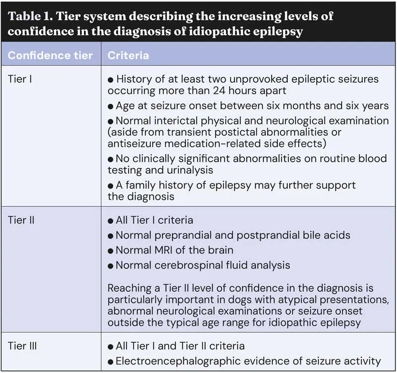
Consequently, IE remains a diagnosis of exclusion and is based on a combination of clinical history, neurological assessment and diagnostic investigation. To help standardise the diagnostic approach, the International Veterinary Epilepsy Task Force developed a three-tier system describing increasing levels of confidence in the diagnosis (Table 1).
Managing epilepsy in dogs is often a lifelong commitment requiring close collaboration between the veterinary team and the owners.
Successful long-term seizure control depends not only on selecting the appropriate antiepileptic drug (AED), but also on having realistic expectations, regular monitoring and maintaining a balance between seizure control and side effects of the medications.
Owners should be prepared for the demands associated with chronic epilepsy management, including daily medication administration, periodic blood testing and follow-up consultations. In some cases, emergency treatment may also be required, for management of cluster seizures or status epilepticus.
The decision to initiate AED therapy should be individualised and based on several factors, such as seizure frequency and severity, underlying aetiology, risk of recurrence and the potential impact on the patient’s welfare. Current recommendations suggest treatment should be considered in dogs presenting with:
Early therapeutic intervention may improve long-term seizure control. Importantly, the goal of therapy is not always complete seizure freedom. In many cases, treatment success is defined as a reduction in seizure frequency of at least 50% while minimising adverse effects. However, depending on the seizure frequency, a 50% reduction might not reflect a significant improvement in the dog and owners’ quality of life.
For dogs with newly diagnosed epilepsy, monotherapy remains the preferred initial approach whenever possible. However, combination therapy may eventually become necessary. Drug selection should be individualised according to seizure type, patient characteristics, comorbidities, owner’s compliance and financial considerations.
Phenobarbital remains a first-line AED in veterinary medicine for its efficacy, availability and relatively low cost.
Several studies have demonstrated good long-term efficacy, with more than 80% of dogs achieving at least a 50% reduction in seizure frequency, while approximately one-third may become seizure-free.
The drug is rapidly absorbed following oral administration and undergoes hepatic metabolism. A typical starting dose is 2.5mg/kg to 3mg/kg orally every 12 hours.
In emergency settings, to rapidly achieve therapeutic serum concentrations, a loading dose can be administered. This is done by administering multiple oral, intravenous or intramuscular doses up to a maximum of 24mg/kg (3mg/kg every 4 hours up to a total of 6 to 8 doses). Subsequent doses should not be given more frequently than every 30 minutes to reduce the risk of causing respiratory depression.
Therapeutic drug monitoring is essential during phenobarbital therapy, as hepatic enzyme induction can alter drug clearance over time. Serum concentrations are generally evaluated two to three weeks after initiating treatment, or changing the dose, then every three to six months during long-term management, together with haematology, biochemistry and bile acid stimulation test.
Monitoring is also recommended whenever worsening of the seizure control or side effects are observed. Phenobarbital serum levels should be maintained at less than 35µg/ml to reduce the risk of hepatotoxicity.
Common adverse effects include sedation, ataxia (usually improving in first few weeks of therapy), polyphagia, polyuria and polydipsia. Elevations in liver enzymes (particularly alkaline phosphatase) are also frequently observed and do not necessarily indicate clinically significant hepatopathy.
Potassium bromide is commonly used as an adjunctive therapy in dogs with inadequate seizure control on phenobarbital alone, although it may also be used as monotherapy in selected cases.
Its use can be particularly useful in dogs with underlying hepatic disease, as bromide is excreted unchanged by the kidneys and does not undergo hepatic metabolism.
The recommended starting dose, administered once daily or divided into two doses, is 40mg/kg/day when used as monotherapy and 30mg/kg/day when combined with phenobarbital. However, for achieving an appropriate potassium bromide serum level quicker, it can be loaded administering 600mg/kg, usually divided into multiple daily doses over three to five days then reducing it to the described maintenance dose.
Care should be taken regarding possible gastrointestinal secondary signs, which can be limited by giving potassium bromide with food.
As bromide has a very long half-life, steady-state concentrations are only reached after several weeks of treatment. Serum bromide concentrations should, therefore, be monitored approximately 12 weeks after starting therapy or changing the dose, then every 6 to 12 months. Target serum concentrations are generally 1mg/ml to 3mg/ml for monotherapy, and 1mg/ml to 2mg/ml when used alongside phenobarbital.
An important clinical consideration is the influence of dietary chloride intake on bromide concentrations. High-chloride diets tend to increase renal bromide excretion, resulting in reduction of bromide serum levels and potentially severe seizures. For this reason, sudden and frequent dietary changes and access to sea water should be avoided.
Common adverse effects include sedation, ataxia, pelvic limb weakness, polyphagia, polyuria and polydipsia and vomiting. Less common adverse effects include megaoesophagus, behavioural changes and dermatological reactions.
Imepitoin has the potential to be used as a first-line AED in dogs with recurrent single epileptic seizures, tends to have a more favourable side effect profile compared to other AEDs, and does not require monitoring. However, it is not recommended as an add-on AED or for treatment of cluster seizures and status epilepticus. Imepitoin is administered twice daily at dosages ranging from 10mg/kg (starting dose) to 30mg/kg.
Levetiracetam is widely used as an adjunctive AED and is increasingly selected in patients with hepatic compromise because it undergoes minimal hepatic metabolism and has a rapid onset of action.
This drug can be rapidly and completely absorbed after oral administration, and it has a wide safety margin.
The standard dose is approximately 20mg/kg to 30mg/kg every eight hours. A starting loading dose of 60mg/kg is often administered. Levetiracetam appears to be effective in preventing cluster seizures when administered as “pulse therapy”. It is given intravenously (in hospital settings) or orally (at home, as soon as the dog can safely swallow following the first seizure) at a dose of 60mg/kg, and then administered every 8 hours at the dose of 30mg/kg for 48 hours before treatment is discontinued (as long as seizures have stopped).
Levetiracetam is generally well tolerated, although some dogs may experience mild sedation or rarely gastrointestinal side effects or behavioural changes.
Zonisamide is a newer-generation AED that appears both effective and well tolerated in many epileptic dogs – particularly those refractory to first-line therapies. The recommended starting dose is 5mg/kg every 12 hours, but the dose should be increased to 10mg/kg every 12 hours when combined with phenobarbital.
Potential adverse effects include sedation, ataxia, vomiting, inappetence and keratoconjunctivitis sicca.
Overall, zonisamide has a relatively high safety margin and is often well tolerated, even during long-term administration. However, it is metabolised by the liver, and when administered in combination with phenobarbital, it can slow down its hepatic metabolism, leading to an increase in phenobarbital levels. For this reason, it is essential to monitor phenobarbital serum levels two weeks after starting treatment with zonisamide or increasing its dose.
Benzodiazepines play a central role in the emergency management of seizures – particularly cluster seizures and status epilepticus. However, their oral chronic use is not advisable due to their short half-life.
Diazepam remains one of the most commonly used emergency anticonvulsants in veterinary medicine and can be administered intravenously at 0.5mg/kg, or per rectum at approximately 0.5mg/kg to 2mg/kg, making it the most commonly used emergency treatment at home. The dose may be repeated up to 3 times within 24 hours if necessary, although repeated boluses are better avoided, and midazolam CRI should be preferred; in fact, most diazepam formulations should not be administered as a CRI due to its tendency to adsorb into plastics, reducing the actual dose delivered of the patient. Midazolam is increasingly favoured in emergency settings, as it can be administered as a bolus intravenously, intramuscularly or intranasally at approximately 0.3mg/kg, and as a CRI up to 0.5mg/kg/hr.
Prolonged seizure activity can result in severe systemic complications including hyperthermia, hypoxia, cardiovascular complications and permanent neuronal injury.
First-line emergency treatment at home involves the use of intranasal midazolam and intrarectal diazepam. These should be administered promptly, and the aim is to reduce the length and severity of the seizure activity the patient is experiencing, and to prevent further seizures occurring.
Cluster seizures and status epilepticus are neurological emergencies, and should instead be managed in a hospital setting. Intravenous administration of midazolam or diazepam remains the first-line treatment in these patients.
If seizure activity persists or recurs after the initial benzodiazepine bolus, a second bolus may be administered after an interval of about two minutes. The need of repeated boluses should lead to prompt initiation of a midazolam CRI. Administration of longer-acting AED should also be considered promptly; levetiracetam and/or phenobarbital are often used in emergency settings and continued as maintenance treatment.
Third-line therapy involves the use of anaesthetic agents for refractory seizure activity. Treatment protocols may include boluses, followed by CRI administration, of ketamine, dexmedetomidine or propofol. Inhalant anaesthesia can also be considered. Appropriate airway control and cardiovascular support is essential in these situations.
Therapeutic drug monitoring plays an important role in epilepsy management and can help clinicians optimise treatment while minimising adverse effects.
Serum drug concentrations are generally assessed:
Although many dogs initially respond well to antiseizure medication, long-term seizure control can be challenging in some cases. Treatment failure may result from several different factors, including disease-related factors (such as progression of intracranial pathology, incorrect diagnosis, comorbidities, newly developed structural brain disease), drug-related factors (such as inadequate drug selection, inadequate dosing, development of pharmacological tolerance or altered drug metabolism, drug interactions), and patient or owner-related factors (such as poor compliance).
Approximately 20% to 30% of canine epileptic patients are considered refractory to treatment, highlighting the importance of regular monitoring and reassessment throughout the course of the disease.
Elisa Falco graduated from the University of Teramo in 2022. Since graduating, she has gained experience in multiple veterinary centres across Europe and Australia, including roles in France, Queensland and the UK, where she completed a rotating internship in a multidisciplinary referral centre in the east midlands. Elisa is currently undertaking a neurology and neurosurgery internship at DWR Veterinary Specialists. She aims to pursue specialist training in veterinary neurology, with a particular interest in neurosurgery.