23 Jun 2026
Ian Wright BVMS, BSc, MSc, MRCVS provides a comprehensive overview of the key disease risks vets need to be aware of – particularly when assessing imported pets
Ian Wright
Job Title
Image: CineLens/peopleimages.com / Adobe Stock
Over the past few years, the number of legally and illegally imported pets entering the UK has steadily increased. Part of this trend has been driven by pets being rescued from abroad and then being rehomed in the UK.
This, in combination with expanding global parasite distributions, increases the risk of exotic parasites being introduced.
This has led to a veritable rogues’ gallery of exotic parasites being identified in UK dogs. Some of these are curiosities, some pose significant risk to the individual patient and some are zoonotic.
Leishmania species are intracellular protozoan parasites of a wide range of animals. Different Leishmania species are endemic in parts of Europe, South America, Africa and Asia, with canine leishmaniosis being predominantly caused by Leishmania infantum in Europe.
L infantum can also infect humans and felids. Transmission occurs primarily through the bites of infected phlebotomine sand flies, but a range of other non-vectorial routes of transmission have also been demonstrated. These include blood transfusion, congenital and venereal routes.
Dog bites and close contact with ulcerative lesions are also suspected as routes of transmission (Naucke et al, 2016; McGrotty et al, 2023).
Reviews of UK veterinary records have indicated a prevalence of between 0.007% and 0.04% of dogs confirmed with clinical leishmaniosis, and given the difficulties in diagnosis and subclinical carriers, this is likely to be significantly underestimated (Shaw et al, 2009; Silvestrini et al, 2016).
A more recent paper examining Small Animal Veterinary Surveillance Network (SAVSNET) data on Leishmania testing in UK cats and dogs found Leishmania antibodies detected in 39.7% of tested dog samples.
In 368 dogs with leishmaniosis were identified from clinical narratives and, of these, 189 had either visited or were rescued/imported from Spain, Greece, Cyprus and other southern European countries.
Dogs aged between three and six years of age were found to be 4.71 times more likely to have leishmaniosis than those two years or younger, and increased risk between 2017 and 2022 was found compared to 2014 (Checa et al, 2025).
Horizontal transmission has been recorded in the UK in the absence of the sand fly vector (McKenna et al, 2019; Wright and Baker, 2019; McGrotty et al, 2023).
The signs associated with Leishmania infection are immune mediated and in dogs commonly include lymphadenopathy, anorexia, weight loss, dermatological signs (alopecia, dermatitis, hyperkeratosis, dermal ulcers), ocular signs (conjunctivitis, uveitis) and epistaxis without thrombocytopenia. Thrombocytopenia and anaemia, polyarthritis and splenomegaly can also occur. Immune complexes in the kidneys lead to glomerular nephritis.
Many Leishmania infections are subclinical, with highly variable periods occurring from initial infection to development of clinical leishmaniosis. Clinical signs can develop months or years after initial exposure.
Zoonotic transmission occurs via the sand fly vector, and the risk canines pose to humans from non-vectorial transmission routes is extremely low.
Good hygiene should be practised, however, around infected dogs with active skin lesions, and close contact with the severely immunosuppressed should be avoided. Care should also be taken with contaminated needles from blood draws or cytology sampling.
Risk of onward transmission and UK establishment
Despite the absence of the sand fly vector, the risk of UK Leishmania transmission and transient endemic foci developing due to non-vectorial routes is significant.
Breeding infected animals is the most likely source of this transmission, but the potential for horizontal transmission through close association or dog bites also needs to be considered. If large numbers of dogs are kept together in the UK or are kept in densely canine-populated areas, then the odds of transmission by these routes will increase.
This is a concern, as many of the Leishmania infected dogs in the UK are in urban areas of the south of England, with large numbers of positive dogs socialising together (Shaw et al, 2009; Silvestrini et al, 2016).
As the climate warms, the possibility also exists of sand fly vectors establishing in the UK in the future (Koch et al, 2017).
Minimising the risk of dogs becoming infected and onward transmission requires a combination of preventive measures.
Health checks and screening relevant dogs with relevant histories. These include dogs that have been imported and those with clinical signs and dogs that have links with imported dogs (breeding or close contact). Dogs with a travel history and relevant clinical signs should also be tested. Quantitative serology is useful as a screening tool and to then track courses of infection. PCR from affected skin and lymph nodes is also useful. Blood PCR can carry a relatively low sensitivity, but conjunctival swab PCR may be useful as an adjunct to serology. Because Leishmania infections are known to have long incubation periods, and no test is 100% sensitive, owners should be advised to re-test their imported pets for infection six months after importation and ideally for at least two years after importation.
Neutering positive dogs. This prevents congenital or transplacental transmission.
Screening blood donors. This should be focused primarily on dogs with travel history, but an argument can be made for screening all dogs to be used as donors, as breeding and travel histories may be lost.
Protecting dogs travelling to endemic countries. The use of products containing pyrethroids with licensed sand fly repellent will reduce the risk of transmission. Treatment should be applied one week before travel, as pyrethroids take time to reach full distribution and activity. No fly repellent, however, is 100% effective, and so other preventive measures, such as avoiding activity outside at dawn and dusk, sleeping upstairs and camping in breezy locations, will also reduce bite risk. Vaccinations are available in some European countries to reduce the risk of disease, but currently none are available in the UK.
Dirofilaria immitis is a filarial heartworm primarily of dogs, but also can infect ferrets and felines. Transmission occurs via the bites of infected mosquitoes, and the parasite is present in parts of Africa, Asia, North America and South America.
In Europe, it has been limited to southern and parts of eastern Europe, but due to climate change and increased dog movements, is now spreading further north and eastwards.
Although D immitis is not endemic in the UK, cases are being seen by vets in dogs with travel history. European Scientific Counsel Companion Animal Parasites (ESCCAP) case recording maps currently show 1.95% of UK canine samples submitted to IDEXX testing positive (tinyurl.com/4jp49zct).
In a large group of illegally imported dogs seized by the RSPCA, 4.1% were found to be positive for D immitis (Wright et al, 2023).
D immitis is a significant cause of heart disease. Infections may remain subclinical for months or years before potentially severe signs develop associated with chronic heart changes, adult worm death, thromboembolism, and inflammatory responses associated with larval worm migration.
Although aberrant migration of larvae can occur in human hosts, zoonotic exposure is through the feeding of infected mosquitoes; therefore, no zoonotic risk from D immitis exists in the UK unless it becomes endemic in the future.
Mosquitoes capable of transmitting D immitis can be found throughout the whole of Europe, including the UK, but the climate in northern and central Europe has been too cold to allow D immitis to complete its life cycle in the mosquito year on year.
Climate change in combination with increased movement of pets, however, is allowing the parasite to spread. A 2024 paper highlighted cases in untravelled dogs from Estonia, the furthest north in Europe that they have been reported (Mõttus et al, 2024).
It is likely that if positive dogs continue to enter the UK, then endemic foci will establish in the next few years.
Pets visiting endemic countries require a monthly licensed macrocyclic lactone to prevent infection. Rapid diagnosis in imported pets is important to manage infection and, as temperatures warm, to minimise mosquito exposure and the risk of UK establishment.
Diagnostic tests available for screening pets entering the UK include antigen serology and the Knott’s test for microfilariae detection in the blood. Because these tests require adult worms to be present, and these take time to develop post-infection, it is advised that pets be tested again six to nine months after UK entry.
Dirofilaria repens is a closely related parasite to D immitis, but with adult worms living subcutaneously. It is currently not endemic in the UK, but cases have already been reported in dogs with a travel history (Wright, 2017; Agapito et al, 2018; Panarese et al, 2023).
Many infections are well tolerated, but disease can occur, with dermatological signs being most common. These can include nodular or papular dermatitis, pruritus, erythema, papules, alopecia, crusting, distinct nodules, acanthosis and secondary pyoderma.
Ocular signs may also be present.
D repens has zoonotic potential and can cause creeping eruptions and conjunctivitis in people. Zoonotic exposure occurs through bites from infected mosquitoes, so is not a concern in the UK unless endemic establishment occurs.
Mosquito vectors capable of transmitting D repens are currently present in the UK and can complete their life cycle at lower temperatures than D immitis (Morgan, 2016).
As a result, if infected dogs continue to enter the UK and are not treated promptly, a likelihood exists that endemic establishment will occur.
Pets visiting endemic countries require a licensed monthly macrocyclic lactone to prevent infection. Rapid diagnosis in imported pets is important so treatment can be initiated to minimise mosquito exposure and the risk of endemic establishment.
Dogs with import history should, therefore, be screened for the parasite. If adult worms are present, they should be sent to the APHA’s exotic worm surveillance scheme (tinyurl.com/mrxwp8mn). Otherwise, the Knott’s test should be used to look for microfilariae in the blood.
Thelazia callipaeda is a vector-borne nematode that resides in the conjunctival sac, lacrimal duct and tear glands in a wide range of mammalian hosts including cats, dogs and humans.
The worm has rapidly spread across Europe this century, driven by increased pet movement and the spread of its fruit fly vector Phortica variegata.
The fly transmits the parasite when feeding on lacrimal secretions. Although the worm is not currently endemic in the UK, cases in UK dogs with a history of travel have been recorded (Graham-Brown et al, 2017), most recently from the Balkans (tinyurl.com/bddkar6n).
Infection can be well tolerated, but if left untreated can lead to conjunctivitis, keratitis, epiphora, eyelid oedema, corneal ulceration and, in serious cases, blindness.
T callipaeda has zoonotic potential and can cause ocular complications in human hosts. Zoonotic infection occurs through exposure to infected fruit flies, so is not a concern in the UK unless endemic establishment occurs.
P variegata has been recorded in the UK, with climatic modelling suggesting conditions favourable for spread and, if exposed to infected dogs, could lead to endemic establishment.
No licensed product is available for the prevention of T callipaeda when travelling to endemic countries, but it is likely that macrocyclic lactones employed for D immitis prevention will provide some protection.
Rapid diagnosis in imported pets is important so treatment can be initiated to minimise fruit fly exposure and improve prognostic outcomes. Dogs with import history should be checked for signs of ocular discomfort and pathology, and if present should be examined carefully for worms. If adult worms are present, they should be sent to the APHA’s exotic worm surveillance scheme.
Onchocerca lupi is a filarial nematode affecting dogs, with potential to also infect felids and humans. It has a widespread distribution, being found in continental Europe, Africa, the Middle East and North America. Adult worms are typically found in periocular nodules, while microfilariae are located in the skin. The vector for this species of Onchocerca is uncertain, but Simulium species (black flies) are vectors for other species and, therefore, likely to play a role.
Cases have rarely been recorded in the UK, but two recent cases demonstrates the need to be vigilant for the parasite in imported dogs.
Some infected animals remain subclinical, with infection well tolerated. However, in some patients, a wide range of ocular signs may develop, including conjunctivitis, periorbital swelling, exophthalmia, blepharitis, photophobia, uveitis and corneal oedema and/or ulcers.
Enucleation is sometimes required in severe cases.
Zoonotic infections with orthopaedic, spinal and ocular lesions in humans have been reported. These cases arise from exposure to infected vectors, however, so this is not a concern in the UK unless endemic establishment occurs.
Assuming Simulium species are vectors, then these are present in the UK. It is not known what conditions are required for transmission, but it should be assumed that UK transmission and endemic establishment are possibilities.
No licensed product is available for the prevention of O lupi when travelling to endemic countries. It is possible that macrocyclic lactones may provide some protection. Rapid diagnosis is essential in imported pets and pets with travel history and relevant clinical signs. This will improve prognostic outcomes for the patient and minimise the risk of vectors being exposed.
Dogs with import history should be checked for signs of periocular swelling, ocular signs and discomfort. Worms may be visible in, on or around the eye, and may be extracted from nodules.
Skin snips are required for microfilariae visualisation, but this is impractical as a routine screening test.
Linguatula serrata (also known as “tongue worm”) is a nasal pentastomid that resides in the nasopharynx of carnivores including canids and humans.
Eggs are passed in nasal secretion and faeces, where they may be consumed by a wide range of intermediate hosts including humans. Encysted nymphs may then develop in a range of tissues such as lymph nodes, liver and lungs.
The parasite is found widely throughout eastern Europe and the Middle East. Cases have been reported in UK dogs in recent years, with one having no history of foreign travel (Mitchell et al, 2016; Villedieu et al, 2017; Macrelli and Macintosh, 2022).
Many cases in dogs and cats are subclinical. However, large burdens can lead to rhinitis and nasopharyngitis, with associated clinical signs such as sneezing, coughing, purulent nasal discharge and epistaxis.
Humans living in or visiting endemic countries can act as definitive hosts through consumption of undercooked meat or offal.
In the UK, zoonotic risk comes from humans acting as intermediate hosts if eggs from nasal secretions or faeces are accidentally consumed. This risk is greatest in those living with infected pets.
A recent untravelled UK case and historic reports of L serrata in UK wildlife has led to the suggestion the parasite may have endemic foci in the UK (Campbell and Jones, 2023). This case, however, had been fed imported food and, therefore, the source of infection may not have originated in the UK.
If intermediate hosts such as wildlife or livestock, however, are exposed to eggs shed from infected pets, then endemic establishment is a likely scenario in the future.
Rapid identification of infected dogs will minimise both zoonotic risk and the exposure of intermediate hosts to eggs. Dogs with relevant clinical signs – especially those with travel history – should be examined for the parasite. Nasal secretions or faecal samples can be examined using flotation techniques for eggs, but are relatively insensitive.
Diagnosis is most commonly made by gross identification of adult parasites expulsed or discovered by rhinoscopy. If adult worms are present, they should be sent to the APHA’s exotic worm surveillance scheme.
Of a wide range of exotic tick-borne pathogens found in UK dogs with travel history, Ehrlichia canis (transmitted by Rhipicephalus species ticks, not currently endemic in the UK) is the most commonly reported to ESCCAP, and Babesia canis (transmitted by Dermacentor reticulatus ticks, currently focally endemic in the UK) is the most likely to become persistently endemic.
An outbreak of babesiosis occurred in dogs from Essex between November 2015 and February 2016 when four cases of babesiosis caused by B canis were identified in untravelled dogs. An endemic focus of B canis in Essex was subsequently confirmed (Phipps et al, 2016; Hansford et al, 2016).
A recent survey of D reticulatus ticks in the UK, however, has found the Essex foci not to have persisted, but a positive tick was found in the south-west of England (Sands et al, 2022). B canis is, therefore, currently thought to be absent or transiently endemic in the UK.
A recent study looked at 76 dogs diagnosed with tick-borne diseases in the UK between 2005 and 2019 (Silvestrini et al, 2023). Twenty-five were diagnosed with ehrlichiosis and 23 with babesiosis. All of these dogs had travel history with the exception of three dogs with ehrlichiosis and one with B canis. The sources of these infections are unclear.
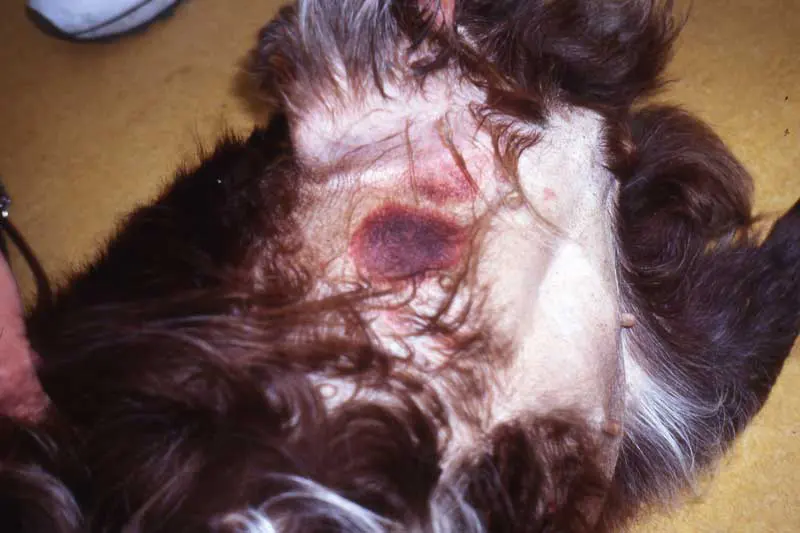
Many clinical tick-borne infections will present acutely with lymphadenopathy and pyrexia, including E canis and B canis. Babesia species infection can lead to immune-mediated haemolytic anaemia in dogs, with severe cases presenting with dark brown urine associated with haemoglobinuria (Figure 1).

Immune-mediated thrombocytopenia can also occur with spontaneous bleeding or bruising (Figure 2).
Infection with E canis can lead to acute disease with pyrexia, anorexia and lymphadenopathy. A subclinical period follows where the organism is either eliminated or a chronic infection develops with the potential for chronic disease to develop. The chronic form carries a poorer prognosis, with leukopenia, thrombocytopenia and associated immune suppression and bleeding disorders. Both acute and chronic ehrlichiosis may present with thrombocytopenia and central nervous signs including ataxia, seizures, paresis, hyperaesthesia, cranial nerve deficits and vestibular signs. Chronic ehrlichiosis may occur months or years after UK entry.
B canis is not considered a zoonosis. The risk to humans from E canis is unclear (Ebani, 2026), but even if it is a significant pathogen, the Rhipicephalus vectors are not currently endemic in the UK.
If R sanguineus establishes in UK households, however, then transmission of a range of zoonotic pathogens including Rickettsia species may occur and this possibility needs to be considered.
D reticulatus ticks are the European vector for Babesia canis. Field surveys conducted in England and Wales during 2009-16 confirmed the presence of D reticulatus in four main areas: western Wales, north Devon, south Devon and Essex. Unlike mainland Europe, these populations have remained mainly static, although a paper last year suggested some localised expansion in south-eastern foci (Biddlecombe et al, 2025). The presence of D reticulatus endemic foci in England and Wales makes the UK susceptible to endemic pockets of B canis developing.
R sanguineus is not currently endemic in the UK, as the climate is currently not favourable for establishment. The tick is endemic in southern and eastern Europe, and moving northwards as climatic conditions become more favourable.
This means that, in the future, endemic populations may establish, with the potential for the transmission of a wide range of tick-borne pathogens including E canis.
R sanguineus has the potential to establish in centrally heated households in a similar way to fleas. Cases of R sanguineus ticks imported on dogs from Spain, Greece and Africa with subsequent household infestations have all been reported to ESCCAP UK and Ireland in the past few years, so owners of pets with travel history need to be vigilant for this possibility.
Vigilance for relevant clinical signs of babesiosis is important in dogs entering the UK from abroad and those from areas endemic for D reticulatus ticks.
Similarly, E canis should be considered as a differential in dogs exhibiting relevant clinical signs with a travel history.
Tick prevention is essential for pets travelling abroad and those that have recently arrived in the country.
In addition, pets should be checked for ticks after outdoor activity and as soon as possible after arrival in the country.
Ticks found in homes should also be kept for identification. All ticks can be sent to the UK Health Security Agency’s tick identification scheme (tinyurl.com/4t6cz84t), where they can be identified and also contribute to surveillance data.
Further information on parasites and distribution maps can be found on ESCCAP UK and Ireland’s website (www.esccapuk.org.uk). Case recording maps are also available on the ESCCAP website to see which parasites are being recorded in what numbers in different parts of the UK (tinyurl.com/3nt53ptx).
Further information on diagnosis, treatment and management of Leishmania and heartworm can be found via the LeishVet (www.leishvet.org) and the American Heartworm Society (www.heartwormsociety.org) websites, respectively.
The trend of importation of rescue dogs from abroad with exotic parasites is only likely to grow over the coming months and years.
While as a profession, vets have a role to play in educating the public regarding the disease risks of importing dogs from abroad, veterinary professionals also have a vital role in assessing imported dogs for evidence of parasitic infection and putting effective preventive measures in place.
Ian Wright is a practising vet and co-owner of The Mount Veterinary Practice in Fleetwood, Lancashire. He has a master’s degree in veterinary parasitology and is chairman and director of the European Scientific Counsel Companion Animal Parasites (ESCCAP), as well as guidelines director for ESCCAP Europe.