
22 Sept 2026
Bryn Jones BVM, BVS, CertAVP, DipECVIM-CA, MRCVS defines and differentiates two serious diseases in cats, in the first part in this series.

Bryn Jones
Job Title
Image: Krakenimages.com / Adobe Stock
In cats presented with chronic gastrointestinal (GI) signs, after exclusion of extra-GI causes, the two key differentials are chronic inflammatory enteropathy (CIE) and low-grade intestinal T-cell lymphoma (LGITL).
Differentiating the two diseases is challenging, sometimes even when extensive investigations are performed.
In 2023, an ACVIM-endorsed statement was released suggesting guidelines on how LGITL and lymphoplasmacytic enteritis (LPE, the most common form of CIE) can be diagnosed and distinguished, adding some clarity to a complex topic1.
However, many difficulties remain in managing cats with chronic enteropathies, either because investigations are not available or pursued, because results are still hard to interpret or because achieving a diagnosis does not always predict how a patient will respond to treatment.
In the first part of this article, we will look at what these two diseases are, and the various tests we can perform to try to separate them in our patients.
The term chronic enteropathy (CE) is inconsistently applied, but is most commonly used for cats that have been showing suspected GI signs (such as weight loss, altered appetite, vomiting or diarrhoea) for at least three weeks and where extra-GI, metabolic, infectious and/or large structural disease (such as solid tumours) have been excluded as much as possible2,3,4,5,6.
It is an umbrella term applied to patients that have not undergone biopsy and received a histopathological diagnosis, but some degree of investigations (such as basic blood tests with or without abdominal imaging) have been performed.
What is embraced under the term is debatable, but usually includes CIE, food-responsive enteropathy (FRE), inflammatory bowel disease (IBD)/steroid-responsive enteropathy (SRE) and LGITL (Figure 1). The term CE should, therefore, be understood as a syndrome rather than a specific diagnosis, implying that further testing would be required to delineate the underlying cause.
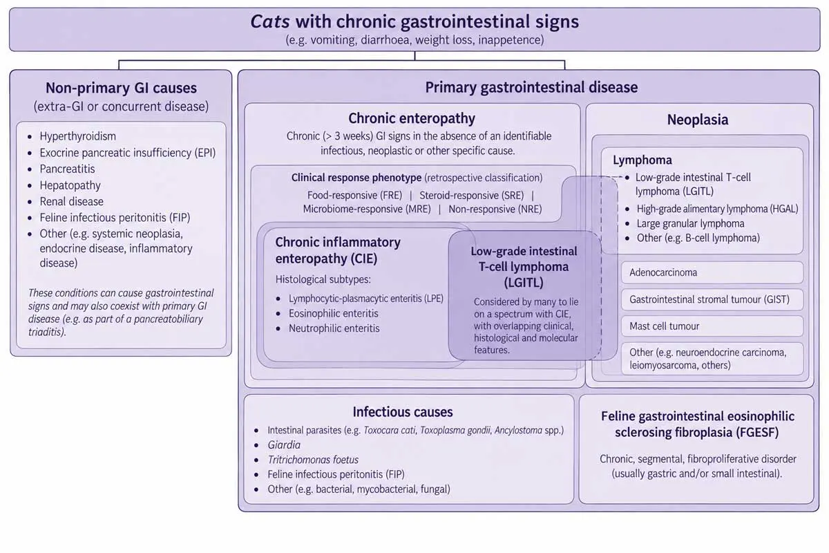
CIE is a disease that can affect cats of any age. Just like CE, CIE is unlikely to be a single disease process and, therefore, can be subcategorised, too, typically based either on its histopathological features, response to treatment or both. The aetiology is not fully understood but, similarly to human IBD, likely involves a complex interplay between dietary antigens, immune dysfunction/hypersensitivity and dysbiosis, potentially in predisposed patients due to genetic or other inciting factors5. Treatment strategies may focus on targeting any or all of these components.
Histopathologically, the most common form of CIE seems to be LPE, but eosinophilic and neutrophilic enteritis can also be appreciated, with many cats often sharing multiple histological subtypes.
Lymphoplasmacytic inflammation is a non-specific response to chronic stimulation. This can be primarily immune-mediated, or a response to antigenic stimulation associated with dietary components (particularly proteins) or luminal microorganisms. Whether predominantly eosinophilic or neutrophilic enteropathies should be approached differently to LPE is not always clear.
For most cats, histopathology largely comprises lymphoplasmacytic inflammation and is not much recognised to tell us anything about the prognosis or best treatment. For these reasons, and crucially because treatment is frequently initiated without histopathology, CE is also often categorised phenotypically by its response to treatment. Accordingly, disease can be classified as food-responsive (FRE, the most common form), microbiome-related manipulation responsive, immunosuppressive/steroid responsive or non-responsive.
Some consider FRE and CIE/LPE/SRE/IBD to be different diseases, but they are typically indistinguishable other than through response to treatment. Some classification systems also use the term “IBD” to refer either to SRE or to CIE more generally. Recent efforts have tried to dissuade the use of this term in veterinary medicine2,7.
The terms CE/CIE were coined in part to avoid confusion with IBD in human medicine, used to cover Crohn’s disease and ulcerative colitis. While many similarities exist between these diseases and feline CE, human IBD much more often requires immunomodulatory medications or surgery2.
Three forms of GI lymphoma are most recognised in cats. Most high-grade alimentary lymphoma (HGAL) in cats is large-cell and B-cell in nature, and has an aggressive behaviour. Large granular lymphoma (LGL) is a rarer large-cell lymphoma originating from cytotoxic T-cells, with 92% of cats having GI involvement8.
Cats with these higher-grade disease will usually be presented for recent development of GI signs and are much easier to diagnose, as imaging will often reveal an intestinal or gastric mass/masses with significant lymphadenopathy. Median survival times of 7 to 10 months have been reported with chemotherapy for HGAL, and likely much shorter for LGL8-10.
LGITL is considered the most common form of GI lymphoma in cats, comprising up to 70% to 75% of known cases5,11. Other terminology used for this condition include small cell lymphoma and low-grade alimentary lymphoma (LGAL). LGAL has been reported either as mostly (more than 90%) or exclusively T-cell in nature (LGITL)5,11,12.
LGITL appears analogous to “indolent T-cell lymphoproliferative disorder of the GI tract” in humans1,13,14. LGITL consists of mature, well-differentiated clonal populations of small T-lymphocytes with low mitotic rates, and consequentially a long course of slowly progressive disease1.
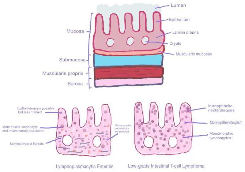
The lymphocytes are associated with infiltration of the lamina propria or epithelium (Figure 2). Nodal involvement may be present in less than half of cases15. Some LGITL may have lymphocytosis due to bone marrow involvement or, more likely, spillover from local disease into the blood16. Hepatic involvement is also reported, but no evidence exists to suggest this affects the prognosis17,18,19.
In contrast to HGAL and LGL, LGITL almost universally has a more indolent behaviour and much better prognosis20,21. Earlier studies recognised that a subset of cats with alimentary lymphoma had unexpectedly long survival times22,23.
Reports following the recognition of this separate disease process have stated survival times with treatment (typically glucocorticoids and metronomic chlorambucil) of 1.5 to 3 years11,17,19,24,25. However, this indolence can also make the clinical picture less dramatic and the diagnosis more challenging, not helped by the fact that concurrent/adjacent LPE is often present, too.
Across multiple studies and experiences, major overlap exists in signalment, duration of illness, clinical signs, clinical pathology and imaging findings. Additionally, LGITL is not diagnosed on fine-needle aspiration cytology from lymph nodes. An expanded population of small lymphocytes in lymphoid tissue is the expected response to chronic antigenic stimulation, but is also the appearance of LGITL, and architectural changes are more important than the cellular infiltrate5.
To add additional confusion to the topic, chronic inflammation and LGITL often co-exist, supporting the possibility of LPE possibly progressing to LGITL as part of a disease continuum1. Progression from chronic enteritis to T-cell lymphoma has been established in people, including in refractory coeliac disease26.
In CE where extra-GI causes, solid/focal tumours and other lesions have been excluded, the leading differentials are CIE (most commonly LPE, but not excluding eosinophilic enteropathies) and LGITL.
Unfortunately, differentiation is not straightforward, and we cannot rely on less invasive measures than biopsy to help us make a definitive call1,27. Numerous differences between LPE and LGITL have been identified at population level (Table 1), but none currently provides sufficient discriminatory accuracy to diagnose an individual cat without biopsy.
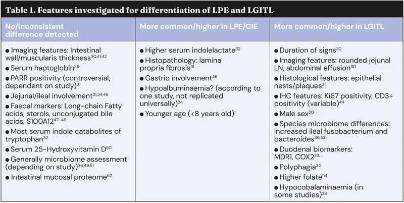
Often, biopsy for diagnosis of LGITL versus LPE is performed in cats either with more severe clinical signs/features, or in those that have failed diet trials and where immunomodulatory therapy is being considered. How many cats with CE at the stage of biopsy consideration have LGITL is not clear, but it may be as many as half, of course depending on criteria used28,29.
A limitation needs to be borne in mind whenever discussing the evidence surrounding LGITL and LPE: the classification of disease may not always be 100% accurate – especially in older studies where our understanding of the delineation between the two diseases was less clear. It has become more obvious that a true “gold standard” for us to base a diagnosis on may not exist, and often the best metric we have is to use a combination of findings.
Many studies now use a consensus between multiple pathologists and internists to try to decide, alongside multiple diagnostic techniques.
While some clinical differences do exist, some statistically significant, without biopsy none are distinctive enough to allow either to be excluded, even when used in combination1,5,28.
The only exception is perhaps age. While CIE is a disease that can affect any age, LGITL is generally considered a disease of middle-aged or older cats, with medians of 12 to 13 years old reported17,24,25,28,30-32. Though significant overlap exists between the diseases, the youngest reported case of LGITL in studies is four years old, so cats younger than this almost certainly do not have the disease25. Studies using modern criteria show a lower range, starting at seven to eight years old24,28,30,31,33,34,35,36.
Cats with LGITL have a longer duration of signs than cats with LPE (median 365 versus 107 days, shortest duration for LGITL 62 days), which again may suggest a disease continuum30. Unlike in dogs, diarrhoea is not the classical presenting sign for CE in cats. Weight loss and vomiting appear to be most common for both CIE and LGITL12,17,19,24,28,30,33,37. Weight loss in cats is often through muscle first, with significant sarcopenia prior to loss of adiposity5.
On multivariable analysis, Freiche et al found that when comparing cats with LPE versus LGITL, the clinical features most predictive of a neoplastic diagnosis when combined were male sex, polyphagia and duration of clinical signs30. Use of these types of models – especially if refined in the future with larger populations of cats – may help us to better select the patients most likely to benefit from biopsy.
“True” PLE with marked hypoalbuminaemia and associated complications appears to be fairly rare as a syndrome generally in cats.
Though many CE will show at least mild hypoalbuminaemia (14% to 100% of cases), in one study no cats with LGITL had hypoalbuminaemia1,24,30. Investigations have shown that, if anything, hypoalbuminaemia is more common in inflammatory disease than LGITL24.
Hypocobalaminaemia is a marker of intestinal or pancreatic dysfunction, and is reported in 18% to 80% of CE (12.5% to 44% of CIE, and 50% to 88% of LGITL)17,30,33,38. Serum cobalamin should be assessed/treated in both diseases, but may be more common in LGITL, reported as 57% versus 19% in one study, while others reported no significant difference30,33,38,39.
Abdominal ultrasonography has been well studied and described in CE, and again definitively distinguishing features are lacking1,28,40.
Ultrasonography is best utilised to identify focal diseases and exclude extra-GI complications6. Although HGAL will typically be associated with intestinal mass lesions and marked lymphadenopathy, LGITL is only rarely24. Abdominal lymphadenomegaly is common in inflammatory enteropathies, not just lymphoma. Some studies have shown no difference in node size between LPE and LGITL, while another suggested a significant difference but still overlap30,41,42.
Diffuse or sometimes multifocal thickening of the muscularis propria, submucosa and/or mucosal layer are the most common findings in both diseases, present in 50% to 95% of cats with CE1,24,27,28,30,39,41,42,43. Diffuse muscularis thickening in particular seems to be an indicator of enteropathy compared to healthy cats (Figure 3).
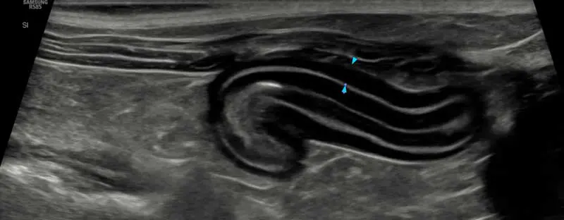
Earlier studies considered muscularis thickening to be a sign most suggestive of LGITL and, indeed, was found in 100% of LGITL in one study41,44. However, it has become clear that cats with both diseases frequently have this feature40,43. Studies have yielded contradictory and, therefore, ultimately inconclusive evidence on the specificity of muscularis thickening for LGITL, or even for CE generally30,41,42,45.
What is also clear is that some cats with clinically significant enteropathy, including LGITL, can have little to no ultrasonographic changes (reportedly 33% to 54% of LPE cats, and 10% to 15% of LGITL)28,39,43. Additionally, some apparently healthy (especially older) cats may have ultrasonographic changes; whether as a normal finding or due to subclinical enteropathy, we do not know42.
Freiche et al found that no single feature or collection were 100% accurate for diagnosis, but did find that combining the presence of rounded jejunal lymph nodes and abdominal effusion (45% LGITL cats versus 14% in LPE) into a model was helpful to differentiate the two30.
Due to limited diagnostic utility of cytology, histopathology has generally been considered the gold standard for differentiation of LGITL and CIE/LPE.
However, the sensitivity and specificity of histopathology has been called into question. The inherent difficulty in distinguishing cases based on histopathology alone has been noted frequently. Discrepancy is noted between pathologists – especially when LPE severity is more marked – with early studies reporting disagreement in 17% of cases with histopathology alone46,55. Another found that in 158 biopsy samples from 85 cats with CE, a uniform diagnosis across three pathologists was only achievable in 34% of samples6.
Biopsy can be pursued via endoscopy or surgery. Further discussion of the advantages, disadvantages and recommendations regarding choice of technique and where to biopsy will be found in the second part of this article.
WSAVA guidelines attempted to standardise recognition and reporting of disease in small animals, but was largely designed for inflammatory disease rather than the difficulty of LGITL versus LPE, and inter-observer variability remains a significant issue56,57.
A new scheme has been proposed and is supported by the ACVIM consensus statement1,31. Hallmark characteristics of LGITL have been described as a monomorphic lymphocytic population and in-depth mucosal infiltration of the lamina propria and epithelium (Figure 2). Intraepithelial lymphocytes (epitheliotropism) can be increased in both LPE and LGITL, but patterns more common in LGITL include epithelial plaques/nests (reported as 100% specific in one study), Ki-67 expression, and CD3+ (T-cell marker) apical-to-basal gradient31,58.
LGITL can rarely become transmural, with the prognostic significance of this uncertain6,11. LPE cases typically have marked fibrosis in the lamina propria30.
It is now recognised that the cellular distribution in LGITL can be variable, with some more subtle forms showing only gradients within the lamina propria or nests/plaques within the intraepithelial compartment rather than more obvious cases involving complete effacement of the lamina propria and loss of architecture. Ambiguity occurs when cases have concurrent neoplastic and inflammatory features (not uncommon), where concerning lesions are scattered, or where monomorphic populations are visible within a polymorphic background.
Expert opinion and even statistical analysis suggest that combining histopathology of biopsy specimens with immunohistochemistry (IHC) and analysis of clonality, in ambiguous but not necessarily all cases, leads to more accurate differentiation of neoplastic versus inflammatory lymphocytes58. It is specifically highlighted that clonality cannot be used as the sole means to reclassify disease13.
Using ancillary tests only in cases of suspected or possible lymphoma has been suggested and is the author’s approach as well, with review and/or re-biopsy if response is poor35,53,58.
Markers can be used to identify T-cells (CD3), B-cells (such as CD79a, CD20) and potentially proliferation (Ki-67). LGITL has been reported to be 100% CD3+ positive, in line with its known T-cell nature, and this was usually extensive and diffuse, with low numbers of B-cells31.
Expression in LPE is more variable, with a more heterogeneous population including B-cells. However, most still agree that the majority of lymphocytic infiltrates in LPE are T-cells, too6.
Another study showed a significant number (12 out of 20) of ostensibly healthy cats can have a CD3+ epitheliotropic lymphocyte population, concerning LGITL59.
Using IHC as an adjunct, 18% to 26% of cases previously classified as CIE may be re-diagnosed as LGITL, including up to 50% of severe LPE29,46,58. Less commonly, cats diagnosed as LGITL can be re-categorised as LPE29,60.
PCR for antigen receptor re-arrangements (PARR) is a test to assess for clonality, a hallmark of neoplasia. This was proposed as a highly specific test to confirm a diagnosis of lymphoma and, therefore, help differentiate LPE from LGITL in cases that were challenging histopathologically61. Indeed, some studies have used clonality as the crucial criteria for a diagnosis.
However, it has become clearer with time that chronic inflammation can also lead to selected expansion of lymphocyte lines and, therefore, PARR positivity. A 2016 study comparing different methods of LPE/LGITL diagnosis on duodenal biopsies found an almost 50% split between LPE and LGITL when using clonality as the ultimate differentiator, compared with 79% and 21% in favour of LPE with histopathology alone29. That study accepted clonality as the decisive marker of LIGITL, interestingly citing an association between PARR positivity and poorer survival (risk of death 2.8 times higher, median survival time 416 days versus not reached), therefore justifying its selection.
Other studies found 53% to 67% of cases originally diagnosed as CIE on histopathology alone were re-defined as T-cell lymphoma when IHC and PARR were used alongside58,62. Some of these cases had been deemed “definitive” CIE on histopathology alone. Earlier studies reported fantastic specificity, but the controls used were often healthy young cats or those without significant GI disease; that is, not representative of the population we are faced with. Subsequent studies have shown specificities as low as 54% and 33% for discriminating inflammation from neoplasia, amid similar concerns in human medicine, too1,31,59,63,64.
One study reported clonality in 70% of LPE versus 86% of LGITL cases, suggesting limited ability to discriminate31. Another showed no significant association between results of histopathology and PARR59.
PARR techniques are non-standardised, with each laboratory likely to have different sensitivity, specificity or accuracy depending on primers used and processing techniques6. In human medicine, only 5% to 15% of cases are considered to benefit from additional clonality testing, and it is clear that clonality is not synonymous with malignancy1.
Accordingly, the use of PARR is now recommended only in conjunction with other tests, where it can be used to support a diagnosis but not give a definitive answer1,6.
As most cats undergoing GI biopsy are those showing chronic GI signs, what constitutes “normal” may be misunderstood, and the WSAVA guidelines based “normal” on much younger cats.
A 2019 study investigating gastroduodenoscopy biopsy results in reportedly healthy cats (median age 9.5 years) used a combination of blinded histopathology, IHC and PARR59. All 20 cats had histopathology considered abnormal by WSAVA grading; 12 were defined as LGITL, one as emerging LGITL, six as LPE and one as “pseudoclonality”. Only three of the 20 cats went on to develop clinical GI signs within two years.
It is possible to draw one of two conclusions: either most healthy cats have subclinical CE, or histopathological evidence of inflammation and clonality is present in the GI tract of healthy cats; the latter is suspected. It highlights the pitfalls of over-reliance on clonality tests for diagnosis, as this was strongly suspected to be the main contributor to an LGITL diagnosis in many65,66.
Machine-learning and AI are being actively researched as a tool to help in our decision making, again with the obvious trouble in defining a gold standard to train models on67-69.
Finally, a novel technique utilising mass spectrometry applied to intestinal biopsies has been developed and may be more accurate than PARR when used in conjunction with histology, but is far from being readily available63.
While a clear gold standard for diagnosis is still lacking, many studies needing inclusion criteria have turned towards not using the specific results of any single set of tests, or even one pathologist, but instead using a panel of board-certified specialists to make a final call63. This just goes to highlight how much of a challenge this subject is, as even then disagreement occurs, and obviously this technique is not available on a daily basis.
Other studies have side-stepped this issue entirely and excluded cases where the pathologists had disagreement, which are exactly the cases that most require studying69.
Clearly, non-invasive tests cannot reliably distinguish LGITL from LPE in our CE patients.
Biopsies are required to help us answer this, but even histopathology and other tests have their weaknesses, either due to genuine limitations with the test or due to biological overlap of the diseases.
Natural considerations would be: how do we best collect biopsies? When are we best to collect them? Is there any reason to do it at all?
We will explore this decision making in the second part of the article, as well as management of enteropathies after, or in lieu of, a histopathological diagnosis.
Bryn Jones qualified from the University of Nottingham in 2014 and moved into small animal general practice in south Wales for four years, where he completed a CertAVP. After completing a small animal medicine residency in Hampshire, he moved to Willows Referral Service in 2023, where he currently works. He has earned recognition as an RCVS and European specialist in small animal internal medicine. Bryn has an particular interest in hepatology, nephrology and especially gastroenterology.