5 Jun 2017
Frances Taylor examines means of performing small animal oncology tests with the aim of tailoring a clinical plan for each patient.
Frances Taylor
Job Title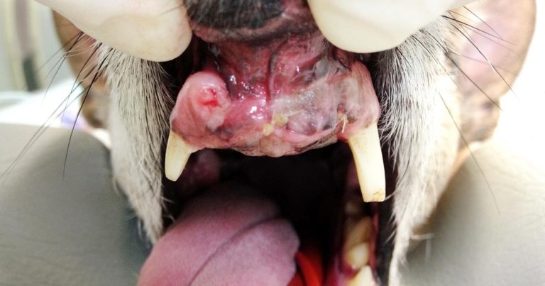
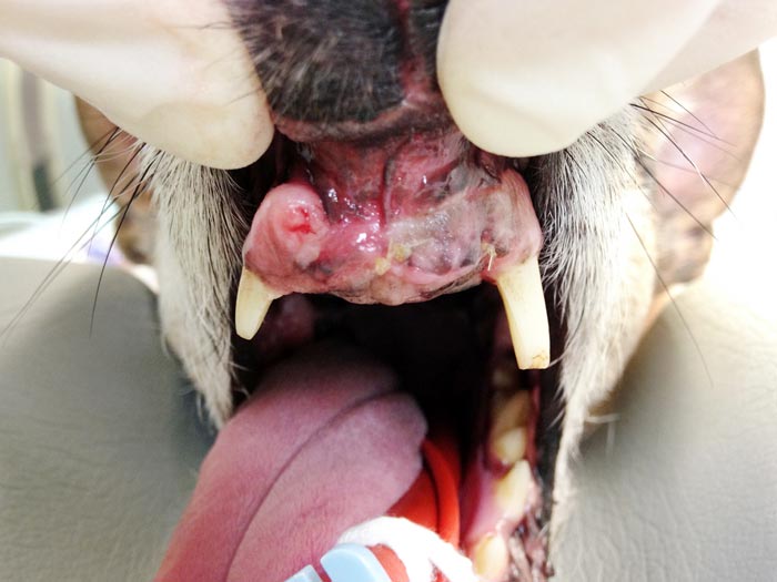
An incisional biopsy in this small oral mass case allowed vets to plan a relatively conservative partial rostral maxillectomy for the dog.
This article discusses guidelines on appropriate sampling techniques for animals with various masses and tumours. This seems a simple task, but, as every patient is different, strict guidelines can be difficult to collate. Generally speaking, cases should be approached as individuals and the clinical plan tailored to each based on clinical judgement, patient comorbidities (if present), available finances and the temperament of the patient.
These are a broad guide to workup and investigate masses based largely on location.
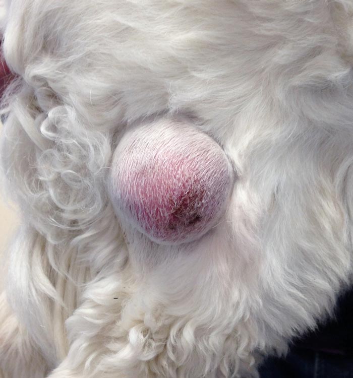
Surface masses can, in most cases, be sampled using fine needle aspirate (FNA) cytology. This can commonly be performed with the patient conscious and be a cost-effective method of diagnosis.
According to one study, cellular samples are retrieved in around 83% of FNA samples of this type of mass and, overall, the cytological diagnosis was in agreement with the histological diagnosis in 90% of cases (Ghisleniet et al, 2006). For diagnosing neoplastic masses, cytology had a sensitivity of 89.3%, a specificity of 97.9%, a positive predictive value of 99.4% and a negative predictive value of 68.7%. Therefore, cytology can be good at ruling in neoplastic disease, but not always as helpful at ruling it out.
Initial samples should be taken without using suction, as cells such as neoplastic lymphocytes are commonly fragile. If a reasonable sample cannot be achieved without suction then it is acceptable to apply suction when taking a sample, as some tumours do not always exfoliate well.
Several factors can reduce the success rates of FNA cytology:
A tissue biopsy is appropriate if a diagnosis cannot be reached cytologically and where additional information, such as tumour grade, will aid planning therapy.
An incisional rather than an excisional biopsy should be performed wherever possible. In some cases, a Tru-cut biopsy under sedation and local anaesthetic may be appropriate, especially with larger surface masses. Tru-cut biopsies can also be helpful for aggressive-appearing masses where biopsy tract wound healing may be compromised by tumour growth.
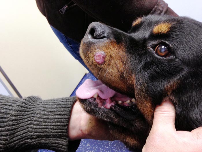
Excisional biopsies are best avoided, as the surgical scar created following excisional biopsy will require a much larger surgery to cleanly excise than the original mass if clean margins have not been achieved. Excisional biopsy where clean margins have not been achieved causes contamination and disruption of local tissue planes, both of which can drastically reduce the chances of achieving a complete excision with a following curative-intent surgery. In addition, in cases where an excisional biopsy has been taken and tumour-free margins not achieved, a much higher risk exists of wound break down, especially with mast cell tumours and aggressive sarcomas.
Instances where excisional biopsy should be avoided include:
It is very uncommon to possibly sample oral masses while a patient is conscious, so, in the majority of cases, patients are anaesthetised or sedated to allow sampling to take place. Therefore, the opportunity to take a tissue sample using a biopsy, rather than obtaining an FNA-obtained cytology sample, should be taken, as a biopsy has a greater chance of providing a diagnosis.
It is important to remember when taking any biopsy not to risk compromising any future curative-intent surgery and some knowledge of potential future surgical procedures is helpful when biopsying any masses. During curative-intent surgery, the entire biopsy tract or scar needs to be removed with a margin; therefore, incisional biopsies from the centre of the mass are appropriate to minimise any impact in surgical planning. Areas of tumour that appear to be necrotic should be avoided.
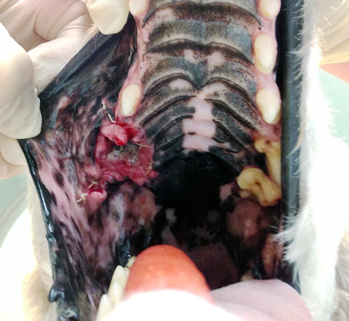
Maxillary and mandibular masses should not be biopsied by cutaneous approach if they can be accessed via the gingiva, because, during a maxillectomy and mandibulectomy, the approach is intraoral via the gingival tissue, and removing the biopsy tract can be challenging, impossible or involve a more extensive procedure if this is made through haired skin.
The most commonly occurring tumour affecting the urinary tract in dogs is a transitional cell carcinoma, and this tumour has significant potential to seed along needle or biopsy tracts. Therefore, sampling of suspected lower urinary tract tumours (including the prostate) should be carried out initially via the urethra.
Cytology of the urine can sometimes be helpful. If tumour sampling is required, this is commonly done using a suction catheter method under ultrasound guidance. Cystoscopic biopsying procedures can also be used. These methods of urinary tract sampling are performed with the patient anaesthetised, as urinary tract tumours – especially prostate tumours – are commonly painful.
FNA sampling using ultrasound guidance is standard for sampling the majority of intra-abdominal masses. It is prudent to check the patient has adequate platelet numbers prior to sampling. The FNA sampling of aggressive-looking or cavitated splenic masses is commonly, at best, unrewarding and these are, in most cases, best removed via splenectomy to obtain a diagnosis.
Where a diagnosis cannot be reached cytologically for hepatic or renal masses, sampling via ultrasound-guided Tru-cut biopsies can be performed following careful assessment of coagulation status. Careful assessment of renal status is imperative prior to renal biopsy being performed, as biopsy can negatively impact on renal function. Where masses are cystic or cavitated, an increased risk of complications and the sample being non-diagnostic exists, so each case should be assessed individually prior to biopsy being performed.
Intestinal masses where a diagnosis cannot be reached cytologically are normally excised with a 2cm to 3cm margin following abdominal and thoracic imaging for staging purposes. Incisional biopsies of gastrointestinal masses should be avoided where possible to reduce the risk of dehiscence.7
Adrenal masses are not normally sampled in the first instance due to the risk of complications associated with phaeochromocytoma stimulation; cases with functional adrenal masses are usually investigated medically. Surgical excision of adrenal masses is normally a specialist procedure due to patient support requirements, and risks of intraoperative and postoperative complications.
Sampling of peripheral pulmonary masses can be possible in many cases using ultrasound-guided FNA sampling. Deeper pulmonary masses cannot be sampled in this way due to difficulty of obtaining an ultrasound image through air-filled lung tissue and the risk of inducing a pneumothorax. Commonly, pulmonary masses are excised to obtain a diagnosis following full staging, typically using CT imaging, as the presence of lymph node metastases or further pulmonary nodules significantly impacts on patient prognosis.
Mediastinal masses are commonly sampled using FNA sampling, although, in many cases, a tissue biopsy, normally via ultrasound-guided Tru-cut sampling, is required to obtain a diagnosis.
The amount of tumour staging carried out at the time of initial tumour sampling is at the discretion of the attending veterinary surgeon and based on informed client choice. The type and amount of tumour staging required for a patient will not often be clear until a diagnosis is reached. However, instances occur where staging is sensible prior to a diagnosis being obtained; for example, imaging of the thorax and abdomen prior to taking a patient to theatre for removal of a splenic mass or an intestinal mass is recommended.
Also, if the patient is sedated or anaesthetised for biopsy of a mass then assessment and aspiration of any local draining lymph nodes and thoracic radiographs can be helpful, especially if a malignant tumour is suspected.