02 June 2025
Sophie Duguid BVMS, MRCVS considers the practical applications of this branch of medicine, weighs up its various methodologies and explores latest innovations.
Sophie Duguid
Job Title
Image: Seventyfour / Adobe Stock
Haematology stands out in veterinary diagnostics for its breadth of application, offering valuable insights across diverse clinical settings.
By analysing blood parameters such as cell counts, haemoglobin levels, and morphological features, haematology serves as a diagnostic pillar for:
With increasing stress levels among veterinary professionals (HealthforAnimals, 2022), advanced diagnostic systems, including haematology tools, offer a practical solution to streamline workflows and reduce pressure in busy clinical environments.
Although recent workforce modelling predicts significant growth in the UK veterinary workforce by 2035, the average full-time equivalent workforce is expected to decline (RCVS, 2024) – making any tool that enhances efficiency a valuable consideration for practice.
This article will examine the role of haematology in veterinary diagnostics, focusing on its practical applications in both routine and specialised settings. It will highlight recent advancements in technology, such as AI-driven image flow cytometry and viscoelastic focusing (VEF), and their impact on diagnostic workflows and patient care.
By incorporating innovation in haematology, clinics can also support their veterinary teams in managing caseloads efficiently and maintaining high standards of practice.
Haematology tests, in conjunction with other data, help to identify the presence and cause of a condition to formulate a list of differential diagnoses, track disease progression and treatment effectiveness.
This involves both quantitative and qualitative analyses for a thorough evaluation of blood. Quantitative analysis, typically done through a complete blood count (CBC) using automated analysers, measures components such as PCV and haemoglobin levels.
On the other hand, qualitative analysis involves examining blood smear morphology to identify changes in cellular structure.
Combining these two methods ensures a comprehensive and accurate laboratory diagnosis, enabling more informed clinical decisions (Villiers and Ristic, 2016). Reasons for performing screening tests include:
Historically, these tests were done at an external laboratory due to a lack of necessary expertise, equipment and supplies to perform them accurately in house. (Harvey, 2012). However, in recent years, technological advancements have helped advance haematological evaluation at the point of care.
The main diagnostic test carried out in practice is CBC analysis (Morissette, 2022). In a sample, an automated CBC analyser measures key parameters such as haematocrit, red blood cell count, mean corpuscular volume and mean corpuscular haemoglobin concentration (El Brihi and Pathak, 2024).
These values help assess red cell mass, cell size and haemoglobin content, while also providing white blood cell and platelet counts.
Reticulocytes are immature red blood cells released by the bone marrow during erythropoiesis. They contain residual RNA and organelles, which are progressively lost as they mature into functional red blood cells.
Monitoring reticulocyte levels can help provide a clear indication of how effectively the bone marrow is responding to anaemia (Riley et al, 2001).
Elevated reticulocyte counts indicate active bone marrow regeneration, commonly seen in cases of blood loss or haemolysis. Conversely, low counts may point to underlying conditions such as bone marrow suppression or chronic disease, where red cell production is insufficient (Riley et al, 2001). Pre-regenerative anaemia also highlights the importance of reticulocyte monitoring over time, as it reflects the initial lag before bone marrow activity increases. Incorporating reticulocyte analysis into routine practice can help ensure veterinary professionals accurately assess erythropoiesis and tailor interventions accordingly.
Blood smear analysis is a vital procedure in veterinary haematology, providing essential qualitative data to complement automated CBC results.
Analysis involves preparing a blood film to examine cellular morphology, assess abnormalities and verify quantitative CBC values, such as white blood cell differentials and platelet counts, which can be impacted by the aforementioned clumping or atypical platelet size.
This allows for the identification of regenerative responses in anaemia, inflammatory leukograms and other haematologic abnormalities that may not be evident in the quantitative results.
By confirming quantitative data and identifying subtle pathological changes, these tools can provide clinicians with a more comprehensive understanding of a patient’s blood profile.
Recent advancements have introduced innovative tools that leverage artificial intelligence (AI) to streamline these processes, enhancing both speed and performance.
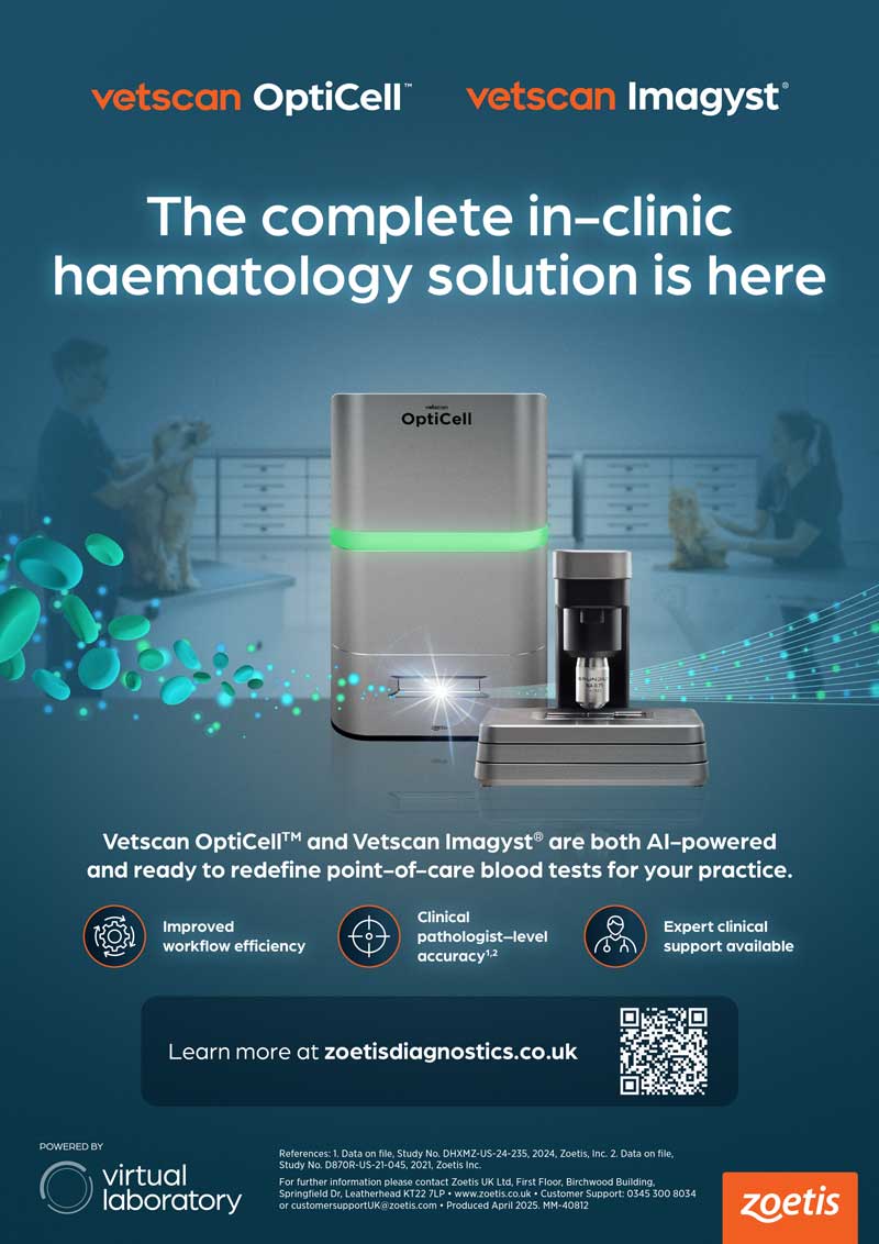
Tools such as Zoetis’ Vetscan Imagyst (a multipurpose analyser) and Vetscan OptiCell (an automated CBC analyser) save veterinary teams valuable time, enabling them to focus on patient care while still gaining the critical data necessary for diagnosis and treatment decisions.
Four primary methodologies exist for generating data in a CBC: manual techniques, quantitative buffy coat analysis (QBCA), automated impedance analysis, and flow cytometry (DeNicola, 2011).
Among these, manual counts of red blood cells, white blood cells, and platelets are largely outdated, time-consuming and requiring the greatest amount of technical expertise (DeNicola, 2011).
QBCA was the first available automated method, using sedimentation in a larger micro-haematocrit tube to count white blood cells and platelets. QBC analysers are efficient for screening, but provide limited haematological data (Giger, 2010).
Impedance counters separate cells by size. They provide rapid results, but cannot deliver a full differential count or reticulocyte data. Their reliance on size separation can lead to inaccurate platelet counts and limited white blood cell differentiation (Giger, 2010).
Flow cytometry is a versatile technique used to analyse multiple properties of cells or particles as they pass through laser beams (Giger, 2010). It processes thousands of cells per second, enabling rapid and detailed multiparametric analysis (Sánchez-Torres et al, 2022).
This technology is applicable to various sample types, including blood, cell suspensions, microorganisms, and inert particles, making it suitable for studying diverse biological and chemical solutions such as serum or plasma (Sánchez-Torres et al, 2022).
Over the past four decades, veterinary in-house haematology technology has made great advances (DeNicola, 2011), with the newest analysers leveraging flow cytometry and AI.
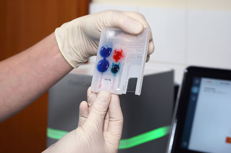
The latest CBC analyser to become available to UK clinics is Vetscan OptiCell (Zoetis). The automated counter combines patented microfluidic VEF technology with advanced AI to count hundreds of thousands of cells, delivering precise and efficient cellular analysis.
Using a self-contained, single-use cartridge, it aligns blood cells into a single-layer plane by guiding them through microfluidic channels. This process minimises issues such as clogging or vibrations.
High-resolution images of these cells are captured and analysed by deep learning AI algorithms, assessing 22 parameters, including reticulocyte count, in three dimensions, including shape, size, colour and density.
Clumped or giant platelets and subtle morphological changes have historically been more difficult for automated analysers to accurately assess.
OptiCell addresses the limitations of traditional machine-generated counters by leveraging its VEF and AI technology.
With these features, the analyser is able to align cells into a single-layer plane before analysis, ensuring cells are evenly distributed and reducing errors caused by clumping or overlapping.
This precision eliminates one of the major issues with platelet counts – particularly in species such as cats and some dog breeds, where giant platelets are common.
By isolating and evaluating individual cells, OptiCell minimises the inaccuracies often associated with standard analysers.
For white cell differentials, Vetscan OptiCell’s AI-powered algorithms analyse cells in three dimensions, assessing parameters such as shape, size, colour and density. These deep-learning models are trained to recognise subtle morphological features and variations, which means that even slight abnormalities are flagged accurately.
The Vetscan OptiCell was rigorously tested using blood samples from canine and feline patients for CBC comparison. The validation study compared its performance across all parameters, demonstrating reliability and precision comparable to that of a veterinary clinical pathologist (Zoetis, 2024).
Overall, OptiCell delivered rapid, accurate (Zoetis, 2024) and repeatable results, proving its capability as a dependable tool for comprehensive blood analysis.
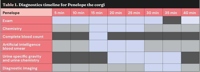
Penelope is a 12-year-old, female, entire corgi who presented for evaluation after experiencing depression, anorexia and polyuria/polydipsia for two days.
On physical examination, she was found to be depressed and dehydrated, with a slightly distended abdomen and swelling in the perineal region.
During the initial evaluation, Penelope’s symptoms indicated the need for further diagnostics to identify the underlying cause. Her physical findings prompted additional tests, including a chemistry panel (Vetscan VS2), comprehensive CBC (Vetscan OptiCell), blood smear (Vetscan Imagyst AI Blood Smear) and a complete urinalysis.
Given her distended abdomen, diagnostic imaging was also performed.
The Vetscan VS2 profile showed azotaemia with elevated blood urea nitrogen and creatinine levels, requiring further assessment to determine whether the cause was pre-renal, renal or post-renal.
Phosphorus levels were also elevated, and total protein was increased due to elevated globulins alongside decreased albumin levels, suggesting chronic inflammation.
Automated CBC results from Vetscan OptiCell revealed normal red blood cells and platelets, but detected neutrophilic leukocytosis with monocytosis.
This inflammatory leukogram indicated a chronic inflammatory process and supported the elevated globulins seen in the chemistry panel as being secondary to inflammation.
The results from the Vetscan Imagyst AI Blood Smear capability visually confirmed the neutrophilic leukocytosis and monocytosis observed in the CBC analysis.
These results further support the diagnosis of chronic inflammation.
Urine specific gravity assessed by a refractometer was 1.012, consistent with renal dysfunction.
Based on clinical history, examination and lab findings, these results pointed to potential pyometra.
Pyometra, often caused by Escherichia coli infection, disrupts renal function by impairing sodium and chloride resorption in the tubules, leading to dehydration and contributing to pre-renal azotaemia.
Following diagnostic imaging, pyometra was confirmed and Penelope underwent surgery.
She recovered well and was discharged with oral antibiotics, which were chosen based on E coli cultured from her uterus and found to be sensitive to the prescribed medications.
Her progress post-treatment was favourable, highlighting the effectiveness of her diagnostics and care.
The timeline in Table 1 highlights how quickly Penelope’s case was handled. Within just 40 minutes, her evaluation, diagnosis of pyometra and preparation for emergency surgery were completed.
This shows the benefits of efficient diagnostic tools and teamwork, helping vets provide prompt care and better outcomes for pets and their owners.
As demonstrated in Penelope’s case, the ability to integrate automated CBC analysers with AI-driven imaging streamlines workflows, supports rapid decision making and ultimately improves outcomes for pets and their owners.
This efficiency is especially beneficial in urgent cases, as it enables faster diagnoses and initiation of treatment plans.
Veterinary haematology has come a long way, evolving from time-intensive manual techniques to the cutting-edge AI-powered systems available today.
Advanced tools such as Vetscan OptiCell and Vetscan Imagyst are steadily becoming a cornerstone of veterinary practice, shaping the standard for efficient and accurate in-house diagnostics.
In addition, Zoetis offers access to expert clinical pathologists for add-on reviews and provides specialist consultants as part of a complementary service for cases requiring specific support.
These resources further enhance diagnostic confidence, empowering veterinary teams to make informed decisions and enhance patient outcomes.
By embracing these innovations and leveraging the available expert support, practices can stay ahead of the curve, advancing diagnostic precision and operational efficiency in an ever-demanding field.