10 Jun 2024
Having outlined the basic pathophysiology of head trauma in part one of this series, Gerardo Poli discusses ways of lowering cerebral blood volume to tackle this issue.
Gerardo Poli
Job Title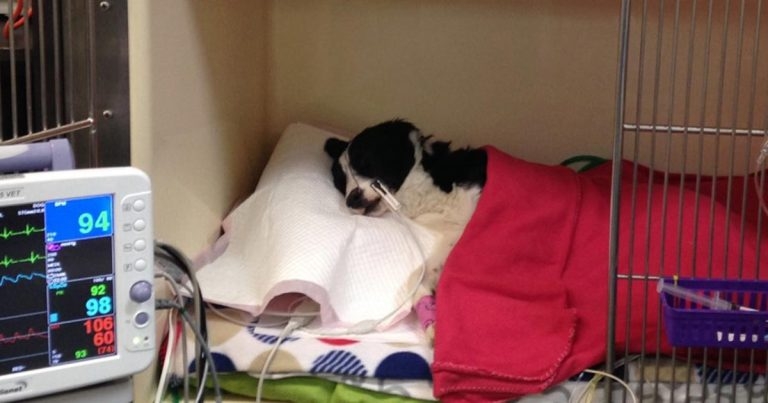
Patient elevated on a 15% incline.
Part one of this series discussed the equation CPP = MAP – ICP.
Cerebral perfusion pressure (CPP) is the difference between the mean arterial pressure (MAP) and the intracranial pressure (ICP), which it is pushing against.
Part one discussed optimising MAP – therefore, part two will look at reducing ICP.
Because the brain is in a fixed space and encased in bone, ICP increases if it swells as a result of trauma.
Apart from reducing brain swelling, a reciprocated reduction in either cerebral blood volume or CSF must occur for ICP to remain the same. CSF volume is difficult to influence safely, so efforts are targeted at reducing cerebral blood volume.
The two main factors that influence cerebral blood volume are vascular tone and venous drainage.
Effects of vascular tone can be dramatic – if vasodilation occurs, cerebral blood flow and volume increase, with a resultant increase in ICP. If vasoconstriction occurs, cerebral blood flow and volume can reduce, and, therefore, reduce ICP. However, excessive vasoconstriction can lead to ischaemia and death of brain tissue, which is not desirable.
Factors that effect vascular tone include:
Increased carbon dioxide levels result in cerebral vasodilation (and increased ICP); this can be measured by venous or arterial blood gas tests, or capnography.
The goal is have CO2 levels within normal ranges of between 35mmHg and 40mmHg. Unfortunately, controlling CO2 levels is intensive, and requires intubation and mechanical ventilation; therefore, it is not readily available in many hospitals.
Excessive reduction in CO2 pressure can result in excessive cerebral vasoconstriction; therefore, it is not advised to drop below 30mmHg.
Optimisation of the partial pressure of oxygen (PaO2) in the blood stream is important, as hypoxaemia causes cerebral vasodilation.
Maintaining pulse oximetry readings greater than 95% are ideal – this equates to a PaO2 of 80mmHg. Increasing inspired oxygen levels via oxygen therapy is the simplest method of addressing this. Mask oxygen, or an oxygen box, should be considered safer than intranasal oxygen lines, as they can trigger sneezing – which causes an increase in ICP.
If the patient is anaemic, blood transfusions may be required to increase haemoglobin levels and, therefore, the oxygen-carrying capacity of the blood.
Cerebral metabolism results in the release of CO2 and hydrogen ions, and, therefore, can result in cerebral vasodilation. The focus here is suppressing brain activity and controlling seizure activity.
Suppressing brain activity is achieved through the use of barbiturates to induce a coma-like state. Barbiturates are also thought to have some additional cerebroprotective properties. Seizure activity, if present, is controlled via the use of standard antiseizure medications.
Cerebral metabolism can also be suppressed through hypothermia. No clear guidelines exist, but allowing patients to remain mildly hypothermic – 1°C or 2°C below normal – and avoiding actively rewarming them may help reduce cerebral metabolism, as long as it does not result in detrimental effects.
Promoting venous drainage, through preserving jugular blood flow, also assists in reducing cerebral blood volume.
Practically, this can be achieved by placing the patient on a 15% to 30% incline; however, it is important the whole body is on an incline – not just the head – as this can bend the neck and occlude jugular blood flow. Placing the patient on an inclined board is the easiest and safest way.
Additionally, avoid jugular blood sampling, as this can lead to a blood clot, impeding reduced venous drainage.