19 Jul 2023
Olivia Munro outlines how the VN can help in an emergent situation, including triage, stabilising patients and dealing with distressed owners.
Olivia Munro
Job Title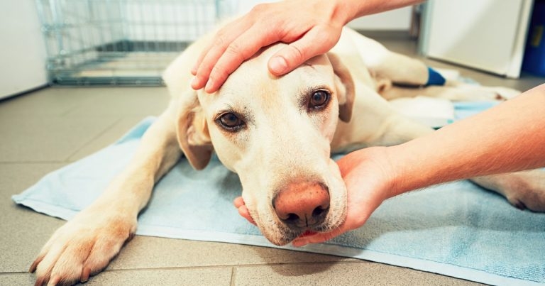
Image: © Chalabala / Adobe Stock

Most emergencies will be become apparent through an owner telephoning the surgery. This is where, if good communication skills are used, we can gather substantial information to prepare for the arrival of the patient, as well as an estimated arrival time.
Owners will often be distressed and need to be calmed, so closed questions and closed loop communication can be extremely helpful to receive correct and precise information.
Closed questions are usually given for short and simple answers that don’t require a detailed response; for example, is the patient conscious? Has the patient had access to any toxins? Closed loop communication involves someone repeating what has been said to them to reduce any misunderstandings.
In most cases, it is more important to get the patient to the practice, rather than the owner administering first aid, to allow for correct treatment to take place.
Very few owners have medical training and their interpretation of clinical signs may be misleading (Boag and Nichols, 2011).
However, owners can help in trauma emergencies; for example, by applying pressure to haemorrhaging wounds with a clean towel or cloth, or lightly covering open wounds with a clean towel during transportation to the practice.
Foreign objects should not be removed by the owners in case of causing further damage to surrounding tissue or worsening haemorrhage. It will also be useful to get the patient’s name, age, breed and any pre-existing conditions at this stage to help with preparation for their arrival, as well as a contact number for the client in case they are not within their estimated arrival time so further communication can be made with them.
It is also important to advise the owner that their pet may be painful and anxious – and, therefore, may react out of character, so care must be taken in handling their pets.
Once initial information has been gathered and the veterinary team informed, it is usually the role of the veterinary nurse to prepare for the arrival of the patient.
A treatment area should be decided on and equipment that may be needed, dependent of the type of emergency.
It is always best to prepare for the worst and have everything readily available. An IV catheter tray can be set up, alongside fluid therapy equipment and syringes for blood sampling. An oxygen source should be readily available and suitable for the patient, as well as intubation equipment and suction.
Resuscitation equipment and drugs should be at hand. In larger hospitals, crash trolleys are useful as all equipment, drugs and monitoring apparatus are together, and can be moved around the practice to allow for CPR across the building. In smaller practices, having a drawer or toolbox designated for CPR can be helpful.
Diagnostic equipment can be set up ready, such as an ultrasound machine for a point-of-care ultrasound, which can be useful for the secondary survey once the patient is stable.
Theatre can also be set up ready; for example, in the cases of surgical emergencies, such as a caesarian or gastric dilatation and volvulus, to save time once the patient has arrived.
On arrival, the patient should be triaged by a trained member of the team and a brief assessment of the major body systems to identify urgent life-threatening problems performed. This is called the primary survey.
The algorithm “ABC” can initially be evaluated:
Some patients may, unfortunately, arrive in cardiopulmonary arrest (CPA) and, therefore, CPR should be started immediately.
Patients with catastrophic problems (airway obstruction, respiratory failure and circulatory failure) can die within seconds (Spreng, 2002), so it is vital to assess these as soon as possible.
The major body systems assessed in the primary survey are the respiratory and cardiovascular systems (responsible for ventilation and perfusion, respectively), alongside the neurological and renal status of the patient, which can then be further evaluated in the primary survey.
The respiratory system should firstly be visual, seeing how the patient is breathing – is increased inspiratory and/or expiratory effort evident? Are the nostrils flared? Is the patient abducting the elbows and stretching out the neck? Are they open-mouth breathing?
A respiratory rate can be counted, and then the whole thorax and trachea auscultated to hear for any crackles or dull lung sounds.
For feline patients, it is worth weighing up this clinical exam if they are in respiratory distress to minimise stress and handling. A pulse oximetry reading can be taken, but bear in mind those patients in respiratory distress may not tolerate equipment around their mouth, so the prepuce vulva or ear can be used.
It is also worth remembering that poor perfusion can give false readings, but oxygen supplementation should be provided to any emergency patient if respiratory compromise is evident or suspected (Brown and Drobatz, 2018), and can be provided by the following:
Again, it is important to assess which option will cause the least amount of stress to the patient as some of these options are more intrusive than others. Flow-by and the use of an oxygen kennel has little contradiction and handling, whereas nasal prongs and catheters are less well tolerated in stressed patients, and are more invasive to place.
The cardiovascular system can then be assessed – again with auscultation. A heart rate can be counted and an arrhythmia noted if present. Pulses should be assessed alongside auscultation by how they feel and whether a pulse deficit is present (does the pulse rate match the heart rate?).
Pulses can be described as “strong”, “good”, “weak”, “bounding” or “thready”, but it is a skill that takes practice to determine the difference. A pulse deficit can indicate a potential arrhythmia and the type of pulse could give you an indication on a patient’s perfusion, so should be assessed regularly throughout the patient’s survey.
Continuing the evaluation of the cardiovascular system, mucous membrane colour and capillary refill time (CRT) should be checked. A CRT should normally be one to two seconds; anything rapid or prolonged should be noted, and could be signs of poor perfusion and/or shock. Some dog breeds will have pigmented membranes, so you may need to assess conjunctival, vulval or preputial membranes instead.
Table 1 shows different mucous membrane colours and which each could be a sign of.
| Table 1. Different mucous membrane colours and which each could be a sign of | ||||
|---|---|---|---|---|
| Mucous membrane colour | Potential cause | |||
| Pink | N/A | |||
| Pale pink or white | Hypovolaemic shock, blood loss, anaemia, hypothermia | |||
| Blue | Hypoxia | |||
| Bright red | Distributive shock, toxicity, sepsis, severe hyperthermia | |||
| Yellow | Liver disease, haemolysis | |||
The patient’s temperature can also be taken at this stage and can be a helpful tool in emergency triage. A low body temperature could indicate poor perfusion and shock, whereas a higher body temperature could be due to an infection, hyperventilation, heatstroke or seizures.
Active warming and cooling should be done carefully and slowly, to not worsen any clinical signs. Mentation can be improved by correcting the patient’s temperature, so it is important to regularly check this parameter – especially in neonatal and geriatric patients, or when active warming or cooling has been implemented.
Care must be taken when using warming devices to not cause any skin burns; therefore, patients should not be left unsupervised and turned regularly to avoid this.
For hyperthermic patients, wet towels should also not be used due to the heat being trapped between the patient and the towel itself, which can, in fact, make the hyperthermia worse. It is important to ascertain when a patient’s temperature is increased – whether it is a pyrexia or hyperthermia – as both are treated differently.
If pyrexic, the source of the pyrexia needs to be treated and active cooling is usually ineffective, whereas a hyperthermia should be rectified with active cooling with the use of cool mats and cool water. Surgical spirit should not be used on hyperthermic patients in case defibrillation is needed.
A brief evaluation of the patient’s neurological status should then be assessed. Mentation can be classified as alert, obtunded, stuporous or comatose.
Many factors exist that can alter a patient’s mentation and it is, again, something that should be assessed regularly throughout the survey, as well as during the patient’s hospitalisation; any change, however subtle, should be notified to the vet.
If any suspicion exists of increased intracranial pressure – for example, either post-seizure or trauma – the patient (if recumbent) should have the head and neck placed elevated at a 30° angle to aid venous return while minimally impairing cerebral perfusion (Hopper, 2008).
The patient’s posture and ability to stand should be evaluated. If plegia is present, it is important for the presence or absence of deep pain to be assessed as this can indicate a spinal cord issue, and a guide to where the damage to the spinal cord may be. The lack of anal tone can also be an indicator to an issue of the spinal cord.
Within the neurological primary exam, the eyes should be checked for any nystagmus, unequal pupil size, reaction to palpebral reflex and light reflex. A head tilt may also be seen.
The renal status can be as easy as asking the owner when the patient last passed urine. It is especially important to ask cases of a collapsed male cat, in case of urinary obstruction. Anuric patients must be seen immediately. As part of the exam, the bladder should be palpated.
The primary survey highlights life-threatening emergencies and allows for initial stabilisation, which should then be followed by the secondary survey.
A full clinical history can be taken from the owners, which can often highlight areas of concern that can then be looked into in more depth. A resuscitation code can also be discussed with the owner at this time.
IV catheters can be placed – most commonly in the peripheral veins (cephalic and saphenous). Blood samples can also be collected at this time to run an initial database. Care must be taken in those patients with suspected coagulopathies or raised intracranial pressure; if any doubt exists about the status of these, peripheral samples should be taken. Cannulas for longer term use – that is, central lines – can be placed once the patient is stable.
For neonatal or severely collapsed patients where peripheral access is not possible, intraosseous catheters can be placed to allow for rapid administration of fluids and drugs.
A minimal database for every emergency patient should include PCV, total protein/total solids, blood glucose, electrolytes and a examination of a blood smear. Most practices now have portable machines for blood work that give rapid, but more in-depth results for these types of patients; for example, epoch or iStat machines, which require only a small volume of blood, but can give renal parameters, a lactate reading and electrolytes, as well as blood gases.
Fluid therapy can also be given under veterinary surgeon instruction – often given in boluses in emergency medicine. It is important after each bolus to repeat the primary survey to look for any improvement or changes to the patient.
Understanding whether fluid therapy is being used to treat a perfusion abnormality, a hydration abnormality, a combination of the two, or to prevent development of one or both of these is crucial to providing good fluid therapy (Boag and Hughes, 2018).
Further diagnostic imaging can then take place where a diagnosis can hopefully be found and a treatment plan followed.
Emergencies can also happen within the hospital itself for patients already in the practice; events such as drug reactions, respiratory aspiration and CPA.
It is vital for all members of the practice to understand and follow their protocol for dealing with a CPA, and as previously stated, having a crash cart, drawer or box is vital for dealing with a CPA in the hospital.
It is also important to have a emergency drug dosing chart to allow for quick administration of drugs and the CPR algorithm to hand with the crash box. Firstly – and the most important part – is raising the alarm that a patient has gone into CPA, whether that be a paging or Tannoy system, or simply shouting for help.
CPR should be started without hesitation; it takes only four minutes from when the heart stops for irreversible ischaemic damage to take place. Compressions should be started with the patient in (typically) right lateral recumbency at a rate of 100 to 120 compressions a minute, regardless of species, and intubated with intermittent positive-pressure ventilation started at 10 breaths a minute.
This cycle should be done every two minutes, with a brief pause to check the ECG trace and capnography, and then restarted if no return of spontaneous circulation (ROSC) is seen.
Compressions are tiring, so this role should be alternated between staff at each round of cycle. A leader should be nominated for the CPR who is an experienced member in the team, but not necessarily the vet, who should take charge of the cycles, monitor for ROSC and assign roles to other team members. Other members required for CPR are a compressor, a ventilator, a drug handler and a recorder.
It is helpful to have trained members within the team, with RECOVER Initiative being the leading veterinary CPR organisation in training veterinary CPR.
For any emergency situation, it is important to remain calm and professional, and to flag any change to the veterinary surgeon – no matter how small you think it may be. Thorough assessments and records should be maintained, as well as clear protocols set in place within the practice.
It is also important to remember mental welfare for not just patient, but also client and staff. Emergency situations can be frightening and stressful for all involved, but always provide reassurance and comfort to the patient, thinking of any other additional needs they may require outside of the emergency situation; for example, if they are blind, keep talking to them and allow them an area they can get used to.
To add, owners will be upset and worried; the stability of their pet can change quickly, so keep them up to date, making sure you have accessible contact information for all times.
Some emergency situations can be upsetting and distressing for staff involved, and can have a mental impact, so it may be beneficial to have a debrief at a suitable time about the patient and the case with the whole team to discuss what went well and areas that could be improved on for future cases.
Emergency situations can be physically, emotionally and mentally challenging for the whole veterinary team involved. Being able to work and communicate together can help give you the best possible outcome for the emergency situation; or in less positive outcomes, allow for learning and development.