
6 Oct 2026
Ferran Valls Sanchez DVM, DipECVIM‑CA, MRCVS shares his advice for when you might encounter this condition in dogs and cats.

Ferran Valls Sanchez
Job Title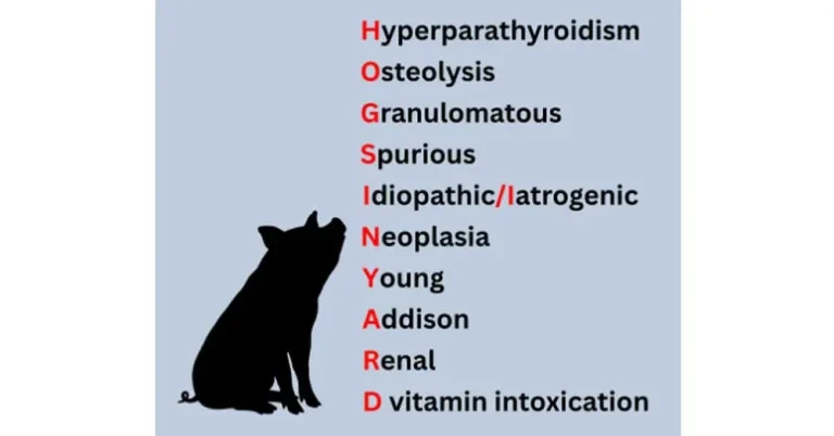
Figure 1. “Hogs in yard” – a mnemonic to remember all causes for hypercalcaemia.
Calcium participates in many metabolic functions and, consequently, hypercalcaemia can cause a wide range of clinical signs:
Therefore, you may encounter hypercalcaemia when investigating patients with these clinical problems.
Alternatively, you should consider adding total calcium and/or ionised calcium to your diagnostic work-up when a dog or cat present to you with these issues, if your standard biochemistry panel does not include them; for example, I often measure calcium in patients presenting with PU/PD or when calcium oxalate urolithiasis has been confirmed or is suspected.
Urolithiasis. Hypercalcaemia is associated with hypercalciuria, which predisposes to the formation of calcium oxalate uroliths. This occurs more commonly with primary hyperparathyroidism, as hypercalcaemia can be present for a prolonged period. In contrast, other causes of hypercalcaemia may have a more rapid clinical course, leading to earlier intervention or deterioration before there is enough time for uroliths to develop.
PU/PD. Calcium interferes with the binding of antidiuretic hormone (ADH) to its receptors in the kidney. If ADH cannot bind to these receptors, water cannot be adequately reabsorbed. From a pathophysiological perspective, hypercalcaemia, therefore, causes a secondary nephrogenic diabetes insipidus.
Lethargy/weakness. These clinical signs occur due to hyperpolarisation of cellular membranes as a result of the increased calcium concentration. This hyperpolarisation can cause a range of neurological signs.
Three main hormones are involved in calcium metabolism:
As we will discuss, calcium metabolism is closely related to phosphate metabolism.
PTH. This hormone is produced by the parathyroid glands, and its net effect is increased calcium and decreased phosphate. A low concentration of ionised calcium stimulates the parathyroid glands to produce more PTH. PTH acts on bone, the kidneys and indirectly on the intestine. At the level of the kidney, it promotes the activation of vitamin D, which increases intestinal absorption of calcium.
Vitamin D. Dogs and cats obtain vitamin D through their diet. Vitamin D becomes active in the kidney, and PTH is required for this process. The net effect of vitamin D is increased calcium and increased phosphate.
Calcitonin. Calcitonin is produced by the parafollicular cells of the thyroid gland, and its net effect is decreased calcium. It inhibits bone resorption, and its production is stimulated by a high concentration of ionised calcium.
Now that we have refreshed our knowledge of calcium physiology, it will be easier to understand and remember the different conditions that can produce hypercalcaemia.
To make things a little easier, there is also a mnemonic: hogs in yard (Figure 1).
An adenoma or adenocarcinoma can cause inappropriate overproduction of PTH. It mainly affects older dogs, and a genetic predisposition has been demonstrated in the Keeshond.
Between 20% and 50% of dogs are subclinical1. Urinary tract signs, PU/PD and weakness occur in approximately 50% of cases1.
The diagnosis is based on demonstrating an inappropriate PTH concentration in the presence of hypercalcaemia. Surgical treatment is very successful, although ethanol ablation is also an option. Long-term prognosis is good and similar regardless of the histological type.
Primary hyperparathyroidism is rare in cats2.
For example, lymphoma and multiple myeloma can cause local bone resorption through the production of cytokines and PTH-related mechanisms.
Bone metastases from solid tumours can also cause hypercalcaemia, whereas primary bone tumours such as osteosarcoma rarely cause hypercalcaemia.
Cytology or histology is usually performed to confirm the diagnosis.
Examples include angiostrongylosis, leishmaniasis and granulomatous skin disease3.
The underlying mechanism is suspected to be related to altered vitamin D metabolism.
It is very important to confirm that the hypercalcaemia is genuine – particularly when the result does not fit the clinical picture – as laboratory errors and artefacts can occur.
Idiopathic hypercalcaemia is one of the most common causes of hypercalcaemia in cats, although the underlying mechanism remains unknown.
It is a diagnosis of exclusion, and treatment is based on dietary management and/or oral bisphosphonates. These drugs inhibit osteoclast activity at the level of bone, reducing local bone resorption and the subsequent release of calcium.
Iatrogenic hypercalcaemia can occur due to excessive supplementation with intravenous calcium or vitamin D metabolites such as calcitriol.
Cancer can cause hypercalcaemia through several different mechanisms. Osteolysis is one of them. Another important mechanism is the production of PTH-related protein (PTHrP).
PTHrP has a similar net effect to PTH, resulting in increased calcium concentrations. This mechanism is known as humoral hypercalcaemia of malignancy, and T-cell lymphoma is responsible for approximately 80% of cases of hypercalcaemia associated with malignancy1.
In cats, approximately one-third of lymphoma cases have hypercalcaemia1. Imaging, cytology or histology, together with PTHrP measurement, may be necessary to reach a diagnosis.
Other tumours commonly associated with hypercalcaemia include anal sac apocrine gland adenocarcinoma, thymomas and several types of carcinoma. In cats, pulmonary carcinoma is the most common tumour associated with hypercalcaemia2.
In growing animals, increased concentrations of both calcium and phosphate are expected.
Hypoadrenocorticism is associated with hypercalcaemia in approximately one-third of cases.
It usually causes an increase in total calcium, while ionised calcium is normal in most cases. The mechanism is probably multifactorial and may include volume contraction and decreased glomerular filtration rate.
A small proportion of dogs and cats with chronic kidney disease (CKD) develop hypercalcaemia. This is often associated with parathyroid hyperplasia. The prevalence of hypercalcaemia in dogs with CKD increases with the severity of the kidney disease. The presence of hyperphosphataemia makes renal disease more likely to be the underlying cause of hypercalcaemia.
Psoriasis creams and colecalciferol rodenticides are common causes of vitamin D intoxication.
In these cases, hyperphosphataemia is also usually present.
Neoplasia and hyperparathyroidism are the most common causes of hypercalcaemia in dogs, accounting for 12.9% and 4.6% of cases, respectively4. In cats, the most common causes are neoplasia, acute kidney injury and idiopathic hypercalcaemia, accounting for 22.7%, 13.4% and 12.6% of cases, respectively4.
Although this is a useful starting point, every case is different.
Basal cortisol. if hypoadrenocorticism is a possibility.
Imaging
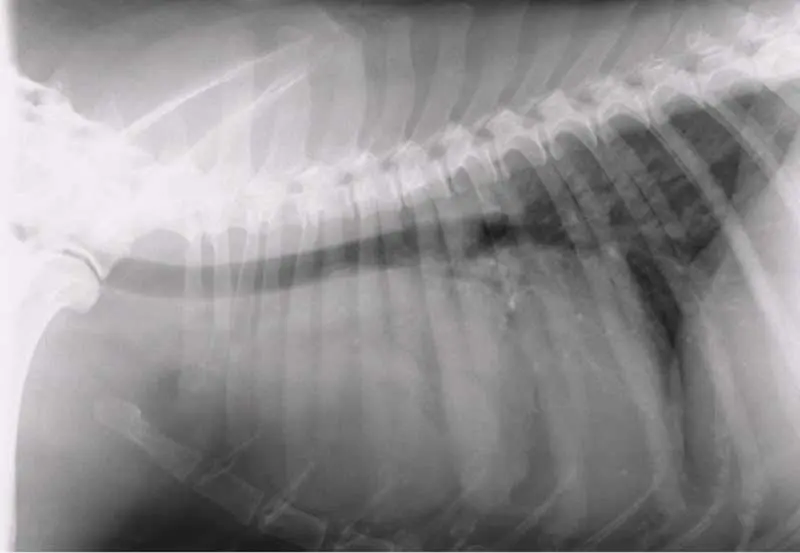
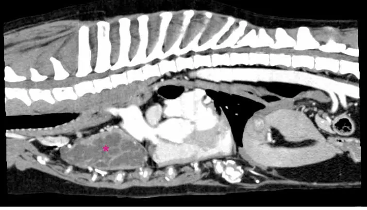
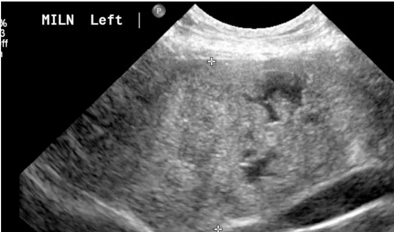
Depending on the imaging findings, fine-needle aspiration or biopsy may be indicated.
PTH/PTHrP measurement. At the same time, blood can be submitted for measurement of vitamin D metabolites, or this can be performed at a later stage if all other tests are negative and there is a possible exposure, or if further investigation is warranted.
If there is generalised peripheral lymphadenomegaly, a reasonable first step is to perform fine-needle aspirates. Depending on the results, the next diagnostic steps can then be planned and discussed with the pet parent.
Idiopathic hypercalcaemia is a diagnosis of exclusion, and it can sometimes be difficult to determine whether it is the primary cause or whether renal disease is responsible when there is concomitant azotaemia.
Fluid therapy is often sufficient to control hypercalcaemia, or at least its clinical signs. I usually choose physiological saline and determine the fluid rate after assessing the patient’s hydration status and estimating the volume of ongoing losses. Some textbooks recommend a rate of 2.5× maintenance, which may be appropriate in some cases, but this should not be applied as a blanket approach.
If there is no concomitant azotaemia or acute kidney injury, furosemide can also be considered if there is an inadequate response to fluid therapy. An intravenous infusion of bisphosphonates is another option (for example, pamidronate). This type of drug inhibits osteoclast activity and induces osteoclast apoptosis. Possible side effects are gastrointestinal signs and renal injury. Ideally, corticosteroids should not be used initially, as they can make the diagnosis of some underlying diseases, such as lymphoma, much more difficult.
In addition to the specific treatments discussed, supportive treatment should be provided as needed:
The prognosis depends on the underlying cause.
Ferran Valls Sanchez is a European diplomate in internal medicine and has worked in multiple referral hospitals in the UK, Ireland, Hong Kong and Australia. He is the founder of WeYouVets – mobile specialist company in the UK – and Thinkifyer 360 – an online free internal medicine and medical diagnostic imaging platform for education and case discussion for vets and nurses around the world: visit www.skool.com/thinkifyervetmed/about