2 Sept 2025
Iga Sieraj RVN discusses the opportunities in diagnostics open to VNs, the benefits for practices and how everyone can comply with the codes of conduct
Iga Sieraj
Job Title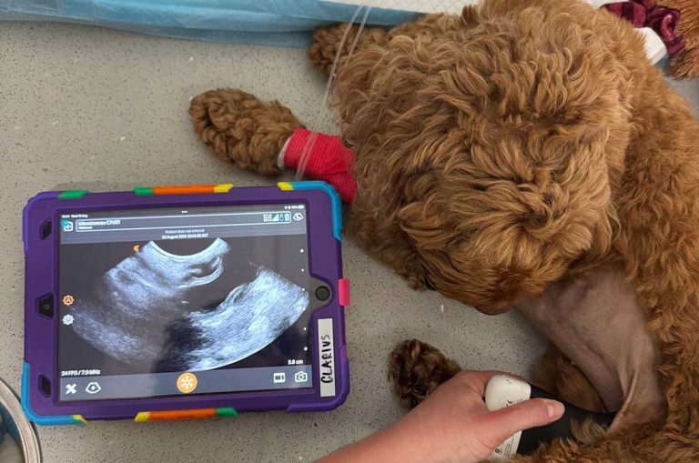
The author performing an ultrasound.
The availability of diagnostic imaging equipment within veterinary practice has come a long way, especially in the past few years, with more and more daytime clinics being able to offer further imaging investigations in-house, without the need for external referrals.
This begs the question: how do RVNs fit in? Especially with the ongoing recruitment challenges across the professions, meaning that staff members are often required to complete more tasks and assist each other in more ways than before. So how can RVNs help loosen the load by getting involved with diagnostic imaging procedures?
As per the RCVS Code of Professional Conduct for Veterinary Nurses, RVNs are able to perform certain procedures under the direction and supervision of a veterinary surgeon that do not involve entering a body cavity. The good news is neither ultrasound nor computed tomography (CT) are met with this limitation – unless image-guided biopsies or aspirates are required, which can be performed only by a veterinary surgeon (RCVS, 2025).
Therefore, it can be safely concluded that RVNs, and by extension SVNs, can perform, if competent and comfortable, ultrasound and CT scans. This can, however, be discussed further – specifically regarding ultrasonography, as RVNs and SVNs are unable to make diagnoses based on ultrasonographic findings, as per the code of conduct, and a veterinary surgeon is still required to review the images or videos taken to make a formal diagnosis and discuss the findings with owners. So, what are the benefits and advantages of involving RVNs in imaging procedures?
There are plenty of benefits to RVNs carrying out ultrasonographic or tomographic assessments, mainly including freeing up veterinary surgeons to perform other tasks that an RVN may not be able to – consultations, reporting results or surgeries – as a lot of imaging procedures can be very time-consuming.
The ability for RVNs to be involved in, or even leading, imaging procedures can be extremely morale boosting, allowing for their comprehensive set of skills to be used.
Additionally, it can improve job satisfaction, potentially leading to improved job retention and advancing the progression of the profession. The BVNA (2024) continuously urges for changes to the current legislations and the way RVNs are used in clinical practice – with, hopefully, more changes to be seen towards the end of 2025.
Just as with veterinary surgeons, nursing specialities and referral opportunities have vastly expanded in the past few years, with more and more certificates and further education being available for many nurses, diagnostic imaging being one of them. In 2023, Improve International released news that an innovative certificate for nurses in diagnostic imaging would be rolled out, with the first cohort starting in May 2024. This was groundbreaking news for all the nurses and technicians out there wanting to further their knowledge in this subject, as the only predecessor was the BSAVA Merit in Diagnostic Imaging, which a lot of the newer qualified RVNs missed out on.
The ability to showcase skills, particularly within such a specific field as imaging, can greatly enhance the way that nurses get involved in clinical discussions; helping make decisions regarding a patient’s care, streamlining a patient’s journey through a clinic or hospital and, most importantly, it can increase the confidence that nurses have in themselves. This therefore greatly increases the collaboration and teamwork between veterinary surgeons and the nursing team.
Ultrasonographic examination can seem extremely daunting at first, as all that can be seen on the screen is black and white “blobs” (or blue, or orange, depending on your machine and the clinician’s preference).
It can take a while before the brain starts registering what all those different black and white “blobs” are and what they’re supposed to look like, but no one will expect an RVN to find the adrenal glands on an overweight Labrador on the first try.
Ultrasound uses the acoustic ability of soft tissues to absorb and reflect sound of varying frequencies to produce an image. The transducer probe produces sound waves that are propelled into the body, reflected by tissues and returned towards the probe to be read and translated into a digital image (Manzi, 2025).
Each machine varies slightly, with the layout of different knobs and buttons, varying default settings and general way of operation, so it is crucial any RVN wanting to pick up a probe gets comfortable handling the machine – in some cases, it’s not as easy as placing the probe on a patient’s abdomen and getting the perfect diagnostic image. As a rule of thumb, the three settings that will be used most frequently are frequency, depth and focus – these are the settings that determine the quality of the image.
The frequency establishes the penetrative power of the ultrasound waves, but also the image resolution; the higher the frequency, the better the image quality, but less wave penetration into deeper tissues, and vice versa.
The depth adjusts the distance that the waves travel, whereas the focus shows the machine where to concentrate the waves to achieve the clearest image at that point.
As nurses, the first and foremost organ that should be confidently identified is the bladder. It’s usually always in the same place, and there is no other organ similar, making it the easiest one to get to grips with. Just being able to complete a quick “bedside” bladder check using an ultrasound scanner will provide a heap of crucial information about the patient’s well-being. Is it big? Is it small? Are there visible floaties or material of mineral origin (liths)? Is the bladder even visible? This is key in cases of known trauma or any urinary tract blockages.
Calculations can even be used to estimate the volume of urine within the bladder. Although not standardised, this can still provide useful information regarding patient status. Carrying out serial bladder checks at scheduled intervals can provide information regarding the patient’s urinary output, whether the patient has voided or whether they are producing any urine whatsoever, which is key in patients with kidney disease.
Fast assessment using sonography for trauma (FAST) can be easily utilised by RVNs wanting to gain more experience with ultrasonography; it is a standardised way to acquire images during triage evaluations (Boysen and Lisciandro, 2013). FAST scans allow for the identification of free fluid within the thoracic and abdominal cavities – remember that RVNs cannot make diagnoses based on sonographic images as per the RCVS, and any perceived abnormalities or anomalies should be reported to the vet immediately, in order for them to make a decision based on the findings.
Getting comfortable with the machine, and being able to identify even one organ, is the first step to expanding an RVNs knowledge and confidence with ultrasonography.
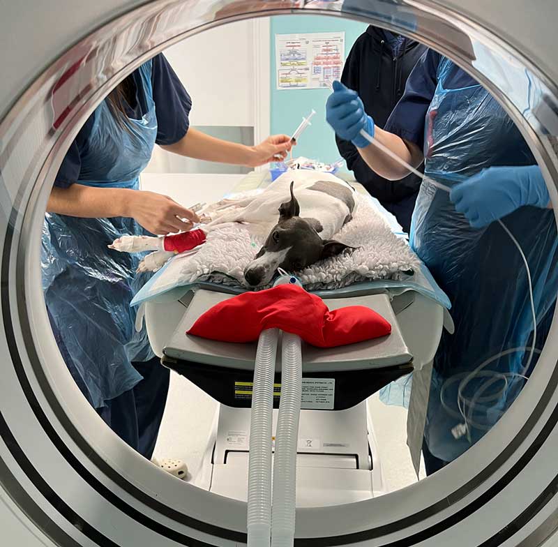
CT has become much more widely available for veterinary patients everywhere; it is a highly detailed and comprehensive imaging modality with a multitude of uses, especially for thoracic imaging, vascular studies and, in cases of trauma, oncologic disease or orthopaedics. While still not available internally for the majority of first opinion practices, it can still be important to understand how an RVN can prove useful during these procedures.
Similar to standard radiography, CT uses ionising x-ray radiation; however, instead of creating a two-dimensional image, CT takes advantage of several rows of detectors and a generator that spins in a helical motion around the patient to produce a three-dimensional image, showcasing the individual structures without any overlap.
CT scanning involves a complicated system that can be tricky to master. Many more variables, settings and considerations are involved when compared to radiography. Furthermore, the individual settings used will vary even more depending on the size of the patient and the areas of interest. Additionally, due to much higher exposure factors, radiation safety is of utmost importance when dealing with this modality. In fact, according to Harvard (2021), a thoracic CT delivers around 70 times more radiation than an x-ray.
As it has been long established that RVNs can, and should, take radiographic exposures, it can therefore also be assumed that RVNs can acquire CT images if they are competent and trained to do so, as CT image can be considered as a more intricate radiograph. Acquiring tomographic images is a form of data collection, similar to ultrasound images, blood samples and basic physical parameters like heart rate and blood pressure, which can then be reviewed by the veterinary surgeon to conclude a diagnosis.
When carrying out a CT study, the RVN must be aware of all the possible complications that can occur; for example, accidental exposures, anaesthetic complications or technical errors.
Due to the high exposure factors, no staff members are permitted inside of the controlled area while an exposure is taking place; personal dosimeters should be worn at all times when in the vicinity of controlled areas.
Any exposures to staff members need to be reported to the Health and Safety Executive as soon as possible, as their ability to work with radiation may need to be prohibited or limited depending on the level of exposure. In extreme cases, medical attention should be sought.
The majority of CT studies, specifically to assess soft tissue anatomy, will require the use of intravenous contrast; the use or disuse of a contrast media will ultimately be the clinician’s decision as this injection needs to be prescribed by the vet, and weighed out against the possible side effects that may occur with its use; anaphylaxis or acute kidney injury are the ones to look out for in patients, despite being rare.
Once the contrast has been prescribed by the veterinary surgeon, the RVN can safely administer the solution under their direction as with any other medication.
Other factors, like positioning and collimating the scanner for the designated areas of interest, can be the RVNs decision as the designated “radiographer” in the scenario, allowing for the RVN to achieve some autonomy when involved in imaging procedures, which can be highly rewarding. However, any imaging that is carried out on any patient must be clinically relevant, recommended by the clinician, and consented to by the owner, especially in cases where ionising radiation is involved – this not only complies with the code of conduct, but also with the Ionising Radiation Regulations (2017).
The use of diagnostic imaging, in any form, can be a daunting process for RVNs to get involved in, and the perceived ambiguity in the RCVS Code of Professional Conduct for Veterinary Nurses and Schedule 3 regarding the scope of an RVN’s role may create some apprehension in our colleagues. However, RVNs still have many ways to support their clinicians, either via clinical discussions or even conducting imaging procedures as part of clinical data collection.
Even though getting confident and comfortable with imaging modalities takes significant training, it can be an extremely proactive way for RVNs to use their hard-earned skills, both increasing job satisfaction and improving morale.
An ever-increasing number of imaging-based webinars and courses specifically aimed at RVNs are being announced across the country, with more and more clinics advocating for their RVNs to improve their skills in this subject; so why not jump on the train and give it a go?