28 Jan 2021
Ian Wright discusses the importance of regular deworming, routine testing, and prevention in canine and feline patients.
Ian Wright
Job Title
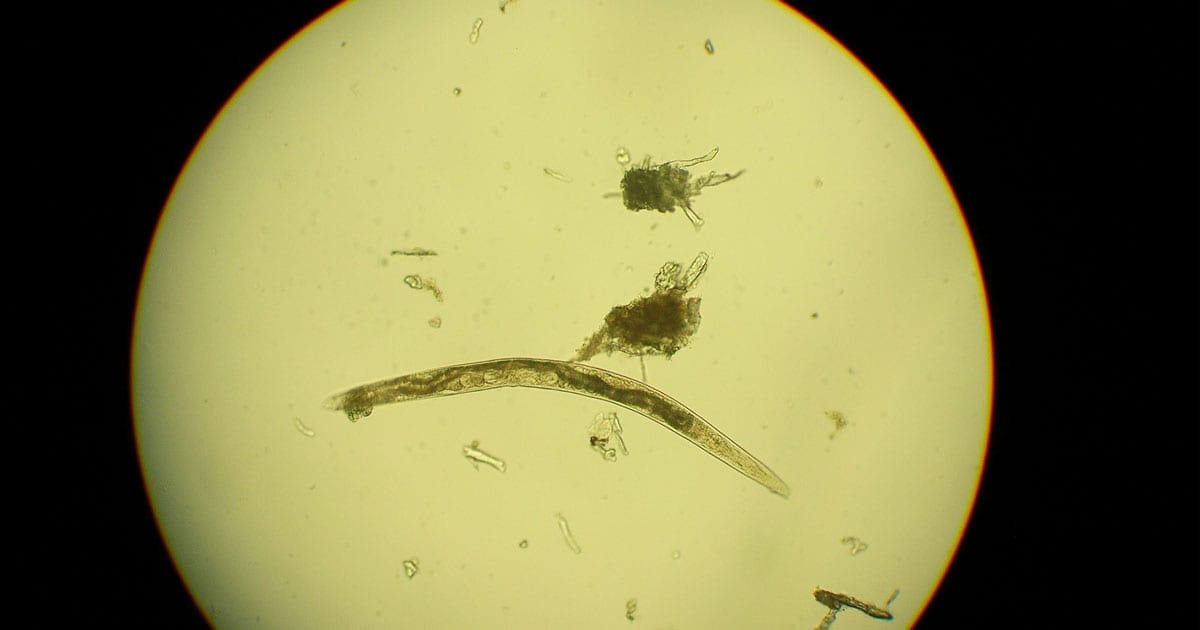
Figure 1. A free-living soil nematode.
Regular deworming of cats and dogs is a fundamental component of preventive health programmes for UK pets, and has been the focus of helminth control rather than testing. This is because testing is often viewed as unnecessary if routine preventive treatment is occurring and should, therefore, only be performed in clinical cases or as an alternative to treatment.
Routine testing alongside prevention for worm infections in cats and dogs is important to demonstrate the effectiveness of treatment plans, and gather data on parasite distributions. It also represents an opportunity to improve health plans and revenue in practice.
Regular deworming of cats and dogs is a fundamental component of preventive health programmes for UK pets.
Treating intestinal roundworms four times a year is likely to keep worm burdens low and reduce zoonotic Toxocara species ova output (Wright and Wolfe, 2007). As a result, it is the minimum treatment frequency for intestinal roundworms recommended by the European Scientific Counsel Companion Animal Parasites (ESCCAP).
Monthly treatment, however, is often required to minimise human and animal health risk. This is the case to prevent Toxocara species shedding in cats and dogs likely to have high worm burdens, or that live with people at high risk of infection. It is also required in dogs whose geographic location and lifestyle puts them at high risk of Echinococcus granulosus or Angiostrongylus vasorum infection.
A recent survey showed 97% of UK dogs and 68% of cats have a relevant risk factor that would make monthly deworming necessary (McNamara et al, 2018). Concerns regarding over treatment, the development of anthelmintic resistance and environmental contamination have led to the question of whether regular testing of pets could replace monthly preventive treatment in these cats and dogs. This approach has a number of drawbacks.
These arguments mean that for testing to replace monthly treatment in high-risk groups, it would need to be performed at least quarterly and ideally monthly to avoid prolonged human exposure to zoonotic life stages and harm to pets through prolonged exposure to pathogenic nematodes such as A vasorum.
Testing alongside routine treatment, however, still has a number of benefits, which are often overlooked.
A recent large-scale survey in the UK showed that cat and dog owners only gave their pets routine treatment for worms 2.5 times a year (McNamara et al, 2018). This is well below ESCCAP recommendations and a concern, given the large percentage of pets falling into high-risk categories requiring monthly worming.
Routine diagnostic demonstrates value in routine treatment for the client and improves compliance. Regular negative tests in pets on routine preventive treatments shows good efficacy of the treatments being used and compliance on the part of the owner. This results in positive reinforcement, and builds confidence in both the current recommendations and the owner’s administration of the product.
Positive results in pets on preventive regimes demonstrates a need to investigate compliance on the part of the owner and potential causes of treatment failure (vomiting after tablet application, tablets not being eaten in food, spot-on applications being washed off, inadequate treatment frequency, drug resistance and so on).
Parasites diagnosed in untreated pets demonstrates a need for treatment if the life stages found are zoonotic (for example, Toxocara species, E granulosus) or highly pathogenic (for example, A vasorum).
Some evidence exists to suggest a minority of dogs may be responsible for a disproportionately high volume of Toxocara egg shedding during their lifetime. This is likely to be a combination of immunological and lifestyle factors.
If pets can be identified through routine testing as high or low egg shedders over the course of months and years, this does allow a more selective deworming regime to be adopted.
Whether routine deworming is required in dogs for A vasorum lungworm and E granulosus tapeworm depends on both geographic and lifestyle factors.
A vasorum has spread rapidly over the past 20 years from endemic foci in Wales, and the south-west and south-east of England, across the whole of the UK. Increased reporting of cases has been seen in domestic dogs, with 20% of practices across the country having seen at least one case over a 12-month period (Kirk et al, 2014).
Postmortem surveys carried out on foxes, which act as wildlife reservoirs, in 2005 (Morgan et al, 2008) and in 2014 (Taylor et al, 2015) showed an overall rise in prevalence and extension to regions previously clear of infection, such as northern England and Scotland. The spread of A vasorum is therefore genuine, but not uniform, with focal areas of very high prevalence and other areas remaining free of infection.
Case reporting sites by IDEXX and Bayer Animal Health (now Elanco) have been useful in mapping areas of high prevalence and the introduction of A vasorum into new areas. Reporting of cases is only voluntary, however, and distribution of infection very fluid with new foci forming.
In areas known to be endemic foci, monthly preventive treatment should be seen as routine – especially prior to surgery. In areas where endemic status is less certain then testing of dogs prior to surgery, those with relevant clinical signs, and routine testing at wellness checks will rapidly build up a picture of whether A vasorum is endemic in an area and whether routine preventive treatment for dogs is required.
Similarly, for E granulosus, routine prevention in its known endemic areas of Herefordshire, mid-Wales and the Western Isles of Scotland is essential. Surveys of abattoirs and hunting dogs in Britain, however, suggest endemic foci exist in other parts of the country.
The location of these is currently uncertain and, therefore, outside of these known endemic areas, lifestyle factors (access to raw offal or livestock carcasses) alone are currently recommended to inform the need for preventive treatment.
The increasing availability of faecal PCR testing alongside the ongoing development of faecal antigen tests means that, in the future, routine testing will help to indicate if the parasite is present in an area and whether dogs require preventive treatment.
Routine testing for endoparasites is, therefore, of benefit to practices in terms of reinforcing good practice among pet owners, identifying which parasites are present in a local area, and generating revenue through the testing itself and subsequent treatment. It can be performed at annual wellness checks, six-monthly or quarterly heath checks, and, in the case of A vasorum, before surgery.
A variety of diagnostic tests for canine and feline helminths are available, which can be run in practice or sent to external labs.
Many parasites intermittently pass ova, cysts or larvae in the faeces, which can then be detected by faecal analysis. Shedding of larvae and ova is often intermittent, so ideally, samples should be collected over three consecutive days for increased diagnostic sensitivity.
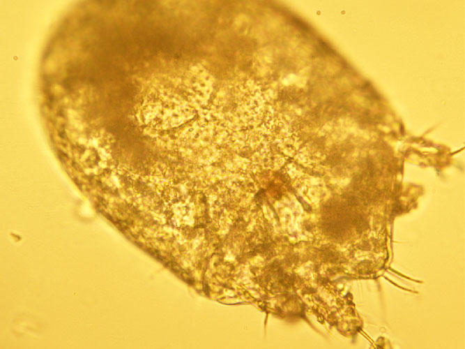
If the sample is collected by the client, it must be picked up as soon as it is passed. If left on the ground, faeces rapidly become contaminated with free-living nematodes (Figure 1) and mites (Figure 2). These may be confused with parasitic life stages and will also obscure the field of view during microscopic examination if present in large numbers.
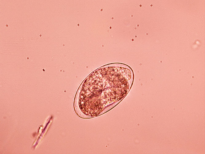
Once collected, samples should either be examined immediately, or stored at 4°C to prevent hatching of ova or larval development. Hookworm eggs will rapidly larvate (Figure 3) and hatch if faeces are left at room temperature.
Faeces can be examined for parasite ova and larvae by the following methods.
Direct smear is a very simple technique and can easily be performed in practice. Direct smears only analyse very small volumes of faeces, and, as a result, are considered too insensitive for the detection of helminth ova, which are passed in relatively few numbers.
Lungworm larvae such as Crenosoma vulpis and A vasorum may be detected, and is useful as an initial screen for these parasites, but the low sensitivity (54% to 61%) for the detection of lungworm by this method means it should not be relied on as a sole test if negative (Humm and Adamantos, 2010).
A faecal sample approximately 1mm3 (the size of the head of a match) is mixed with a drop of water on a microscope slide and examined with a cover slip under the microscope. The addition of a drop of lugol’s iodine will aid in the detection of Giardia cysts, which will be stained yellow.
Faecal flotation allows much larger volumes of faeces to be examined by concentrating ova present in the faeces in small volumes of liquid while eliminating debris.
This technique may still have diagnostic sensitivities as low as 60% for some roundworm ova such as Toxocara species and has poor sensitivity for tapeworm egg detection (Wolfe et al, 2001). Sensitivity increases with pooled samples over three days.
It is important to know whether dogs are coprophagic prior to testing as this can lead to false positive results. Strongyle eggs in ruminant and horse faeces will pass through the digestive tract of cats and dogs unchanged, giving the impression that the pet is infected with hookworm. Similarly, Toxocara cati eggs may be found in dogs that have eaten cat faeces.
Many different flotation methodologies are described in the literature but an example is described here.
The flotation solution can either be sugar solution or zinc sulphate. Zinc sulphate is less sticky and easier to handle, but slides tend to dry out and crystallise very quickly. Distortion of ova can also occur, making identification more difficult. Centrifugation of samples increases test sensitivity.
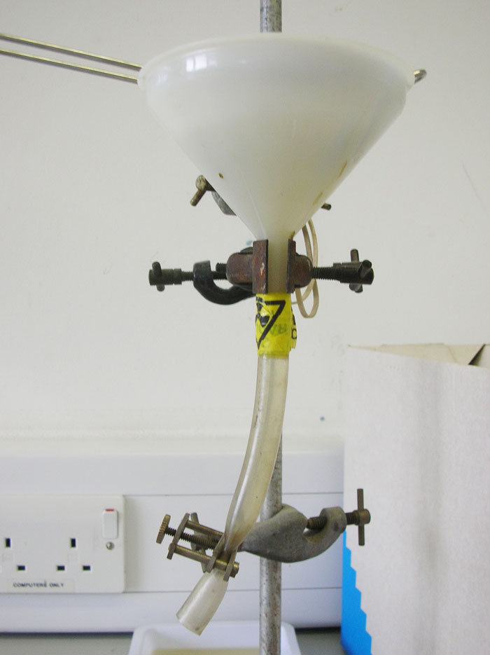
The Baermann technique is used for the detection of larvae in faeces. One of the advantages of the Baermann technique is that it requires inexpensive equipment – much of which can be reused for further tests (Figure 4).
As well as A vasorum, the larvae of other lungworms such as Oslerus osleri and C vulpis may also be detected using this method.
Antigens of some parasites can be detected in faeces, aiding in their diagnosis. Patient side testing for Giardia faecal antigens is a highly sensitive and specific test, as are recently commercially launched test kits for intestinal roundworms, whipworms and hookworms.
These tests allow infections to be detected when ova shedding is not occurring and avoids false positive results due to coprophagia. They give no indication, however, to what extent ova shedding is occurring.
Coproantigen testing is also being developed for commercial Echinococcus species testing, and PCR testing of faeces is now commercially available.
Testing for parasite antigens in blood is useful for A vasorum and Dirofilaria immitis heartworm detection. While Baermann apparatus remain the gold standard for A vasorum infection, the patient side blood test Angio Detect has revolutionised A vasorum testing.
Only a small volume of blood is required, and results are rapidly obtained at relatively little cost. This has made the screening of large numbers of suspected cases of angiostrongylosis more achievable in first opinion practice.
It is highly specific, but less sensitive than a Baermann test carried on pooled faecal samples collected over three days. If A vasorum infection is suspected and the Angio Detect test is negative then the Baermann technique is warranted.
Routine testing for D immitis is not currently required in the UK as it is not endemic, but is essential in dogs imported from endemic countries. The test should be performed after the dog has been in the country for six months and on arrival if the dog is at least six months old.
Traditionally in the UK, routine treatment of cats and dogs for worms has been the focus of worm prevention in cats and dogs. As a result, a prevailing feeling often exists that routine testing is made redundant by these strategies and that testing should only be used to replace them.
Treatment and testing, however, are not mutually exclusive, and routine testing supports and emphasises the need for the treatments employed to control helminth infections. Routine testing also produces data for individual practices, local regions and nationally to help inform geographic risk when deciding which parasite control regimes are necessary.