4 Nov 2019
Kate Parkinson discusses common parasitic worms, and why anthelmintic use and client education are vital in lowering the risk of infection.
Kate Parkinson
Job Title
Virtually all companion animals have, or are at risk of, parasitic infections.
Many of these are zoonoses. Common zoonotic worm species in the UK include roundworms, tapeworms (for example, Dipylidium species and Taenia species) and lungworms (Angiostrongylus vasorum); although exotic parasites – such as Echinococcus tapeworms, heartworms, eyeworms, whipworms and hookworms – are being seen increasingly in the UK.
Many parasitic worms have complicated life cycles and may cause serious disease in the intermediate or primary host. Zoonotic disease is an important public health concern, with infection being especially common in children.
Although it is impossible to eliminate worm eggs from the environment, treatment of the infection in primary hosts and good hygiene reduces transmission and zoonotic risk. Many effective and cheap anthelmintics are available, which should be used according to European Scientific Counsel Companion Animal Parasites guidelines.
Veterinarians are ideally placed to educate clients about the risks of zoonotic infection.
Cats and dogs have lived with humans for thousands of years – and parasites have been with us even longer1.
In the modern world, pets are family members and share many human activities. This close relationship has undeniable benefits – including improved quality of life, increased empathy and reduced physician visits1.
However, keeping pets comes at a price – an increased risk of zoonotic disease.
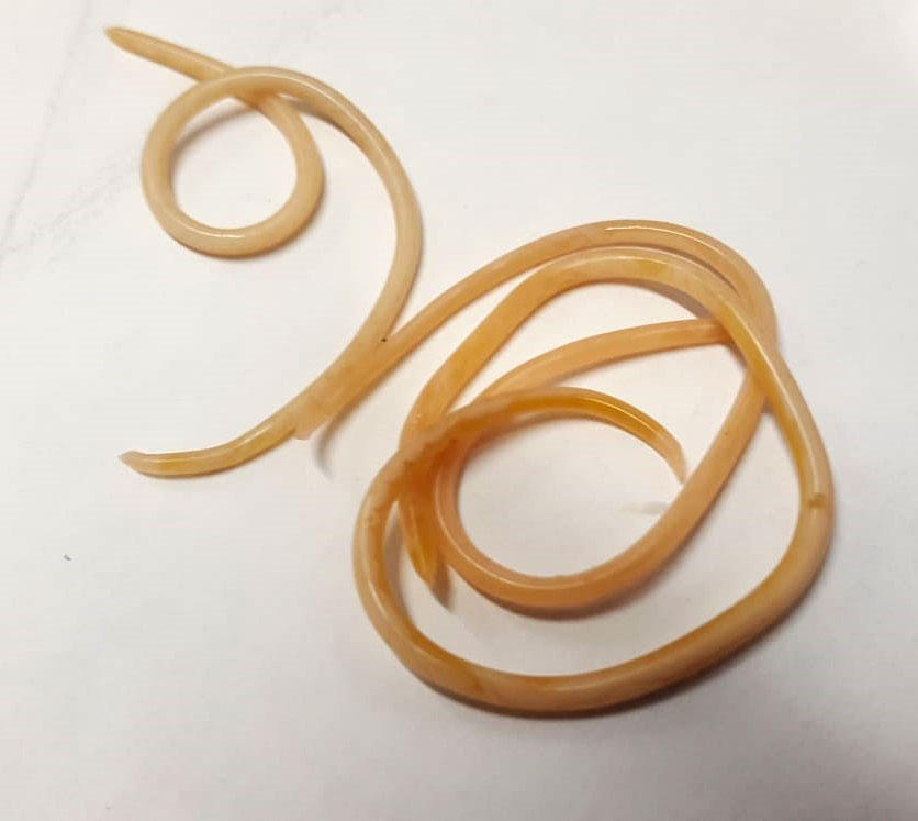
Zoonotic intestinal helminths have complicated, elegant life cycles. Here, the author describes common species in the UK – including ascarids (roundworms; Figure 1), tapeworms and lungworms – and exotic species.
Toxocara canis infection occurs when a dog ingests a roundworm egg or eats a paratenic host (mouse or rat) containing encysted larvae.
In adults, the larvae hatch in the intestine, mature and reproduce, laying eggs that are excreted in the faeces. Other larvae penetrate the dog’s tissues and encyst there.
In bitches, larvae also migrate to the mammary glands. Once the animal becomes pregnant, encysted larvae mobilise and infect puppies in utero. Due to this prenatal transmission, T canis eggs may be present in puppies of only two to three weeks of age5. Larvae are also transmitted in the milk.
The eggs hatch in the puppies’ intestine and larvae migrate through the liver to the lungs6 – where they develop and are coughed up and swallowed, re-entering the intestines.
Infection rates in dogs vary from 3.5% to 34%, depending on the study7.
Toxocara cati infects cats in a similar way, although cats often ingest eggs from the environment by grooming.
Toxocara leonine affects both dogs and cats. Infection is only possible by ingesting larvae in the environment or via paratenic hosts.
Although between 49% and 87% of urban foxes are infected with T canis, the contribution of urban foxes to ascarid infection in humans is estimated to be less than a tenth of the contribution of dogs8.
Mild ascarid infections are often asymptomatic. In young animals with high levels of infection, larval migration to the lungs may cause coughing, nasal discharge, pneumonia and pleural oedema2 that may prove fatal.
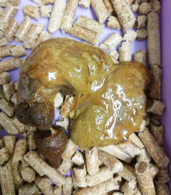
Adult worms in the intestines produce gastrointestinal symptoms, such as enteritis, vomiting, diarrhoea, ascites, pot belly, poor coat, poor body condition and failure to thrive5 (Figure 2).
Dipylidium caninum is transmitted to dogs and cats via ingestion of infected fleas or lice9.
The egg hatches in the intestine and the tapeworm attaches to the gut lining, producing segments called proglottids.
Older proglottids progress to the end of the tapeworm and are passed in faeces. The segments open and the eggs are consumed by flea larvae in the environment10.
Taenia tapeworms are common and have similar life cycles to Echinococcus and Dipylidium species.
Animals are infected by consuming infected intermediate hosts – such as cattle, sheep, rodents, pigs and goats, depending on tapeworm species. The clinical effects are often more severe in intermediate hosts than final hosts6.
Taenia taeniaeformis commonly infects cats that consume rodents6.
Echinococcus granulosus and Echinococcus multilocularis are found in parts of Europe.
Infection occurs when an intermediate host (rodent, cattle, horse, swine or human) ingests an oncosphere. When the oncosphere hatches, the larvae penetrate the host’s intestines and migrate through the body, forming a fluid-filled cyst.
Once the intermediate host has been ingested by a primary host, the larvae in the cyst develop into tapeworms in the primary host’s gut, where they pass millions of immediately infective eggs11 into the environment.
Angiostrongylus vasorum is a common parasite of foxes and dogs12.
Larvae develop in intermediate hosts, such as slugs and snails. Once ingested by the primary host, larvae develop as they migrate through pulmonary tissue – maturing to adult worms in the pulmonary artery13 and right side of the heart6, where they mate and lay eggs. Larvae penetrate the alveoli, where they are coughed up and swallowed.
The fox lungworm, Crenosoma vulpis, has a similar life cycle.
Both A vasorum and C vulpis may cause coughing, retching, nasal discharge and dyspnoea. Infection may mimic allergic respiratory disease16. A vasorum may also cause clotting deficits and haemorrhage.
Rapid environmental change and increased international travel have increased the likelihood of parasites or vectors colonising new areas17. As a result, veterinarians practising in the UK are increasingly likely to encounter exotic parasite species.
Culicidae mosquitoes – the heartworm vector – are not present in the UK, but common in south-eastern Europe and the southern US states.
Cats and dogs may be infected. Infection causes coughing, weakness and dyspnoea, progressing to right-sided heart failure and death.
Although several treatments are available, worm death – and the subsequent inflammation and pulmonary thromboembolism – may be fatal, making the parasite challenging to treat18.
In cats, the severe inflammatory response causes an obstructive pulmonary condition known as heartworm-associated respiratory disease19.
Thelazia callipaeda is enzootic in areas of Europe, although rare cases are seen in the UK.
Its vector, the fruit fly Phortica variegata, is present in southern England. Thelazia is found in the lacrimal duct, conjunctival sac and beneath the nictitating membrane20.
Trichuris vulpis has a direct life cycle. Ingested eggs hatch in the intestine of animals6 and are excreted in faeces.
Adult worms inhabit the colonic mucosa and may cause haemorrhagic diarrhoea23.
Hookworm is found in European dogs and cats. Adult worms inhabit the small intestine, and cause symptoms ranging from enteritis to haemorrhagic diarrhoea and fatal anaemia.
Eggs are passed in the faeces and develop to third-stage larvae in the environment, where they are ingested.
Larvae in soil may penetrate skin and cause skin damage, often on the feet6.
Parasitic worms infest animals of all ages, but infection is most common in puppies, kittens, and animals younger than six months of age7.
Infection is more likely to be serious in younger or immunocompromised animals.
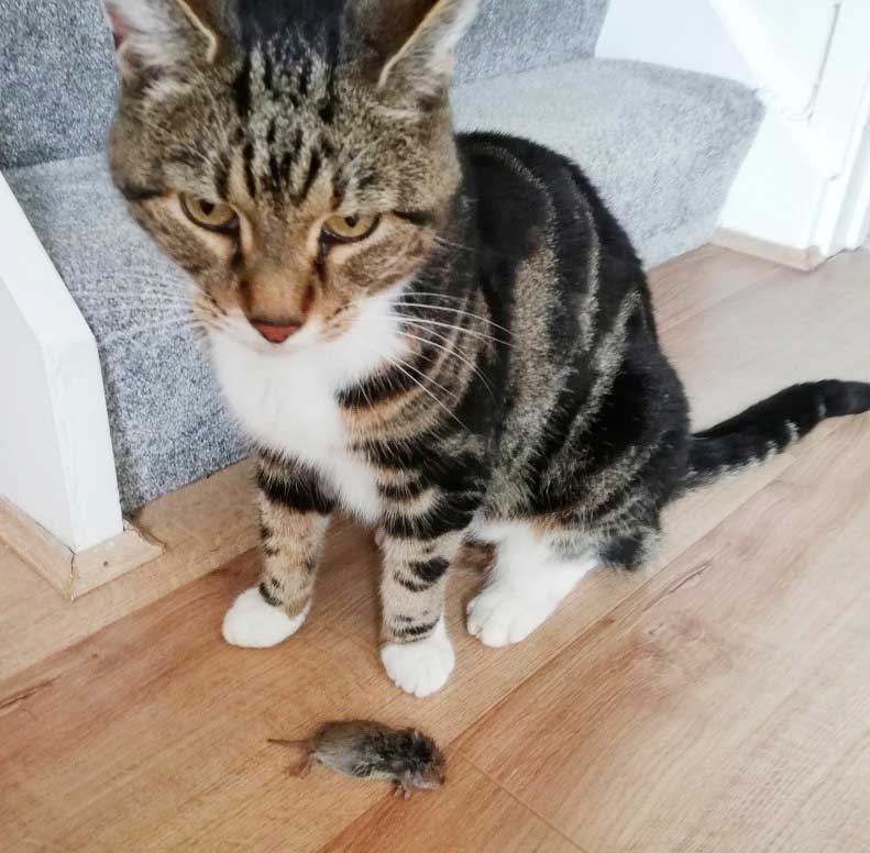
Free-roaming animals with uninhibited access to prey or offal are at increased risk of infection (Figure 3).
Toxocara infections in humans are relatively common, and vary from between 3% and 19% in various European countries5.
Owners may be exposed to ascarid eggs on pets’ fur, though infection via this method is uncommon7. When eggs hatch in the intestines, the larvae penetrate the intestinal wall and migrate through the body without ever maturing.
Most infections are asymptomatic5, but larvae may encyst in tissues and cause local damage. This condition is called visceral larval migrans and is often seen in children, where it causes fever, malaise and pain, as well as locally specific inflammation such as hepatitis, bronchiolitis, pneumonia or meningoencephalitis25.
Ocular larval migrans occurs when larvae migrate to the eye, causing inflammation that may result in glaucoma or blindness. A mass found in this area may mimic retinoblastoma and lead to unnecessary enucleation5.
E granulosus causes hydatid cyst disease in humans and E multilocularis causes alveolar cyst disease, which has 94% mortality within 10 years11, while Taenia tapeworms are hugely important in human and public health.
Dipylidium infection most commonly occurs in children and is usually asymptomatic10.
It is impossible to eliminate worm eggs from the environment. Control is focused on the prevention of contamination by eliminating the infection in hosts.
In dogs and cats, feeding commercial diets or cooked food prevents infections transmitted by raw meat. If possible, pets should not be permitted access to rodents, carcases, placentae or aborted fetuses. Clean water should always be available6.
In humans, parasite transmission may be reduced by hand-washing, disposing of faeces and closely supervising children, who are especially vulnerable to infection. People with weakened immune systems should be especially careful of contact with animals8. Proper food handling procedures reduce the risk of transmission from contaminated food.
A Canadian study28 found 13% of veterinarians recommended deworming protocols consistent with established guidelines for puppies – and 39% for kittens – while 44% of veterinarians discussed the zoonotic risk of endoparasites with owners.
The study concluded increased education for veterinarians on this subject was warranted; however, an English study found veterinarians were the main party from which worming information was sought29.
Veterinarians should provide concise and objective information about the risk of zoonotic infection – via discussions, flyers, workshops and social networks – while veterinary nurses are ideally placed to promote good worming practices to clients30.
Guidelines for the worming of adult cats and dogs, puppies and kittens, professional dogs, and pets sharing homes with young children or immunocompromised individuals are detailed in Panel 1.
Repeated applications of anthelmintics are recommended for puppies (Figure 4) and kittens – and their mothers – in lactation and early life.
Puppies: Milk transmission of larvae continues to at least five weeks postpartum7. Puppies should be dewormed at two weeks, four weeks, six weeks and eight weeks, then monthly until six months of age.
Lactating bitches: Should be treated concurrently with puppies.
Pregnant bitches: To prevent transmission to puppies, pregnant bitches may be dosed with macrocyclic lactones on the 40th and 55th days of pregnancy, or dosed continuously with fenbendazole from the 40th day of pregnancy until the 14th day postpartum6. However, it is impossible to treat encysted larvae in tissue; therefore, it is not advisable to treat pregnant animals to reduce perinatal transmission.

Kittens: Kittens are not infected prenatally. Kitten worming may start at three weeks of age6.
Lactating queens: Should be treated concurrently with kittens.
Older animals should be treated routinely, as per the European Scientific Counsel Companion Animal Parasites guidelines6 or according to the results of regular faecal testing.
Despite public perception, neither annual nor biannual treatments prevent patent infection within a population – a general recommendation of four times a year should be used for adult animals.
These guidelines should be viewed as a flexible template, allowing veterinarians to create individualised treatment and control strategies for each pet.
In professional dogs – such as police or guide dogs – or pets that share homes with young children or immunocompromised individuals, where excretion of worm eggs is to be minimised, pets should be treated monthly.
Lungworm: Monthly treatment with macrocyclic lactones is recommended for dogs at high risk of lungworm infection6.
Dipylidium: Regular ectoparasiticide use prevents Dipylidium transmission.
Treatment and control of exotic worm infestations should be researched on an individual basis.
No proven evidence exists for resistant populations of gastrointestinal worms in pets, except for Dirofilaria immitis in the US6. Resistance may become a future problem, especially if worming treatments are used blindly.
Faecal sampling, which is relatively easy and cheap in a practice accustomed to the procedure, may be used to target anthelmintic use more effectively.
In countries where routine treatments are not acceptable or possible, regular faecal examinations may be performed instead6.
Despite the popularity of herbal anthelmintics such as garlic, no evidence exists to support the use of garlic as a method of preventing intestinal worm burdens9.
As pets become increasingly common worldwide and temperatures increase due to global warming, global worming will become increasingly important in the lives of humans and their animals.
Veterinarians may be particularly susceptible to zoonoses, with 4% of veterinarians in one survey reporting they have acquired a zoonotic disease33.
As zoonotic helminth exposure is virtually inevitable, veterinarians should promote the safe and effective use of anthelmintics to prevent themselves, their staff and clients from infection34.
Zoonoses, although rare, may prove fatal – and although parasitic infection may sometimes be perceived as a historical concern without much relevance to modern life, infection has serious public health implications for our lives and those of others.
The author would like to thank Alison Lumley, HRVN, NCertA&CC, and Tom Southgate, BVetMed, MRCVS, for reviewing the article and photography; Jo at Bayer Animal Health; and Karen Partridge for photography.